Thank you Charlotte for always supporting me and giving me all the help in the lab, for always being available for all my questions and sharing your expertise. Thanks to Manfred Lehner and the entire research team at CCRI for all the discussions and insights you shared with me.
Yeast surface display
With a library size of up to 109 clones, YSD libraries are typically smaller compared to other methods such as phage display where up to 1014 variants can be screened [10]. Finally, important kinetic values such as dissociation rate constants (kof f) or binding affinity can be determined by FACS measurement.

Therapeutic binder’s requirements beyond affinity: The concept of ‘developability’concept of ‘developability’
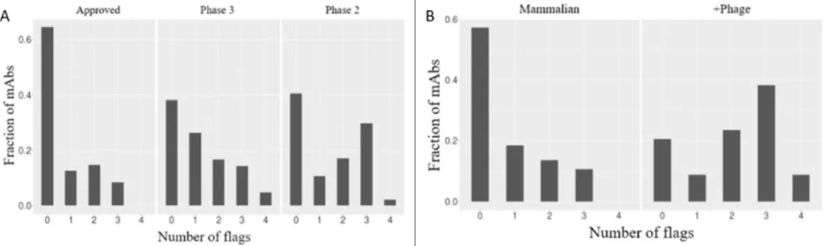
To their surprise, approved drugs consistently received fewer flags compared to earlier-stage antibodies (Figure 1.2A). In addition, antibodies engineered through phage display showed a significantly higher number of flags compared to antibodies derived from mammalian sources, indicating poorer biophysical characteristics (Figure 1.2B) [23].
Major factors that drive non-specific binding
In particular, the positively charged amino acid arginine has been repeatedly reported as a major contributor to non-specific interactions. Unlike positive charge, negatively charged amino acids have been mostly associated with specific antigen recognition in the past.
Magnetic bead based selection as an efficient alterna- tive to FACS for capturing weak protein-protein inter-tive to FACS for capturing weak protein-protein inter-
Recently, a study with synthetic yeast-displayed scFv libraries revealed some important insights regarding common features and motifs that can drive non-specific binding. A modified version of the PSR assay that will be discussed in more detail below (see chapter 5) was used to deliberately enrich non-specific binders in the constructed library. The branched hydrophobic amino acid was found to be significantly enriched in the engineered library, whereby two or three consecutive Val as a motif appeared to have a significant impact on non-specific interactions.
However, two consecutive tyrosines, for example, were also identified as a non-specific interaction-promoting motif by investigators [26]. Evaluation and tuning of non-specific binding behavior is usually only handled after binding discovery, so it is limited to a few prime candidates and can be expensive. Within this project we aimed to create an assay based on bead selection that could help reduce such non-specific interactions in protein libraries.
We hypothesized that proteins exhibiting characteristics proposed in the literature as the main factors contributing to nonspecific binding, namely hydrophobicity and positive charge, immobilized on the surface of the magnetic beads would be able to elicit such interactions and thereby capture and successfully deplete the non - specifically interacting versions from a differentiated library. Consequently, we first wanted to express and characterize a set of proteins that showed non-specific binding properties. Thus, this bead-based assay would represent a cost-effective new tool for reducing the non-specific binding content of a protein library that would be easily implemented in the development process of therapeutic binders.

Materials
PCR and DNA Purification Kit Monarch New England Biolabs Monarch DNA Gel Extraction Kit Monarch New England Biolabs Plasmid Mini Prep Kit Monarch New England Biolabs NucleoBond Xtra Midi/Maxi Kit Macherey Nagel Frozen-EZ Yeast Transformation II Kit Zymo Research EZ-Link Sulfo-NHS-LC- Thermo Scientific LC-Biotin Kit. Wash Buffer 1 5 mM imidazole His-purification TALON Equilibration Buffer – Wash Buffer 2 15 mM imidazole His-purification TALON Buffer Equilibration – Elution buffer 250 mM imidazole. Notl-HF Restriction Enzyme New England Biolabs Restriction Enzyme BamHl-HF Restriction Enzyme New England Biolabs HindIII Restriction Enzyme New England Biolabs Q5 Hot Start High Fidelity DNA Polymerase New England Biolabs NEBuilder HiFi Assembly DNA Master Mix New England Biolabs.
Methods
SDS PAGE
The soluble expression of the proteins to be coupled to the magnetic streptavidin-coated Dynabeads (Thermo Fisher Scientific) was performed in E. Extinction coefficients and molecular weights of the proteins used for the measurements were determined with the online tool ProtParam and are listed in the following Table 3.17. The elution of the sample was performed by applying a linear gradient to the HIS column, where the concentration of elution buffer containing 500 mM imidazole was gradually increased.
Biotinylation of proteins coupled to streptavidin-coated magnetic Dynabeads (Thermo Fisher Scientific) was performed using the EZ-Link Sulfo- biotinylation kit. The column was equilibrated by the application of 5 x 4 mL of PBS followed by the application of the sample. The concentration of biotinylated proteins was determined with a DeNovix spectrophotometer and the proteins were stored at −80◦C.
HA staining was performed by adding a final concentration of 2 μg/mL of anti HA antibody (Alexa Fluor 488 clone 16B12) from BioLegend. Staining of the c-myc tag was performed with a primary staining step using a mouse anti-c-myc antibody (clone 9E10), followed by secondary staining with 1 μg/mL of a goat anti-mouse IgG antibody (conjugated to Alexa Fluor 488). After incubation, a washing step with PBSA was performed to remove any free antigen followed by a resuspension of the beads in 10 µL PBSA.

Selection of the proteins associated to the streptavidin beads
Enriching the two aforementioned YSD libraries for K-Ras-G12D binding yielded a second Sso7d-based protein of our R1 data set (originally named R11.1) [19]. Analytical data from the SEC group show that the protein was partially in an oligomeric state (80% monomer). The aim of the study was to define a practical guide for future antibody drug candidates regarding drug-like behavior, which has recently gained attention as an additional critical feature for the clinical success of therapeutic binders in addition to antigen recognition [23].
The dataset analyzed by Adimab included a sirukumab-based IgG1 scFv that performed poorly in all of the biophysical property assays testing for specificity and aggregation behavior. Furthermore, its increased size (27 kDa vs. 7/10 kDa for Sso7d and FN3-based binders, respectively) compared to the other proteins tested on the streptavidin beads would hint at potential effects of protein size on the performance of our assay. All proteins tested on the streptavidin beads were co-expressed with an N-terminal fusion partner to increase expression levels and stability.
As mentioned above, in the case of the Sso7d & FN3-based proteins, this was the SUMO protein, while the sirukumab-based scFv was expressed in fusion with MBP. Then the selection test was tested with each combination of binders on the yeast cells and protein on the beads. Like the sirukumab-based scFv, this decision was based on the Adimab study mentioned above [23].
![Figure 4.1: Exemplary scaffold structures of expressed proteins. Interacting beta strands of (A) rcSso7d (K-Ras specific mutant shown, PDB download: 5UFQ [19]) and loops of the (B) FN3 (wild-type FN3 shown, PDB download: 1TTG [40]) & the (C) scFv scaff](https://thumb-eu.123doks.com/thumbv2/pubdocorg/274270.42838/39.892.151.746.393.596/exemplary-scaffold-structures-expressed-proteins-interacting-specific-download.webp)
Biophysical characterization of solubly expressed pro- teinsteins
SDS-PAGE
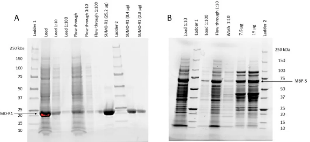
Regarding the expression and purification of scFv, we encountered some problems. Accordingly, consider that the amount of purified protein listed for MBP-S in Table 4.2 corresponds to the fractions shown on SDS-PAGE above (Figure 4.3) rather than the pure fusion protein alone. In addition, protein binders that interact non-specifically with the column material are expected to show delayed elution, indicating sticky characteristics of the binders.
Through the measured MALS data, we were able to assign the eluted peaks to the aggregation states of our proteins. In addition to the determination with the MALS detector, we also determined the molecular weights using a protein standard. Using the retention times of the standard, a trendline was constructed (see Figure 4.6 B) that allowed us to calculate the molecular weights of our soluble-expressed proteins in addition to the masses determined by MALS (included in Table 4.4).
The MW determined by MALS measurement as well as the molecular weight calculated through the standard corresponds to the protein in monomeric form. Since its MALS-determined masses came with such a high uncertainty, we decided to calculate the molecular weights also using the protein standard. However, the MALS data and the masses calculated with the standard suggest that peak 2 could correspond to the fusion protein in a monomeric form.
Evaluation of biotinylation
Due to the fact that R1 could not be eluted from the column when measured by SEC-HPLC in previous projects (orally communicated [19]), the biotinylation of SUMO-R1 was not evaluated. B - SEC-HPLC runs of SUMO-L biotinylated (5 µg) on the left, followed by streptavidin (28 µg) and SUMO-E5 biotinylated (5 µg) + streptavidin (28 µg).
Definition of a suitable displayer to non-displayer ratio
Obviously, the 1:3 ratio of displays to non-displays would not represent very well the ratio of non-specific binders present in a library since their frequency in a library is very low. On the other hand, the 1:30 promoters in the no promoter condition appeared to be quite close to the detection limit, where possible effects caused by the bead selection assay might be less obvious and more difficult to measure. Thus, we concluded from this experiment that a ratio of 10% would best suit our purposes and performed all bead selection analyzes discussed in the next section with that proportion of prominent displays to nondisplays .
Above, a geometric series up to a dilution of 1:300 of showrs in a non-shower population is shown.
Bead selection assay
Then the non-specific binding protein could be separated from the rest of the library along with the beads using a magnet. SEC-MALS measurements of the sticky fusion proteins showed that the binding agents show non-specific binding and aggregation. Therefore, we attempted to link proteins with such characteristics to the surface of the streptavidin dynabeads.
Since we could not rule out that this rigidity of the paratope would inhibit the functionality of the Sso7d-based binders in the bead selection assay, we also examined the performance of SUMO-L and MBP-S on the beads. However, we could not observe the desired effects in the bead selection assay for any of the interaction conditions. The slightly increased size of the scFv compared to the other binding scaffolds also did not seem to favor the selection of non-specific binders.
Both methods potentially use sticky proteins to trap non-specific binders in diversified YSD libraries. In the case of the PSR, these are mammalian proteins extracted from the soluble membrane fraction. In addition, a significant drawback of the FACS-based selection of non-specific binders used in the PSR assay is the need for multiple washing steps prior to detection.
For the bead selection assay, no washing steps are required between incubation and bead separation. In theory, the most important advantage of the bead-selection-based approach compared to a FACS-based screening is the inherent characteristics of the method.

![Figure 1.3: Jain et al. performed 12 biophysical property assays with 137 therapeutic antibodies [23]](https://thumb-eu.123doks.com/thumbv2/pubdocorg/274270.42838/17.892.191.704.153.370/figure-jain-performed-biophysical-property-assays-therapeutic-antibodies.webp)