In einem nächsten Schritt wurde der Einfluss der Expressionstemperatur auf die intrazelluläre B1-scFv-Ausbeute und -Löslichkeit in beiden Stämmen untersucht. Darüber hinaus wurde der Einfluss des Linkerpeptids auf die intrazelluläre Ausbeute und Löslichkeit von B1 scFv in E untersucht. In einem nächsten Schritt wurde der Einfluss der Expressionstemperatur auf die intrazelluläre Ausbeute und Löslichkeit von B1 scFv in beiden Stämmen untersucht.
Aim of the Study
Introduction
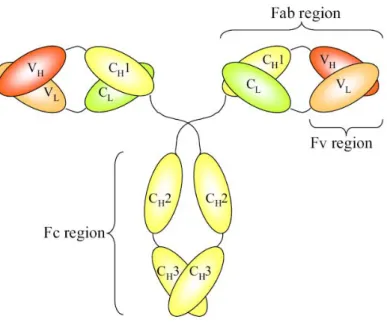
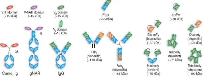
Therapeutic antibody formats
- Single-chain fragment (scFv)
- Antigen-binding fragment (Fab)
- Therapeutic monoclonal antibody H6
- Therapeutic monoclonal antibody B1
- Therapeutic monoclonal antibody A4
The formation of diabodies (60 kDa), tribodies (90 kDa) and tetrabodies (120 kDa) was previously observed when the length of the linker peptide was reduced (Dolezal et al., 2003). Diateles have two functional antigen-binding domains, which can be the same (bivalent diateles) or specific for different antigens (bispecific diateles) (Joosten et al., 2003). Monoclonal antibody B1 (mAb/B1) is a short-half-life humanized antibody directed against 'fibroblast-activating protein alpha' of invasive epithelial growth.
Protein expression in E. coli
- Inclusion body (IB) formation
- Influence of host strains on protein expression
- Influence of codon usage on protein expression
- Influence of temperature on protein expression
- Influence of linker sequence on protein expression
- Extended Linker (E-Linker)
- Linker 218 (L218)
- Linker 205 (L205)
- LLB18
In some cases, disulfide bonds were formed despite the reducing conditions in the cytosol (Miele et al., 1990). IBs are inactive, tightly packed aggregates of pure and homogeneous target protein (Singh et al., 2005). IB formation results from specific aggregation between folding intermediates of the recombinant protein molecules (Speed et al., 1996).
Materials and Methods
Bacterial strains
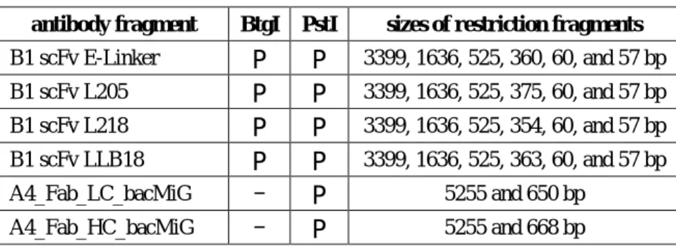
Expression vectors
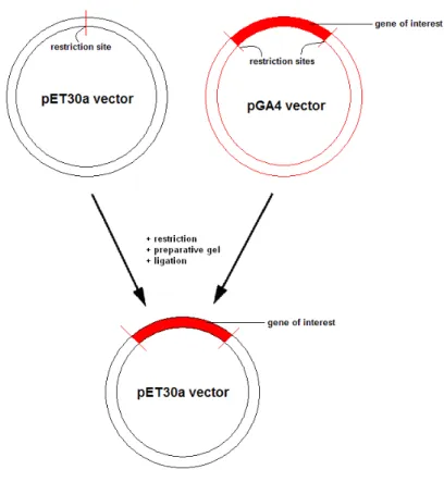
- Cloning
Subcloning of pGA4 to pET30a vectors was done using standard cloning procedures, illustrated in Figure 3-1.
Model proteins
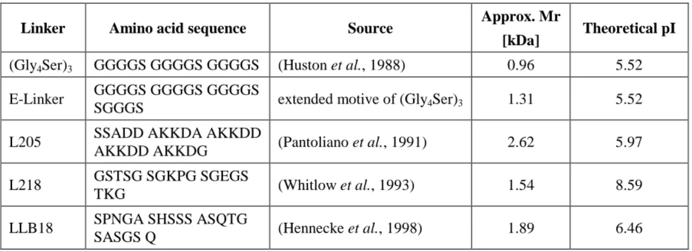
Additionally, different linkers (Table 3-2) were inserted into the existing B1 scFv fragment to investigate the influence of linker peptides on scFv cytoplasmic solubility. The heavy and light chains of the A4 Fab fragment were expressed separately from each other in E.
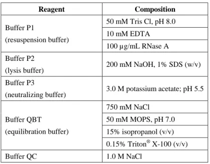
Preparation, purification, and modification of DNA
- Preparation of plasmid DNA
- Miniprep
- Midiprep
- Purification of plasmid DNA
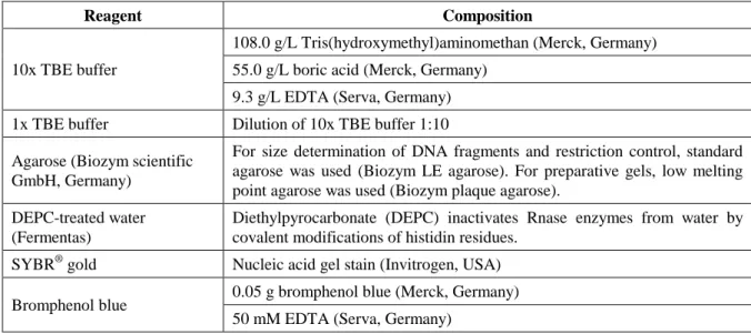
- Agarose gel electrophoresis
- Modification of plasmid DNA
- Restriction of DNA
- Ligation
250 µL of buffer P2 was added and the reaction tubes were carefully inverted until the suspensions turned blue. Then 4 mL of buffer P2 was added and the reaction tubes were carefully inverted until the suspensions turned blue. Then 4 ml of buffer P3 was added and the reaction tubes were inverted until the suspensions became colourless.
Transformation of DNA into E. coli
100 µL of chemically competent cells and 1-5 µL of plasmid solution (10 ng/µL) were mixed in pre-chilled 1.5 ml reaction tubes on ice. For heat shock, the reaction tubes were incubated at 42°C for 45 seconds (Eppendorf® Thermomixer comfort, Germany). 200 µL from all three dilutions were used for plating on selective agar plates containing the appropriate antibiotic.
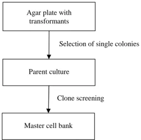
Clone screening and cell banks
- Parent culture
- Clone screening
- Master cell bank
After transformation (see section 3.5), transformants were randomly picked from selective agar plates with an inoculation loop and transferred to test tubes. Precultures were grown in 250 ml shake flasks with 50 ml SLB medium and 50 µg/ml kanamycin sulfate. Main cultures were performed in 1 liter shake flasks with 250 ml SLB medium and 50 µg/ml kanamycin sulfate.
Cultures were induced with IPTG resulting in a final inducer concentration of 1 mM in shaking. The samples were centrifuged at 4°C and 13200 rpm for 15 min (Eppendorf® Centrifuge 5415R, Germany), the supernatant discarded and the pellet cooled at -20°C.
Cultivation
- Media
- Fermentation systems
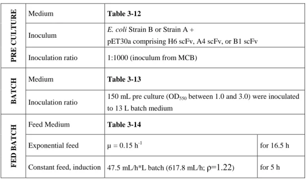
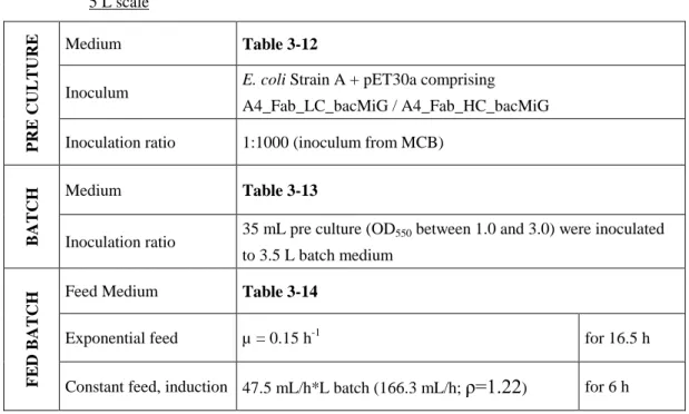
- Pre cultures
- Fermentation conditions
- Sampling
- Microscopy
- Optical density (OD 550 )
- Dry cell weight (DCW)
- Wet cell weight (WCW)
- Glucose
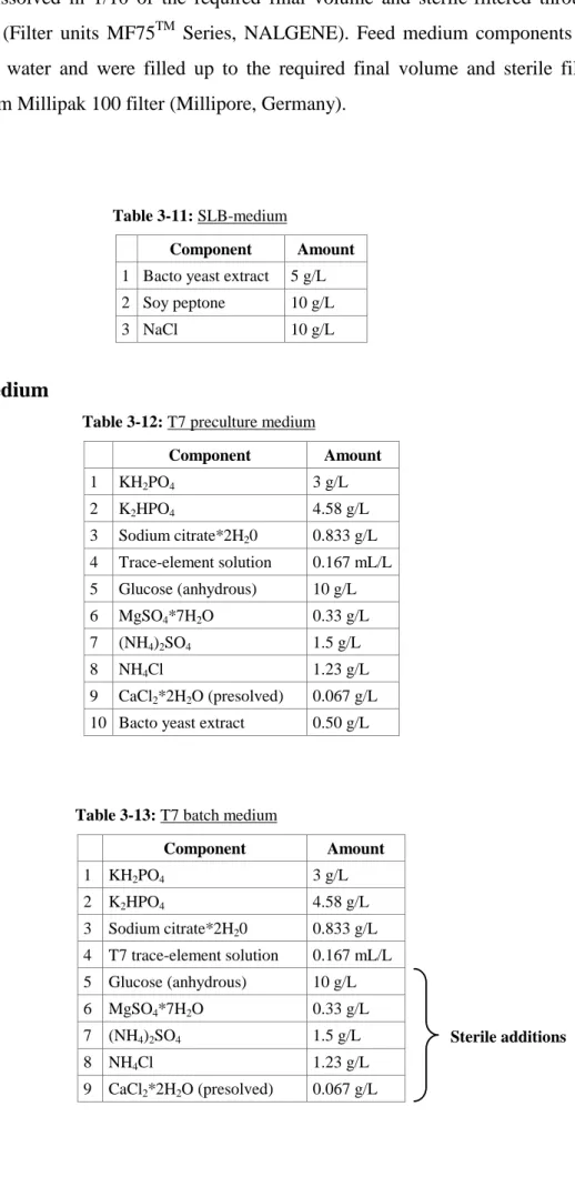
Nutrient medium components were dissolved in hot water and filled to the required final volume and sterile filtered through a 0.22 µm Millipak 100 filter (Millipore, Germany). For complex medium fermentations, 10 g/L bacto yeast extract and 20 g/L soybean peptone were added to the batch medium (Table 3-13) and autoclaved. For complex medium fermentations, 10 g/L bacto yeast extract and 20 g/L soybean peptone were added to the nutrient medium and sterile filtered through a 0.22 µm Millipak 100 filter (Millipore, Germany).
Cultivation took place in an orbital shaker (INFORS® Multitron II, Switzerland) at 300 revolutions per minute and the desired temperature. All reactors were equipped with integrated controllers for pH, temperature, mixing, aeration, pressure and feed pumps. The bioreactors were equipped with probes for pH, pO2 (both Mettler-Toledo, Switzerland) and temperature (Jumo, Austria).
The temperature was measured with a Pt-100 electrode and maintained at the desired temperature with a cooling/heating cycle. All containers were connected to the fermenter with plastic tubing and sterile needles pierced through the fermenter door diaphragms. To determine DCW, 10 ml of fermentation broth was centrifuged in 50 ml reaction tubes at 4000 revolutions per minute for 15 minutes (Eppendorf® centrifuge 5810R, Germany).
Glucose concentration was measured by biochemical analyzer YSI 2700 Select Glucose-analyzer (Kreienbaum, Germany) from 0.22 μm filtered supernatants.
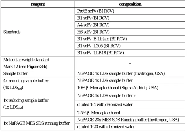
Analytics
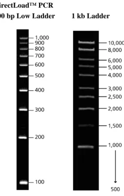
- DNA quantification
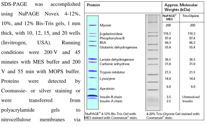
- SDS-PAGE
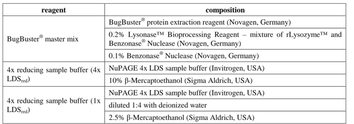
- Ultrasonic disintegration
- Enzymatic disintegration
- Staining methods
- Determination of product purity
- Western blot
1x NuPAGE MES SDS running buffer NuPAGE 20x MES SDS running buffer (Invitrogen, USA) diluted 1:20 with deionized water. 1x NuPAGE MOPS SDS running buffer NuPAGE 20x MES SDS Running buffer (Invitrogen, USA) diluted 1:20 with deionized water. The pellets remained in the original reaction tubes and were considered the insoluble protein fraction.
To minimize soluble protein in the insoluble fraction, 1 ml of deionized water was added to the pellets. The reaction tubes were vortexed vigorously until the pellets were resuspended in the deionized water. Then the gels were placed in deionized water for 30 minutes at room temperature and shaking (150 rpm).
After SDS-PAGE, gels were rinsed with deionized water and fixed in Fixing Solution II for 10 minutes. The gels were sensitized 2x 30 minutes in the sensitizing solution, washed 2x 10 minutes with deionized water and stained in the staining solution for 15 minutes. After SDS-PAGE, the gels were rinsed with deionized water and placed on a nitrocellulose membrane.
6 staining in staining solution at RT until bands can be seen well 7 3x 10 min at 150 rpm and RT in deionized water.
Results and Discussion
Bacterial strains
Expression vectors and model proteins
- Subcloning and transformation
In addition, B1 and A4 scFv fragments were used as model proteins to extend the technology platform to more molecules. In the present work, the feasibility of separate and intracellular expression of the light and heavy chain of the Fab fragment in E. was selected. The best producers were selected by clone screening in shake flask cultures and used to establish cell banks.
Best producers were selected by clone screening in shake flask cultures and used for cell bank establishment.
Expression of scFv fragments in E. coli
- Expression of different scFv fragments at 35°C
- IB formation and soluble expression in the cytosol
- Product purity
- Expression of B1 scFv comprising different linker peptides
- IB formation and soluble expression in the cytosol
- Product purity
For fermentations with B1 scFv (Figure 4-1), DCW values were lower for Strain A than for Strain B at the fermentation point (52 g/L compared to 60 g/L). This is caused by higher specific B1 scFv concentrations in Strain A compared to Strain B (59 mg/g DCW and 52 mg/g DCW). As stated in Chapter 4.3.1, values for specific product concentrations (Table 4-6) as well as volumetric product concentrations (Table 4-7) of B1 scFv only reached about 50% compared to A4 and H6 scFv.
The data presented clearly indicate that expression temperature does not affect intracellular solubility of B1 scFv. Maximum specific product concentrations of B1 scFv comprising L205, LLB18 and E-Linker were reached after 3 hours of induction compared to 6 hours of induction for L218. B1 scFv comprising the linkers L205, L218, LLB18 and E-Linker was detected only in the intracellular insoluble fraction.
The dependence of the specific concentration of B1 scFv [mg/g IB] on the binding peptide applied is remarkable (Table 4-20). Therefore, the B1 scFv comprising L218 not only contains the highest volumetric and specific concentrations [mg/g DCW] among all binding peptides applied, but also exhibits the highest purity within one SDS-PAGE lane (80%) and the highest specific concentration [mg/g IB].
Expression of Fab fragments in E. coli
- Separate expression of light- and heavy chain of A4 Fab
- Product purity
The heavy chain tends to aggregate in the cytoplasm if its folding is not assisted by a sufficient amount of light chain. After induction, the volumetric concentration of the product of both A4 Fab chains increased steadily until the fourth hour of induction (Figure 4-14 and Table 4-22). This is also shown by Coomassie-stained gels of the A4 Fab light and heavy chains (Figure 4-15 and Figure 4-16).
After the fourth hour of induction, the increase in volumetric product concentration weakened, resulting in 14.2 g/l for the light chain and 11.7 g/l for the heavy chain at the end of fermentation. Maximum specific concentrations were reached at the end of fermentation for both Fab chains, yielding 217 mg/g DCW for the light chain and 198 mg/g DCW for the heavy chain. In the present work, significantly higher maximal expression levels were obtained for the individual Fab chains: 14.2 g/L for the A4 Fab light chain and 11.7 g/L for the heavy chain.
However, the volumetric concentrations obtained for A4 Fab chains may not be applicable to Fab chains of other mAbs because expression levels can be highly dependent on the protein used. According to SDS-PAGE variation, the A4 Fab chains have similar purities within the SDS-PAGE bands (Table 4-23). The data presented indicate that intracellular and separate expression of A4 Fab light and heavy chains is a valuable expression strategy.
Further studies on the refolding and reassembly of both chains must be performed to assess its full potential.
Conclusions
This is consistent with previous observations by Wagner, 2008 for Fab fragments, where a 100-fold increase in expression level was observed using codon-optimized genes. As a consequence, the influence of different expression temperatures on B1 scFv yield, intracellular solubility and purity was subsequently investigated. For these reasons, 35°C was found to be the ideal expression temperature in terms of B1 scFv yield and purity.
As described in Section 2.2.5, the linker peptide linking the two variable domains within the scFv fragment is thought to influence aggregation (Whitlow et al., 1993), thermodynamic stability (Robinson and Sauer, 1998) and solubility (Tang et al. al., 1996). However, the expression levels (Tang et al., 1996) and folding properties (Hennecke et al., 1998) of the scFv fragment are more influenced by the domains than by the linker. In order to reveal the influence of different linker peptides on B1 scFv expression efficiency, intracellular solubility and purity, further studies were performed.
Four different linker sequences were introduced into the existing B1 scFv to replace the original (Gly4Ser)3 linker (Chapter 2.2.5). In conclusion, B1 scFv encompassing L218 yielded the highest volumetric concentrations and highest purities of all linkers examined. To evaluate its full potential, further research into the refolding and reassembly of the two chains needs to be done.
Regarding the manufacturing platform, two antibody fragments (scFv and Fab) derived from three therapeutic monoclonal antibodies (H6, B1 and A4) were evaluated in this study.
Appendix
Abbreviations
SDS-PAGE natriumdodecylsulfat polyacrylamid gelelectrophoresis SOC super optimal bouillon med extra glucose.
Cabilly, S. (1989) Growth at suboptimal temperatures allows production of functional antigen-binding Fab fragments in Escherichia coli. Kane, J.F. (1995) Effects of rare codon clusters on high-level expression of heterologous proteins in Escherichia coli.