2 TEM Summer School
3 CNPEM
The lectures are going to be held in the LNLS Auditorium at the Storage Ring Building.
4 TEM Summer School
Program 5
Presentation 7
Organizers | Scientific Committee 9
Speakers 10
Abstracts
5
CNPEM
6 TEM Summer School
7 CNPEM
TEM Summer School
The Electron Microscopy Laboratory (LME), of the Brazilian Nanotechnology National Laboratory (LNNano), which is a part of Brazilian Center for Research in Energy and Materials (CNPEM), Campinas, Brazil, organizes every two years a short course on theory and practical concepts of Transmission Electron Microscopy. This is the sixth edition of the event, between January 11 and 29, 2016.
This school aims to contribute to the advanced training of graduate students and researchers in the fields of engineering, materials science, physics, chemistry and related fields from academic and industrial communities, in Brazil and abroad, on theoretical and practical concepts of TEM techniques applied for materials characterization. We encourage the formation of new users and complement the training of users who has experience in the application of TEM techniques. The school instructors are leading researchers associated to important international and Brazilian research and educational institutions.
The school had its first edition in 2007 with 15 participants. Along its history the number of participants grows. In the last edition, the school was attended by 85 graduated students and PhDs from different areas. For this edition, we count with 90 participants, from Brazil and abroad. The school is organized for a duration of three weeks and in the first week, basic TEM theory are covered. Further, in the second week, several advanced topics including Electron Holography, Electron Tomography, Spectroscopy, and Quantitative analyses are the main focus.
Finally, hands-on activities are scheduled for the third week. Additionally, several changes were implemented for the organization of this new edition. For the first time, a registration fee was charged and a poster section was opened. The school will count with the participation of manufacturers of electron microscopes and detectors and vendors.
We thank the financial support from FAPESP (Grant 2015/11990-7), CNPq (Grant 441899/2015- 3), the sponsorship from JEOL, FEI, Hitachi and Analítica companies, and the participation of Gatan and JEOL during the third week of school.
8 TEM Summer School
Aerial photograph of CNPEM campus with artistic view of the Sirius building CNPEM: A cutting-edge research centerWelcome to the Brazilian Center for Research in Energy and Materials (CNPEM), a world-class center for development of science, composed by four laboratories: Brazilian Synchrotron Light Laboratory (LNLS), Brazilian Biosciences National Laboratory (LNBio), Brazilian Bioethanol Science and Technology Laboratory (CTBE) and Brazilian Nanotechnology National Laboratory (LNNano).
Located in Campinas (SP), CNPEM is a private nonprofit organization qualified by the Ministry of Science, Technology and Innovation (MCTI), whose laboratories have open facilities to the scientific and industrial communities across the country and abroad.
The Brazilian Nanotechnology National Laboratory (LNNano)
The Brazilian Nanotechnology National Laboratory (LNNano) is part of the Brazilian Center of Research in Energy and Materials (CNPEM), an organization certified by the Ministry of Science, Technology and Innovation (MCTI) located in Campinas-SP. The LNNano offers to the academic community and professionals, from Brazil and abroad, timely access to up-to-date equipment operated by experienced personnel within an innovative environment. It is dedicated to the creation, dissemination and application of nanotechnologies aiming at innovation. As a multiuser National Laboratory, it collaborates on multidisciplinary projects with researchers and R&D professionals in universities, research centers and companies and also with the other laboratories of CNPEM.
9 CNPEM
Carlos Alberto Ospina Ramirez (LME/LNNano) - Event Coordinator Alexandre Cassago (CME/LNNano)
Érico Teixeira Neto (LME/LNNano) Jefferson Bettini (LME/LNNano)
Marcelo Alexandre de Farias (CME/LNNano) Rodrigo Villares Portugal (CME/LNNano) Naga Vishnu Vardhan Mogili (LME/LNNano)
Local Committee
Gustavo Martins Moreno (CNPEM) Ildéria Maira dos Santos (CNPEM) Pâmela Fernandes Machado (CNPEM) Organizers | Scientific Committee
Funding agency
Institutional Partners
Sponsors
Participation
10 TEM Summer School
11 CNPEM
International invited speakers
Dr. Alexandre Gloter, Laboratoire de Physique des Solides, Université Paris Sud, France.Lectures: Advanced Spectroscopy III and IV
Prof. Angus Kirkland, Department of Materials, University of Oxford, United Kingdom.Lectures:
Quantitative Electron Microscopy I and II
Dr. Etienne Snoeck, Group NanoMaterials, Centre d’Élaboration de Matériaux et d’Etudes Structurales, Centre National de la Recherche Scientifique, France.
Lectures: Electron Holography I and II
Dr. Yongsoo Yang, Coherent Imaging Group, Department of Physics and Astronomy, University of California, USA.Lectures: Electron Tomography I and II
Prof. Raul Arenal, Advanced Microscopy Laboratory, University of Zaragoza, Spain.
Lectures: Advanced Spectroscopy I and II Brazilian invited speakers
Prof. André Linhares Rossi, Brazilian Center for Research in Physics, Rio de Janeiro, RJ.
Lectures: Guns, Electron Optics and Pumps.
Prof. Conrado Ramos Moreira Afonso, Department of Materials Engineering, Federal University of São Carlos, São Carlos, SP.
Lectures: Electron Diffraction II; Imaging
Prof. Luiz Fernando Zagonel, Institute of Physics, University of Campinas, Campinas, SP.
Lectures: STEM; Introduction to Electron Spectroscopy.
Prof. Paulo Fernando Papaléo Fichtner, Institute of Physics, Federal University of Rio Grande do Sul, Rio Grande do Sul, RS.
Lectures: Interaction Electron – Matter Local speakers
Dr. Alexandre Cassago, Cryo-Electron Microscopy Group (CME), Brazilian Nanotechnology National Laboratory (LNNano).
Dr. Carlos Alberto Ospina Ramirez, Electron Microscopy Laboratory (LME), Brazilian Nanotechnology National Laboratory (LNNano).
Dr. Jefferson Bettini, Electron Microscopy Laboratory (LME), Brazilian Nanotechnology National Laboratory (LNNano).
Dr. Marcelo Alexandre de Farias, Cryo-Electron Microscopy Group (CME), Brazilian Nanotechnology National Laboratory (LNNano)
Dr. Rodrigo Portugal, Cryo-Electron Microscopy Group (CME), Brazilian Nanotechnology National Laboratory (LNNano).
Dr. Vishnu Mogili, Electron Microscopy Laboratory (LME), Brazilian Nanotechnology National Laboratory (LNNano).
Technicians:
Fabiano Emmanuel Montoro, Electron Microscopy Laboratory (LME), Brazilian Nanotechnology National Laboratory (LNNano).
Sidnei Ramis de Araújo, Electron Microscopy Laboratory (LME), Brazilian Nanotechnology National Laboratory (LNNano)
Speakers
12 TEM Summer School
13
CNPEM
14 TEM Summer School
15
CNPEM
16 TEM Summer School
17
CNPEM
18 TEM Summer School
19
CNPEM
20 TEM Summer School
21
CNPEM
22 TEM Summer School
23
CNPEM
24 TEM Summer School
25
CNPEM
26 TEM Summer School
27
CNPEM
28 TEM Summer School
29
CNPEM
30 TEM Summer School
31
CNPEM
32 TEM Summer School
33
CNPEM
34 TEM Summer School
35
CNPEM
36 TEM Summer School
37
CNPEM
38 TEM Summer School
39
CNPEM
40 TEM Summer School
41
CNPEM
42 TEM Summer School
43
CNPEM
44 TEM Summer School
45
CNPEM
46 TEM Summer School
47
CNPEM
48 TEM Summer School
49
CNPEM
50 TEM Summer School
51
CNPEM
52 TEM Summer School
53
CNPEM
54 TEM Summer School
55
CNPEM
56 TEM Summer School
57
CNPEM
58 TEM Summer School
59
CNPEM
60 TEM Summer School
61
CNPEM
62 TEM Summer School
63
CNPEM
64 TEM Summer School
65
CNPEM
66 TEM Summer School
67
CNPEM
68 TEM Summer School
69
CNPEM
70 TEM Summer School
71
CNPEM
72 TEM Summer School
73
CNPEM
74 TEM Summer School
75
CNPEM
76 TEM Summer School
77
CNPEM
78 TEM Summer School
79
CNPEM
80 TEM Summer School
81
CNPEM
82 TEM Summer School
83
CNPEM
84 TEM Summer School
85
CNPEM
86 TEM Summer School
87
CNPEM
88 TEM Summer School
89
CNPEM
90 TEM Summer School
91
CNPEM
92 TEM Summer School
93
CNPEM
94 TEM Summer School
95
CNPEM
96 TEM Summer School
97
CNPEM
98 TEM Summer School
99
CNPEM
100 TEM Summer School
101
CNPEM
102 TEM Summer School Gold nanoparticle based sensor for early breast cancer detection
Vera Katic1, Juliano Alves Bonacin2
State University of Campinas, UNICAMP, Campinas, State, So Paulo [email protected]
Cancer is a disease that alters health status of cells and ultimately leads to malignant tumors.
Late detection of disease increases the number of deaths. Therefore, early detection of cancer can save lives [1]. Our objective is to create highly accurate portable device for early breast cancer detection. The portable device (sensor) would be based on the interaction between gold nanoparticles and biomarkers [2]. Biomarker refers to a measurable indication of biological state or condition. Thus, cancer biomarkers are an indication of cancer and via its detection the existence of specific cancer can be verified. One of the most studied breast biomarkers is CA 15- 3, located in the blood. Therefore our focus is to create sensitive sensor for CA 15-3 detection.
Initially, shape and size controllable gold nanoparticles will be obtained utilizing photoinduced methods. The following steps will include detailed characterization of aforementioned systems via spectroscopy and electron microscopy. Nanoparticles will be characterized with transmission electron microscopy (TEM) in order to investigate morphological, crystallographic and elemental composition. The final step will be focused on the development of electrochemical and plasmonic sensors.
Thanks to LNNano - Laboratório Nacional de Nanotecnologia CNPEM - Centro Nacional de Pesquisa em Energia e Materiais Campinas - SP – Brasil and CNPq grant number (161819/2014-1)
[1] T. N. Seyfried et al. Carcinogenesis, 35, 515-527, 2014 [2] Li et al. Biosensor and Bioelectronics, 43, 25-29, 2013 .
103 CNPEM
Mechanical and microstructural characterization of steels Ultra – High- Strength Thermally and Thermochemically Treated by plasma.
Y. O. Iwaki1, A. S. M. Cardoso2, D.Y. K. Ranzini3
1 Centro Tecnológico da Marinha em São Paulo - CTMSP, Iperó, SP, Brazil
2, 3 Centro Tecnológico da Marinha em São Paulo - CTMSP, São Paulo, SP, Brazil [email protected].
br
The high strength steel with low alloy and ultra-high strength steel are materials with great industrial application, in which its technology enables strategic innovation. The understanding of the medium carbon steel composition modification SAE 4340 [1] enabled the purchase of the 300M Stainless steel, although in the development application of rocket and energy areas, the Maraging 300 steels were also correlated. To assess and compare the mechanical properties of these steels under different treatments, together with its microstructural correlation, would enable an understanding of the improvements or fragilization, which are decisive in the resistance of such materials. As received the Maraging steel 300M and 300 exhibited high mechanical strength values catastrophic failure in the plastic regime, as well as high threshold in fatigue life compared to steel 4340 [2]. As for the thermochemical nitritation treatment and the properties studied, there was, in steels 4340, significant increase in tensile strength, but decrease in fatigue life. Nevertheless Maraging steels 300M and 300 declined in strength in tensile tests and fatigue life, however exhibited higher hardness values and corrosion resistance.
The 300M and Maraging steel showed unfavorable results in fatigue and stretching. Scholars have noted that thermochemical treatments such as nitritation at certain temperatures (acting as tempering / aging in the inner region of the material), can weaken steel with high grades of silicon and titanium [3].
Centro Tecnológico da Marinha em São Paulo and Marinha do Brasil.
[1] MESQUITA, R. A. et al., Tecnologia em Metalurgia e Materiais, v.4, n.4, p. 7-15, (2008).
[2] CARDOSO, A. S. M. et. al, Advanced Materials Research, Vol. 891-892, p. 1507-1512 (2014).
[3] GARRISON JR, W.M., Materials Science and Technology, v. 3, n. 4, p. 256-259 (1987).
104 TEM Summer School Synthesis and characterization of Sr
1-xCu
xTiO
3and SrTi
1-xCu
xO
3compounds applied to
catalysis of the water-gas shift reaction
Vitor C. Coletta1, Francielle C. F. Marcos2, Francisco G. E. Nogueira2, Maria I. B. Bernardi1, Alain Michalow- icz3, Renato V. Gonçalves1, Elisabete M. Assaf2, Valmor R. Mastelaro1
1São Carlos Institute of Physics, University of São Paulo, São Carlos, São Paulo, Brazil
2São Carlos Institute of Chemistry, University of São Paulo, São Carlos, São Paulo, Brazil
3Universite Paris-Est Creteil Val de Marne – ICMPE-CNRS, Thiais, France [email protected]
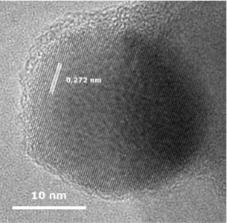
The water-gas shift reaction is carried out in the production of H2 free from CO, necessary in applications such as the use in proton exchange membrane (PEM) fuel cells. [1] Oxides with perovskite structure containing copper are promising for catalysis of this type of reaction for presenting a good chemical stability. [2] This research consists in performing the synthesis and characterization of nanoparticles of composition Sr1-xCuxTiO3 and SrTi1-xCuxO3 looking for the application as catalysts for the water-gas shift reaction. The synthesis was done through the polymeric precursors method with calcination in N2 atmosphere followed by a heat treatment in O2 for carbon removal, resulting in nanoparticles with large surface area in comparison to the conventional method. [3] X-ray diffraction results showed that the copper atoms are segregated from the perovskite phase for x ≥ 0.06 in form of CuO phase. The composition SrTi0.80Cu0.20O3 presented the better activity with 74% of CO conversion at 350°C. Thus, our TEM analyses, at the Chemistry Institute of Unesp, Araraquara, begun with this sample compared to the undoped SrTiO3. Figure 1 shows a high-resolution TEM image of a crystallite of the SrTiO3 sample. The distance of 0.27 nm between the observed crystallographic planes corresponds to that between (110) planes of SrTiO3 cubic phase (PDF 35-0734). As expected from the X-ray diffraction results, the SrTi0.80Cu0.20O3 sample has SrTiO3 as the main phase and also regions rich in CuO phase on the surface as showed in Figure 2a. The high-resolution image of the region in Figure 2b shows planes spaced with a distance close to that reported for (11-1) of CuO phase (PDF 48-1548).
The presence of the phase is also confirmed by the EDS spectrum in the region (Figure 2c). The next steps of our TEM characterization will involve analyzing the difference between substituting strontium and titanium sites for copper in the morphology and copper segregation.
The authors are grateful for the financial support provided by São Paulo Research Foundation – FAPESP (grant 2013/09573-3) and National Council for Scientific and Technological Development – CNPq (grants 304498/2013-0 and 140631/2013-5). The authors are also grateful to Marcelo O.
Orlandi for the use of the transmission electron microscope.
[1] Gradisher, L. et al. Applied Energy, 139, 335-349 (2015).
[2] Maluf, S. S. et al. Applied Catalysis A: General, 413-414, 85-93 (2012).
[3] da Silva, L. F. et al. Materials Chemistry and Physics, 125, 168-173 (2011).
105 CNPEM
Figure 1 – High-resolution TEM image of SrTiO3 sample.
Figure 2 – (a) TEM image of SrTi0.80Cu0.20O3 sample. (b) high-resolution image and (c) EDS spectrum of the region on the surface.
106 TEM Summer School Identification of secondary precipitates present at grain boundaries of Co based
superalloy
A.M.S. Costa1, E. S.N. Lopes2, A. P. Tschiptschin1,3
1Brazilian Nanotechnology National Laboratory, Campinas, Sao Paulo, Brazil
2Faculty of Mechanical Engineering of Unicamp, Campinas, Sao Paulo, Brazil
3Department of Metallurgical and Materials Engineering at Polytechnic School of USP [email protected]
In this study was focused on the characterization of fine precipitates formed on grain boundaries in Co based superalloys. When the precipitates are presented in metallic materials they could improve materials properties such a mechanical, electrical, magnetic and chemical stability as well. Depending on the dimension in size and shape the precipitates can acting such a reforcement of grain boundaries and helps to prevent grain sliding during high temperature performance.
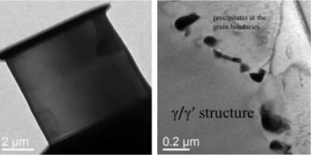
In this way is imperative to know which the chemistry composition, what kind of phase it is (intermetallics, carbides, borides and so on), orientation relationship with the matrix phase, where they could be identified on the microstructure. The transmission electron microscopy technique provides a lot of important information about the more located microstructural characterization what is the main goal of this study. The samples have been studied in this work were prepared by focused ion beam (FIB) technique at the Center for Electron Microscopy and Analysis (CEMAS). The features of the samples were analysed by high resolution transmission electron microscope JEOL model JEM 3010 at LME. It was captured bright field images from grain boundaries region. Figure 1 shows the sample that was prepared by FIB technique. Figure 2 shows a couple of details about the microstructure of the material. It is observed the matrix is microconstituted of γ-Co austenitic phase having (A1) FCC structure and coherent L12 structure precipitates of γ′-Co3(Al,W) inside it. Fine precipitates of minor secondary phase are found at the grain boundaries are showed Figure 2. Because of the additions of C, B, Ni and Al certain kind of precipitates could be formed during the cooling of the material that was processed by arc melting technique. Chemical analysis using XEDS together electron diffraction technique should help to do a proper identification of which kind of phase are decorating grain boundaries.
This study has been supported by The São Paulo Research Foundation (Fapesp – Process Number 2014/13772-4). I would like to thank Dr. Eder Lopes for preparing FIB samples and Dr. Carlos Ospina for acquisition of TEM images.
Figure 1- The sample of Co based superalloy prepared by FIB technique.
Figure 2- Microstructure of Co based superalloys showing matrix structure and secondary minor phases precipitated at the grain boundaries.
107 CNPEM
Vanadium interaction with biological systems
B. C. Costa1, P. N. Lisboa-Filho2
1 Programa de Pós-Graduação em Ciência e Tecnologia de Materiais – Universidade Estadual “Júlio de Mesquita Filho” - UNESP, Bauru, São Paulo, Brazil
2 Departamento de Física, Universidade Estadual “Júlio de Mesquita Filho” - UNESP, Bauru, São Paulo, Brazil [email protected]
Among the most commonly used metallic materials as prosthetic implants are titanium-based alloys, specifically Ti-6Al-4V system, whose use corresponds to 45% of the total production of titanium as biomaterial [1].
In general, metallic materials may suffer corrosion when implanted, leading to release of metal compounds or debris (fragments) into the body. Furthermore, the human body is predominantly composed by water, polymers (proteins) and ceramics (bones) and metals are elements found as necessary only in very small quantities [2]. Thus, when a metallic material is implanted in the human body, the toxicity of their constituents elements should be evaluated before its application. Regarding to the evaluation of toxicological potential of elements of the Ti-6Al- 4V system, aluminum may present acute toxicity at high doses, and is reported as an element related to neurological disorders and mitigating factor of various diseases. Vanadium plays a role that is still poorly understood in the human body and may present ambiguous cellular responses. Furthermore, vanadium compounds may have interactions with proteins within a living organism, changing its functionality [3].
In this context, the present proposal is based on the possibility of long-term exposure to an implant based on Ti-6Al-4V alloy, the adverse effects of debris release from its corrosion in a living system and also the studies related to toxicity of these alloying elements, and the importance of the interaction of this element, especially in its nanostructured form, regarding proteins.
In this way, the in situ liquid TEM technique may allow the orientation and conformation analysis of proteins under the surface of this project samples (Ti-6Al-4V and vanadium debris obtained from tribocorrosion tests and V2O5 powder), and this is the key-role to understanding the interaction and toxicity mechanisms of vanadium in live systems. So a better understanding of this powerful technique is extremely recommended.
The authors would like to thank CAPES and CNPq - Brazilian Funding Agencies for the financial support.
[1] DAVIES, J.R. Handbook of Materials for Medical Devices, Ohio, ASM International, Materials Park, 2003.
[2] CHEN, Q. et al., Materials Science and Engineering R, 87, 1 – 57 (2015).
[3] MUKHERJEE, B. et al., Toxicology Letters, 150, 135 – 143 (2004).
108 TEM Summer School Effect of impurity segregation on intermediate temperature, ductility-dip cracking
(DDC) in high-Cr, Ni-base alloys
Carolin Fink, John C. Lippold
The Ohio State University, Columbus, Ohio, USA [email protected]
The chemical composition of the filler metal has proven to be one of the most important factors controlling DDC in Ni-base weld metals. Alloying elements, such as Nb, determine the precipitation behavior at the end of weld metal solidification, mitigating DDC by grain boundary (GB) pinning and boundary “tortuosity” [1]. On the other hand, impurities and interstitials are also considered to play an important role in DDC susceptibility. Both hydrogen and oxygen embrittlement in Ni-base alloys has been reported [2, 3]. Both elements are known to have a detrimental effect on resistance to DDC. A possible contribution due to a GB decohesion effect or local oxidation at the grain boundary is considered possible [4]. However, on the atomic scale, there is very limited knowledge regarding interactions between interstitial elements at the GBs and at the interface between intergranular precipitates and the matrix. These interactions may not be the primary cause of DDC but may promote further intergranular embrittlement in the DDC susceptible temperature range, potentially helping to explain the variability in susceptibility within the same filler metal specification.
The proposed work will elucidate such interactions using electron microscopy (SEM, TEM) coupled with EDS and EELS measurements to understand their role on the cracking mechanism. Gleeble- based thermo-mechanical simulation will be performed with additions of either hydrogen or oxygen (via shielding gas or enriched atmosphere) for weld metal sample preparation. Samples for electron microscopy will be prepared from GB and crack-tip regions and analyzed for oxygen segregation, oxide formation or oxidation of GB particles. To determine the role of hydrogen migration to the GBs additional in-situ TEM analysis could be performed to study its influence on strain localization. This could be done by in-situ TEM tension [5] to observe and analyze the dynamic process of dislocation motion and crack nucleation.
[1] Ramirez, A.J. et al., Materials Science and Engineering A, 380(1-2), pp. 245-258 (2004).
[2] Woodford, D.A., Energy Materials, 1(1), pp. 59-79 (2006).
[3] Collins, M.G. et al., Welding Journal, 82(10), pp. 288s-295s (2003).
[4] Zheng, L. et al., Critical Reviews in Solid State and Materials Sciences, 37, pp. 181-214 (2012).
109 CNPEM
Low Energy Ion Implanting of Silver or Copper Ion for antimicrobial activity
Cesar Henrique Wanke*, Tatiana Pacheco Soares, Andreia Valim de Souza, Carlos Alejandro Figueroa, Otávio Bianchi, Cesar Aguzzoli
University of Caxias do Sul, Caxias do Sul, Rio Grande do Sul, Brazil
In modern medicine, biomaterials have been widely used as implants. However, orthoses and prostheses infected with post-implanted microorganisms require surgeries and medical interventions that are costly and treatment can take months. To combat infections associated with implants, the best strategy is to prevent them from occurring. One strategy is the silver ion implantation in these biomaterials [1-4]. As the antimicrobial efficacy of the nanoparticle depends on the shape and size of them [5], this can be confirmed by studying the inhibition of bacterial growth by differentially shaped and size nanoparticles. In this work, silver or copper ions were implanted at low energies in medical titanium with the goal to render bactericidal substrate surface and therefore, inhibit biofilm formation. The implantation was done using the Ion Plating equipment. After ion implantation, the samples were analyzed by Rutherford Backscattering Spectrometry (RBS), Glow-Discharge Optical Emission Spectroscopy (GD-EOS) and X-Ray Diffraction. The RBS analysis was performed at the Federal University of Rio Grande do Sul. The results show that the silver was implanted at depths of until 10 nm, corroborating with simulation data. In addition, preliminary tests of antimicrobial activity show that the approach adopted in this work has been effective. However, for a better understanding of how the nanoparticles are distributed in the outer surface, i.e. if are sprayed or in the cluster form, are necessary to use techniques that enable to evaluate this distribution. The most widely used technique for this is the transmission electron microscopy. It is intended to verify how nanoparticles are distributed on the surface (Figure 1a) as well as its distribution in depth (Figure 1b). For this, it needs to analyze the surface and cross section. These analyses will be used to correlate with the process parameter of the equipment and thermal treatment of samples after ion implantation.
We are indebted to the Brazilian agency CAPES, University of Caxias do Sul and Ion Implantation Laboratory (Institute of Physics, Federal University of Rio Grande do Sul, Porto Alegre, RS, Brazil).
[1] L. Zhao et al., Biomaterials, 32, 5706 - 5716 (2011).
[2] A. Melaiye and W. J. Youngs, Expert Opinion on Therapeutic Patents, 15, 125 - 130 (2005).
[3] D. R. Monteiro et al., International Journal of Antimicrobial Agents, 34, 103 – 110 (2009).
[4] K. Hori and S. Matsumoto, Biochemical Engineering Journal, 48, 424 – 434 (2010).
[5] M. Rai et al., Biotechnology Advances, 27, 76 – 83 (2009).
Figure 1. Representation of the formation of nanoclusters of the silver of copper nanoparticles on surface (a) and its distribution in depth (b).
110 TEM Summer School Synthesis and structural analysis of CoFe
2O
4:BaTiO
3nanocomposites
D. Alanis1*, P.N. Oliveira1, R.D. Bini1, D.M. Silva1, G.S. Dias1,I.A. Santos1, L.F. Cótica1
1Department of Physics, State University of Maringá Av. Cololmbo, 5790, Maringá, Paraná, Brazil 87020-900
*e-mail: [email protected]
Multiferroic materials have attracted interest due to the presence of more than one ferroic order, for example magnetic and electric orders, in the same phase. This particular coupling phenomenon is known as magnetoelectric (ME) effect. However, it is difficult to obtain significant ME effect in single-phase materials, since magnetic and electric dipoles have to coexist in the same asymmetric structure. Thus, in order to enhance the ME effect, piezoelectricmagnetostrictive composites have been exploited. Among some different configurations to prepare composite materials, the core-shell nanoparticles are also investigated as a beneficial starting material for magnetoelectric nanocomposite (MENC) materials. This structure provides direct and large surface contact of phases and preserves the individual piezoelectric and magnetostrictive properties of each phase. Particularly, ferrite-barium titanate in a core-shell configuration has been investigated due to appropriate individual (magnetostrictive and piezoelectric) properties of the components at room temperature. In addition, their chemical and mechanical stability and nontoxic properties are also important in applications regarding the environment and biological applications. This work aims to prepare MENC’s in a core-shell configuration with a narrow size distribution. A combination of polymeric and citrate methods was used for the MENC’s syntheses. MENC’s were prepared in the 1:1, 3:2 and 7:3 (CoFe2O4:BaTiO3) molar ratios, respectively. The structural characterizations of the obtained MENC’s were performed by X-ray diffraction and Fourier transform infrared spectroscopy (FTIR). These analyses showed a combination of a cubic CoFe2O4 and a tetragonal BaTiO3 composite. A core-shell morphology of MENC’s was observed by transmission and scanning electron microscopies. The MENC’s atomic compositions were confirmed using energy dispersive X-ray spectroscopy. Finally, the ferrimagnetic character of the MENC’s was observed in the magnetic characterizations obtained in a vibrating sample magnetometer.
We are thankful to CAPEs, CNPq and FINEP.
[1] L. F. Cotica, et al., Journal of Nano Research, 28 (2014) 131.
[2] L. F. Cotica, et al., Journal of Magnetism and Magnetic Materials, 324 (2011) 559.
111 CNPEM
Relation between the crystal structure interfaces of NiTi/Co, NiTi/Ni and FeRhPd films with their magnetic behavior
Diana L. Torres1 and Daniel R. Cornejo1
1 São Paulo University, Physics Institute, São Paulo, SP, Brazil [email protected]
Magnetic control at the nano-scale is interesting for technological applications. Nowadays, several ways to obtain this control have been reported and the development of new materials to improve it is growing up. For instead, it can be observed in ferromagnetic films coupled with materials which show temperature- or field-driven structural phase transition, as well as, ferromagnetic films which show first order phase transitions trough changes in temperature.
More specifically, we study the magnetic behavior of the NiTi/Co and NiTi/Ni films through the structural phase transition of the nearly equiatomic NiTi layer. The films have been grown on silicon (100) substrates by DC magnetron sputtering. In this structure, the magnetization of the ferromagnetic layer may be modified through changes in the stress field at the interface, when the structural phase transition in the non-magnetic layer is carried-out. Also, we study FeRhPd films deposited on MgO (001) which show a first magneto-structural phase transition.
All these functional structures are characterized by X-ray diffraction (XRD) and Rutherford backscattering spectrometry (RBS) to obtain the crystal structure and chemical composition respectively. Magnetic measurements as a function of temperature or magnetic field are performed by using a superconducting quantum interference device (SQUID) and a vibrating sample magnetometer (VSM) to study anisotropy behavior, coercivity, magnetic phase transitions, etc. Finally, we expect to use High resolution-TEM analysis to get a more precise relation between the structure in interfaces of the films and their magnetic behavior.
112 TEM Summer School Use of TEM for Metallurgical Engineering research on hydrogen embrittlement
D. Pérez Escobar1, E. Wallaert2, L. Duprez3, K. Verbeken2, A. Atrens4, M. Andrade11 ISI of Metallurgy and Special alloys, CIT Senai – Campus Cetec, Belo Horizonte, MG, Brazil
2 Department of Materials Science and Engineering UGent, Zwijnaarde, OW, Belgium
3 ArcelorMittal Global R&D Gent, TechnologiePark 935, Zwijnaarde, OW, Belgium
4 Materials Science, University of Queensland, Santa Lucia, Qld 4072, Australia [email protected] (Corresponding Author)
Transmission Electron Microscopy was performed in cast material in order to study the distribution of TiC precipitates in as-cast steels. The study was performed aiming to study the interaction of hydrogen interaction with this precipitates during my doctorate. One paper was published on international journal: D. Pérez Escobar, E. Wallaert, L. Duprez, K. Verbeken, A. Atrens. Thermal desorption spectroscopy study of the interaction of hydrogen with TiC precipitates. Metals and Materials International 19 (2013) 741-748
After casting, the steel (containing (0.025 wt%C-0.09%Ti) was annealed in a furnace protected with hydrogen atmosphere at temperatures between 550 °C and 800 °C in order to produce different ranges of precipitate sizes. Each sample of cold rolled steel sheet was heated in a furnace at 200 °C/h to an annealing temperature of 550 °C, 650 °C, 725 °C or 800 °C; held 2660 min at the annealing temperature; and cooled to room temperature. 100 precipitates in carbon replicas were analyzed using TEM to determine the size distribution of the precipitates and by energy dispersive X-ray analysis (EDX) in order to determine the chemical composition. To prepare the carbon replicas, each sample was polished to 1 μm to a mirror polish, etched for 10 s with 10% nital, washed with methanol, dried with compressed air, a carbon film was deposited on the surface in a carbon evaporator in vacuum, and the sample with the carbon layer was submerged in 4% nital until the carbon replica peeled off the steel surface. Using a Cu TEM-grid and tweezers, the carbon replica was washed in methanol for ~10 min.
113 CNPEM
The authors would like to thank the technical staff at the department of Material Science and Engineering at UGent and OCAS, where the TEM samples were prepared and analyzed.
[1] D. Pérez Escobar, E. Wallaert, L. Duprez, K. Verbeken, A. Atrens. Thermal desorption spectroscopy study of the interaction of hydrogen with TiC precipitates. Metals and Materials International 19 (2013) 741-748.
114 TEM Summer School ZnO Semiconductor Nanowires Gas Sensors
E.S.H. de Menezes1*, C. L. Sombrio1, B.Canto1, R. Reis2 and D. L. Baptista1,
1Instituto de Física, UFRGS, Porto Alegre, Rio Grande do Sul, Brazil
2National Center for Electron Microscopy, Berkeley, California,USA [email protected]
Zinc Oxide nanowires have a good potential for fast and selective gas sensing applications [1- 4]. When in contact with a small gas concentration the nanowire electrical resistance changes drastically. Therefore it is possible to use such behavior to detect very low gas concentrations through a nanowire-based sensing device. Additionally, the nanowire surface can be modified to change the sensor selectivity. The purpose of this project is to develop different gas sensing devices and characterize the electrical response to different gases. The electrical behavior of the nanowire is related to the lattice structure and defect characteristics. In this work, we present the project perspectives and techniques that will be applied to develop nanowire-based gas sensors. Previous as-grown ZnO nanowires samples were characterized using advanced electron microscopy (FEG-SEM, HRTEM, HAADF-STEM) and spectrum-imaging (EDX, EELS) techniques (Fig 1-2). The determination of growth polarity was also achieved by CBED analysis. HRTEM/
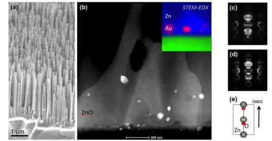
STEM analyses were performed using a XFEG Cs-corrected FEI Titan 80/300 microscope.. The ZnO nanowires were grown by vapor-liquid-solid (VLS) mechanism using sapphire as substrate and Au as catalyst. Dense and vertically aligned ZnO nanowires forest was grown epitaxially on sapphire substrates as showed in Fig. 1 (a). The nanowires were about 50 nm in diameter and its length up to 5 µm. A spatial distribution of Au catalyst nanoparticles at the ZnO/substrate interface is clearly observed in Fig. 1 (b) by HAADF-STEM and EDX-SI. The experimental and simulated CBED patterns presented in Fig. 1 (c-d) indicate a Zn-polar growth (e). New samples with different growth conditions will be synthesized and electron microscopy will be utilized to characterize it.
[1] P. Yang et al., Adv. Funct. Mater., 12 (2002) 323.
[2] M.-T. Chen et al., Nano Lett., 10 (2010) 4387.
[3] C. Soci et al., Nano Lett., 7 (2007) 1003.
[4] A. Janotti et al., Phys. Rev. B, 76 (2007) 165202.
The authors thanks the Materials Metrology Division (DIMAT) for the use of Electron Microscopy facilities at INMETRO. This work was partially supported by Brazilian agencies CNPq and CAPES
115 CNPEM
Fig. 1. Characterization of the as-grown ZnO nanowires. (a) SEM micrograph of an epitaxially grown ZnO forest on c-plane sapphire substrate. (b) HAADF-STEM image of the ZnO/substrate interface and an EDX spectrum image showing the presence of Au seeds at the base of nanowires. (c) Experimental and (d) simulated CBED patterns indicating the Zn-polarity (e) of the ZnO structure.
116 TEM Summer School Synthesis and characterization of Ni supported on Nb
2O
5catalyst to apply in
cellulose conversion reaction
Glauco F. Leal1,2, Dean H. Barret1, Silvia F. Moya1, Antonio A. S. Curvelo2,3, Cristiane B. Rodella1.
1 Brazilian Synchrotron Light Laboratory (LNLS), Brazilian Center for Research in Energy and Materials (CNPEM), Campinas, São Paulo, Brazil.
2 Insti tute of Chemistry of São Carlos (IQSC), University of São Paulo (USP), Departament of Physical Che-Institute of Chemistry of São Carlos (IQSC), University of São Paulo (USP), Departament of Physical Che- mistry, São Carlos, São Paulo, Brazil
3 Brazilian Bioethanol Science and Technology Laboratory (CTBE), Brazilian Center for Research in Energy and Materials (CNPEM), 13083-970, Campinas, São Paulo, Brazil.
The catalytic conversion of cellulose to biofuels and other value added chemicals is one of the most promising routes for the development of economically viable biorefineries. However, the challenge of developing green chemical methods for renewable energy generation based on heterogeneous catalysis relies on a thorough investigation of the physical and chemical properties of the catalyst [1]. Moreover, the correlation of these properties to the functional behavior of the catalyst within the cellulose valorization reaction. Cellulose processing firstly requires hydrolysis to obtain glucose followed by the transformed of glucose to fuels and chemicals [2, 3]. Thus, this PhD project aims to synthesize a new class of bifunctional catalysts composed of Ni and Co dispersed on Nb2O5 for cellulose conversion to obtain platform chemicals such as, sorbitol, HMF and levulinic acid among other products. Solid acid catalysts, such as Nb2O5 can be effective for cellulose hydrolysis. On the other hand, transition metals, such as Ni and Co are catalysts for hydrogenation and hydrogenolysis reactions. The structural, electronic, surface and textural properties will be investigated using ex situ and in situ synchrotron- based techniques (XRD and XAS) as well as N2 physisorption and XPS. Acidic properties of the catalysts will be analyzed by TPD – NH3 and FT-IR using adsorbed pyridine. Physical and chemical characteristics of the catalyst will be correlated with the catalytic performance. It is clear that transmission microscopy is a fundamental technique to study heterogeneous catalysts and it will not be different in this PhD project. TEM will be a powerful technique to investigate morphology, particle size and distribution of the Co, Ni and Nb2O5 from the preparation steps to the post catalytic reaction. These results will be correlated with XRD, XAS and XPS as well as catalytic properties. This will be a step forward in understanding and optimizing the catalyst and reaction parameters to improve specific product yields.
The authors are grateful for the CNPq scholarship and the financial support from the LNLS/CNPEM as well as from IQSC-USP-São Carlos, LNLS and CTBE and to their staff.
[1] R. Rinaldi, F. Schüth, ChemSusChem. 2, 1096-1107 (2009).
[2] I. Nowak, M. Ziolek, Chem. Rev. 99, 3603-3624 (1999).
[3] D. M. Alonso, J. Q. Bond, J. A. Dumesic, Green Chem. 12, 1493-1513 (2010).
117 CNPEM
Bacterial Nanosensor Based in Gold Nanoparticles
J. P. Oliveira1, A. R. Prado2, R.H.A. Pereira1, D.M. Ferreira1, W.J.Keijok1, R. Schuenck3 M.J. Pontes2, M.R.N.
Ribeiro2, B.V. Nogueira1, M. C. C. Guimarães1*
Laboratório Ultraestrutura Celular Carlos Alberto Redins - LUCCAR, Departamento de Morfologia, CCS, Universidade Federal do Espírito Santo, Vitória – ES, Brasil
Laboratório de Telecomunicação, Departamento de Engenharia Elétrica, Centro Tecnológico, Universidade Federal do Espírito Santo, Vitória – ES, Brasil Laboratório de Microbiologia, Departamento de Patologia, Centro de Ciências da Saúde, Universidade Federal do Espírito Santo, Vitória – ES, Brasil
Recently, advances in nanotechnology have brought new perspectives and had been stimulate different areas of research. In perspective, metal nanoparticles due to optical, electronic and magnetic properties has allowed a wide variety of application such as nanosensors, drug delivery system, lubricants, solar cells, catalysis and others [1]. A novel and sensitive colorimetric method to detect Escherichia coli was developed as a model for Gram-negative bacteria by a trypsin modified gold nanoparticles (AuNP’s) sensor. Trypsin (Sigma Aldrich) molecule bound with E. coli via adhesion between positive and negative electricity of trypsin and E. coli respectively. AuNP’s with different sizes were prepared by reduction and simultaneous stabilization with trisodium citrate. Tetrachloroauric acid (HAuCl4, Merk) was used as precursor of AuNP’s and the dihydrate of trisodium citrate (Na3C6H5O7•2H2O, Merck) as reducing agent. All glassware and equipment were cleaned with a solution of aqua regia (HCl:HNO3 = 3:1) and washed with ultrapure water.
To prepare AuNP’s, the trisodium citrate was added to gold precursor solution at 100ºC. The solution was kept under stirring by the time determined by the experimental design [2].
Samples were collected immediately after synthesis and their optical properties were evaluated by UV-vis spectrophotometry (FEMTO 800 XI). The size and morphology were examined by transmission electron microscope (JEM-1400, JEOL, USA). After simple mixing of the trypsin and AuNP’s solution the average diameter is 20 nm under enzyme friendly conditions (pH 8.0). The interaction between trypsin and AuNPs was quantified by measuring the absorbance at 280 nm [3] (Figure 1). Upon addition of increasing concentration of E. coli, as discussed above, an increase in the extinction in 625 nm region along with the concomitant decrease in the intensity of adsorption peak at 520 nm was observed. Besides the spectroscopy UV - visible, the electron microscopy images showed changes in the size of nanopaticulas and interactions of nanoparticles with bacteria (Figures 2 and 3). This bioassay was efficient and promising for bacteria detection.
[1] M.-C. Daniel, D. Astruc. Chem. Rev., 104 (1) (2004), pp. 293–346 [2] Oliveira et al. BMC Proceedings 2014, 8(Suppl 4):P249.
[3] Hinterwirth H1, Lindner W, Lämmerhofer M. Anal Chim Acta. 2012 Jul 6;733:90-7.
118 TEM Summer School
Figura 3: UV-Vis spectrum of gold nanoparticles, with trypsin and trypsin – E.coli (A) and images of nanoparticles complex with E. coli by TEM (120KV) (B,C,D,E).
Figura 2: UV-Vis spectrum of gold nanoparticles (A) and images of AuNP’s by TEM (120KV) (B,C,D,E).
Figure 1 - UV-Vis spectrum and standart curve of trypsin
119 CNPEM
Study of the influence of organic ligands on the catalytic activity of metal nanoparticles
Jhonatan Luiz Fiorio1, Fernanda Parra da Silva1, Liane Márcia Rossi1.
1 Instituto de Química USP, Av. Prof. Lineu Prestes 748, São Paulo, 05508-000, SP, Brasil (11)30919143 [email protected]
Preformed palladium NPs were immobilized on the functionalized magnetic support (˜30 nm silica spheres, Figure 1 a,b) by coordination capture method. 1,2,3 The catalyst contains 1% wt%
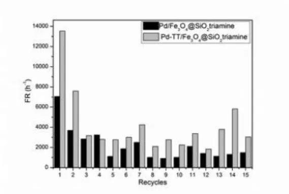
of Pd, as determined by atomic absorption analysis (ICP OES). The sizes of the supported Pd NPs were determined using a transmission electron microscopy (TEM). The Pd NPs (Figure 1 c,d) demonstrated a mean diameter of 3,05 ± 1,46 nm (Pd/Fe3O4@SiO2triamine). In order to study the catalytic activity after the removal of the organic groups, the catalyst were calcined (Figure 1 e,f) at 400 ºC by 2 hours (Pd-TT/F3O4@SiO2triamine). Transmission electron microscopy analysis demonstrated a mean diameter of 4,21 ± 1,64 nm.
The catalytic activity of the Pd catalysts was investigated in the hydrogenation of cyclohexene in solventless conditions. A strong influence of the functional group on the catalytic behavior of the Pd catalyst was observed. The turnover frequency (TOF), for the catalyst functionalized with diethylenetriamine was 7051,86 h-1 (Figure 2). After calcination, to remove the organic ligand, the catalytic rate was improved (13534,22 h-1). Both catalysts, synthetized in this work, were reused in 15 successive runs, and the catalytic activity decreased as the catalysts were reused in successive reactions. These results show that the diethylenetriamine grafted on the magnetite surfaces has a strong deactivating effect on the catalytic activity of the Pd NPs when compared with amine groups or ethylenediamine, reported elsewhere.4,5 The catalyst was calcined its catalytic activity was improved. However calcination causes growth in the nanoparticles.
In summary, we have prepared a magnetically recoverable Pd nanocatalyst with a strong metal- support interaction promoted by ligands grafted on the support surface. The catalytic activity in hydrogenation of olefins was strongly influenced by the organoalkoxysilane present, and the metal-support interaction will be investigated in more detail.
The authors gratefully acknowledge support from FAPESP, CNPq and CAPES.
[1] Jacinto, M. J. et al. M. Appl. Catal. A Gen. 2008, 338, 52.
[2] Rossi, L. M. et al. Appl. Catal. A Gen. 2007, 330, 139.
[3] Silva, T. A. G. et al. Catal. Sci. Technol. 2013, 3, 2993.
[4] Rossi, L. M. et al. Inorg. Chem. 2009, 48, 4640.
[5] Silva, F. P. da et al. Tetrahedron 2013, 70, 3314.
120 TEM Summer School
Figure 1. TEM images of: a) Fe3O4@SiO2triamine, c) Pd/Fe3O4@SiO2triamine and e) Pd-TT/F3O4@SiO2tri- amine; histogram showing particle size distribution of: b) Fe3O4@SiO2triamine, d) Pd/Fe3O4@SiO2triamine and f) Pd-TT/F3O4@SiO2triamine.
Figure 2. Catalyst recycling in the hydrogenation of cyclohexene. Conditions: 6 atm H2, 75 °C, 17,5 mmol of cyclohexene and 50 mg of supported catalyst (2500 mol of substrate/mol of catalyst).
121 CNPEM
Brittle behavior of friction stirred welded joints in pipeline steel API-5L-X80
Julián Arnaldo Ávila Díaz1,2,*, Andre Paulo Tschiptschin2,3, Antonio José Ramirez1,2,4.
1 School of Mechanical Engineering, University of Campinas, Campinas, Sao Paulo, Brazil
2 Brazilian Nanotechnology National Laboratory, Campinas, Sao Paulo, Brazil
3 Polytechnic School, University of São Paulo, Sao Paulo, Brazil.
4 The Ohio State University, Columbus, Ohio, USA
*Corresponding author: [email protected].
The fracture toughness of friction-stirred welded (FSW) joints at the temperature range of 0 C to -40 °C is a critical issue for decisions about its application in API-5L-X80 steels; however, brittle fracture behavior of FSW weldments at -20 °C has been reported [1,2]. Previous results from the microstructural characterization of FSW joints have been shown in Figure 1 [3]. The brittle behavior was associated with crack initiation at the second dispersed phases (martensite- austenite microconstituent –M-A) and (TiNb)(C,N)-type inclusions [1]. In addition, the same mechanism involving M-A particles has been associated with brittle fracture initiation and crack propagation in the coarse-grained heat affected zone of arc welding joints [4–5]. However, these microstructural features do not completely explain the brittle behavior within the stirred and hard zones of friction stir welded joints, overall, arc-welded and FSW microstructures within the joint are dissimilar, although they both have the suspect M-A microconstituent and Ti-N inclusions.
The aim of the present proposal is a deeper microstructural characterization that would pinpoint the key factor that are involved in the reported brittle behavior, more specifically at the stirred and hard zones of FSW welded joints in API-5L-X8 steel, see Figure 1. The focus of this proposal is the determination of the crystal structure, distribution, shape and hardness of the different elements belonging to the bainitic matrix and second dispersed phases over those critical regions. Therefore, transmission electron microscopy (TEM) is the most suitable characterization technique for this proposal.
122 TEM Summer School
Figure 1: Microstructures of different regions within the friction stirred welded joints: a) Cross section Macro photo of the joint, microstructure of b) BM, Base material; c) ITHAZ, intermediate temperature HAZ;
d) HTHAZ, high temperature HAZ; e) SZ, stirred zone and f) HZ, hard zone Etched with Nital 2%. Abreviation:
F, ferrite; BF, bainitic ferrite; GB, granular bainite and SP, secondary phases. The arrows indicate the position of some specific microstructure.
FSW, API 5L X80 steel, brittle fracture, welded joints, TEM
The authors would like to acknowledge the financial support of Colciencias by the scholarship No. 512 from 2010. We would like to thank the following institutions: Petrobras and LNNano/CNPEM for providing eco- nomic funding and laboratory facilities where this work will be developed; TenarisConfab for the materials donation.
[1] Fairchild D.P. et al., Trends Weld. Res. 9th Int. Conf., 193–200, 2012.
[2] Ávila, J.A.D., Eng. Fract. Mech., October, 147, 176–186, 2015.
[3] Ávila, J.A.D et al., E., Metall. Mater. Trans. A, submitting, xx-xx, 20xx.
[4] Mohseni P., University of Science and Technology, PhD thesis, 2012.
[5] Fairchild D.P., ASTM STP 1058, 117–141, 1990.
123 CNPEM
Development of nanostructured lipid carriers containing a phenolic monoterpene as an alternative to leishmaniasis treatment
Juliana G. Galvão1, Raquel L. Santos1, Tainara Santos1, Silvio S. Dolabella2, Rogéria S. Nunes1
11Pharmacy Department, Federal University of Sergipe, São Cristóvão, Sergipe, Brazil
2Morphology Department, Federal University of Sergipe, São Cristóvão, Sergipe, Brazil Corresponding Author: [email protected]
Leishmaniasis consists in a neglected infectious disease caused by protozoa of the genus Leishmania in the family Trypanosomatidae. It is estimated that occurs 1.3 million new cases and 20,000 to 30,000 deaths every year in the world. Since the control of insect vector is difficult and no effective vaccine exists, chemotherapy is the main means of dealing with this disease. In the past 70 years, pentavalent antimonials, amphotericin B and miltefosine have been used as gold standard treatment of leishmaniasis. However, the leishmaniasis conventional treatment presents some undesirable adverse effects such as gastrointestinal disorders, kidney and liver toxicity. Since these drugs are too expensive and Leishmania species have been demonstrated resistance against these drugs, natural products can provide an important alternative to leishmaniasis treatment. A phenolic monoterpene (natural product obtained from essential oils) has been recently reported as a potent leishmanicidal agent. Furthermore, some studies also have been reported the use of nanostructured lipid carriers as drug delivery systems of leishmanicidal drugs. Thus, the aim of this project focuses on the development of Nanostructured Lipid Carriers (NLCs) containing a phenolic monoterpene as an alternative leishmaniasis treatment. Firstly, the NLCs with and without monoterpene will be obtained by solvent diffusion method and evaluated for particle size, polidispersity index, zeta potential. NLCs were characterized by DSC, XRD and Transmission electron microscopy (TEM) as well. Moreover, the analytical methodology for quantify the monoterpene by high performance liquid chromatography (HPLC/UV) will be validated. Afterwards, it will be determined the encapsulation efficiency and in vitro release profile of the drug from the NLCs. Finally, it will be performed the cytotoxicity assay, promastigote viability, as well as, antiamastigote activity evaluation.
The authors acknowledge CAPES and CNPq for supporting funds.
[1] WHO. Leishmaniasis Fact Sheet N° 375 Updated February 2015.
[2] TIUMAN et al., International Journal of Infectious Diseases, 15, 525-532 (2011).
[3] PASTOR et al., Acta Tropica, 145, 31-38 (2015).
[4] DOMINGO et al. Analytica Chimica Acta, 744, p. 8–22 (2012).