Diagnosis and management of nonvariceal upper gastrointestinal hemorrhage: European Society of Gastrointestinal Endoscopy (ESGE) Guideline
Authors Ian M. Gralnek1, 2, Jean-Marc Dumonceau3, Ernst J. Kuipers4, Angel Lanas5, David S. Sanders6, Matthew Kurien6, Gianluca Rotondano7, Tomas Hucl8, Mario Dinis-Ribeiro9, Riccardo Marmo10, Istvan Racz11, Alberto Arezzo12, Ralf-Thorsten Hoffmann13, Gilles Lesur14, Roberto de Franchis15, Lars Aabakken16, Andrew Veitch17, Franco Radaelli18, Paulo Salgueiro19, Ricardo Cardoso20, Luís Maia19, Angelo Zullo21, Livio Cipolletta22, Cesare Hassan23
Institutions Institutions listed at end of article.
Bibliography DOIhttp://dx.doi.org/
10.1055/s-0034-1393172 Published online: 0.0.
Endoscopy 2015; 47: 1–46
© Georg Thieme Verlag KG Stuttgart · New York ISSN 0013-726X Corresponding author Ian M. Gralnek, MD, MSHS Institute of Gastroenterology and Liver Diseases, Ha'Emek Medical Center
Rappaport Faculty of Medicine, Technion-Israel Institute of Technology
Afula, Israel 18101 Fax: +972-4-6495314 ian_gr@clalit.org.il
This Guideline is an official statement of the European Society of Gastrointestinal Endoscopy (ESGE). It addresses the diagnosis and management of nonvariceal upper gastrointestinal hemorrhage (NVUGIH).
Main Recommendations
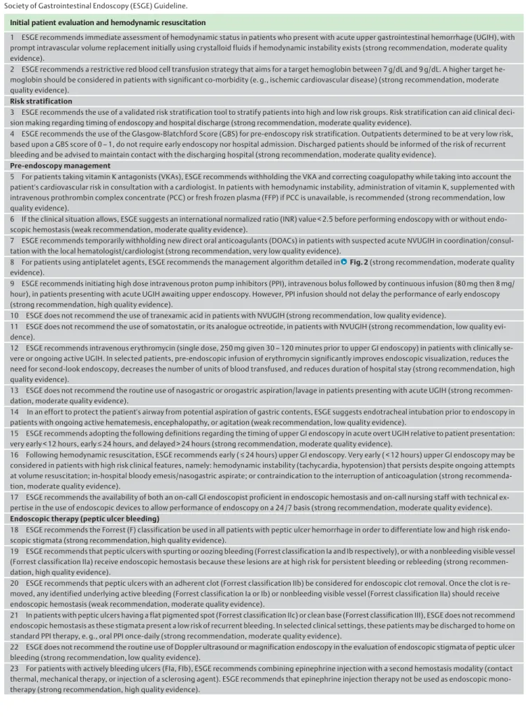
MR1.ESGE recommends immediate assessment of hemodynamic status in patients who present with acute upper gastrointestinal hemorrhage (UGIH), with prompt intravascular volume replacement initially using crystalloid fluids if hemodynamic instability exists (strong recommendation, mod- erate quality evidence).
MR2.ESGE recommends a restrictive red blood cell transfusion strategy that aims for a target hemo- globin between 7 g/dL and 9 g/dL. A higher target hemoglobin should be considered in patients with significant co-morbidity (e. g., ischemic car- diovascular disease) (strong recommendation, moderate quality evidence).
MR3.ESGE recommends the use of the Glasgow- Blatchford Score (GBS) for pre-endoscopy risk stra- tification. Outpatients determined to be at very low risk, based upon a GBS score of 0–1, do not re- quire early endoscopy nor hospital admission. Dis- charged patients should be informed of the risk of recurrent bleeding and be advised to maintain contact with the discharging hospital (strong re- commendation, moderate quality evidence).
MR4.ESGE recommends initiating high dose intra- venous proton pump inhibitors (PPI), intravenous bolus followed by continuous infusion (80 mg then 8 mg/hour), in patients presenting with acute UGIH awaiting upper endoscopy. However, PPI in- fusion should not delay the performance of early endoscopy (strong recommendation, high quality evidence).
MR5.ESGE does not recommend the routine use of nasogastric or orogastric aspiration/lavage in pa- tients presenting with acute UGIH (strong recom- mendation, moderate quality evidence).
MR6.ESGE recommends intravenous erythromy- cin (single dose, 250 mg given 30–120 minutes prior to upper gastrointestinal [GI] endoscopy) in
patients with clinically severe or ongoing active UGIH. In selected patients, pre-endoscopic infu- sion of erythromycin significantly improves endo- scopic visualization, reduces the need for second- look endoscopy, decreases the number of units of blood transfused, and reduces duration of hospital stay (strong recommendation, high quality evi- dence).
MR7.Following hemodynamic resuscitation, ESGE recommends early (≤24 hours) upper GI endos- copy. Very early (< 12 hours) upper GI endoscopy may be considered in patients with high risk clini- cal features, namely: hemodynamic instability (ta- chycardia, hypotension) that persists despite on- going attempts at volume resuscitation; in-hospi- tal bloody emesis/nasogastric aspirate; or contra- indication to the interruption of anticoagulation (strong recommendation, moderate quality evi- dence).
MR8.ESGE recommends that peptic ulcers with spurting or oozing bleeding (Forrest classification Ia and Ib, respectively) or with a nonbleeding visi- ble vessel (Forrest classification IIa) receive endo- scopic hemostasis because these lesions are at high risk for persistent bleeding or rebleeding (strong recommendation, high quality evidence).
MR9.ESGE recommends that peptic ulcers with an adherent clot (Forrest classification IIb) be consid- ered for endoscopic clot removal. Once the clot is removed, any identified underlying active bleed- ing (Forrest classification Ia or Ib) or nonbleeding visible vessel (Forrest classification IIa) should re- ceive endoscopic hemostasis (weak recommenda- tion, moderate quality evidence).
MR10.In patients with peptic ulcers having a flat pigmented spot (Forrest classification IIc) or clean base (Forrest classification III), ESGE does not re- commend endoscopic hemostasis as these stigma-
Abbreviations
!
APC argon plasma coagulation
ASA American Society of Anesthesiologists DAPT dual antiplatelet therapy
CHADS2 congestive heart failure, hypertension, age≥75 years, diabetes mellitus, and previous stroke or transient ischemic attack [risk score]
CI confidence interval DOAC direct oral anticoagulant
ESGE European Society of Gastrointestinal Endoscopy FFP fresh frozen plasma
GBS Glasgow-Blatchford Score GI gastrointestinal
GRADE Grading of Recommendations Assessment, Development and Evaluation
HR hazard ratio
INR international normalized ratio NBVV nonbleeding visible vessel NNT number needed to treat NOAC non-VKA oral anticoagulant
NVUGIH nonvariceal upper gastrointestinal hemorrhage PAR protease-activated receptor
PCC prothrombin complex concentrate PICO patients, interventions, controls, outcomes PPI proton pump inhibitor
OR odds ratio
PUB peptic ulcer bleeding RBC red blood cell
RCT randomized controlled trial RR relative riskorrisk ratio
TAE transcatheter angiographic embolization UGIH upper gastrointestinal hemorrhage VCE videocapsule endoscopy
VKA vitamin K antagonist
Introduction
!
Acute upper gastrointestinal hemorrhage (UGIH) is a common condition worldwide that has an estimated annual incidence of 40−150 cases per 100 000 population [1, 2], frequently leads to hospital admission, and has significant associated morbidity and mortality, especially in the elderly. The most common causes of acute UGIH are nonvariceal [1, 2]. This includes peptic ulcers, 28
%–59 % (duodenal ulcer 17 %–37 % and gastric ulcer 11 %–24 %);
mucosal erosive disease of the esophagus/stomach/duodenum, 1 %–47 %; Mallory–Weiss syndrome, 4 %–7 %; upper GI tract ma- lignancy, 2 %–4 %; other diagnosis, 2 %–7 %; or no exact cause identified, 7 %–25 % [1, 2]. Moreover, in 16 %–20 % of acute UGIH cases, more than one endoscopic diagnosis may be identified as the cause of bleeding. The aim of this evidence-based consensus guideline is to provide medical caregivers with a comprehensive review and recommendations on the clinical and endoscopic management of NVUGIH.
Methods
!
The ESGE commissioned this guideline on NVUGIH and appoin- ted a guideline leader (I.M.G.) who in collaboration with the Chair of the ESGE Guidelines Committee (C.H.), invited the listed au- thors to participate in the guideline development and review.
Key questions were prepared by the coordinating team (I.M.G.
and C.H.) and reviewed and approved by all task force members.
The coordinating team formed four task force subgroups, each with its own coordinator, and divided the key topics/questions amongst these four task force subgroups (seeAppendix e1, on- line-only). Task force members included gastroenterologists/gas- trointestinal endoscopists, an interventional radiologist, and a surgeon. Clinical questions were formulated using the PICO (pa- tients, interventions, controls, outcomes) methodology.
Each task force subgroup performed a systematic literature search to identify the relevant literature that was subsequently used to prepare evidence-based, well-balanced statements on each of their assigned key questions. The Ovid MEDLINE, EM- BASE, Google/Google Scholar, and the Cochrane Database of Sys- ta present a low risk of recurrent bleeding. In selected clinical set-
tings, these patients may be discharged to home on standard PPI therapy, e. g., oral PPI once-daily (strong recommendation, moder- ate quality evidence).
MR11.ESGE recommends that epinephrine injection therapy not be used as endoscopic monotherapy. If used, it should be combined with a second endoscopic hemostasis modality (strong recom- mendation, high quality evidence).
MR12.ESGE recommends PPI therapy for patients who receive endoscopic hemostasis and for patients with adherent clot not re- ceiving endoscopic hemostasis. PPI therapy should be high dose and administered as an intravenous bolus followed by continuous infusion (80 mg then 8 mg/hour) for 72 hours post endoscopy (strong recommendation, high quality evidence).
MR13.ESGE does not recommend routine second-look endoscopy as part of the management of nonvariceal upper gastrointestinal hemorrhage (NVUGIH). However, in patients with clinical evi- dence of rebleeding following successful initial endoscopic hemo- stasis, ESGE recommends repeat upper endoscopy with hemosta-
sis if indicated. In the case of failure of this second attempt at he- mostasis, transcatheter angiographic embolization (TAE) or sur- gery should be considered (strong recommendation, high quality evidence).
MR14.In patients with NVUGIH secondary to peptic ulcer, ESGE re- commends investigating for the presence ofHelicobacter pyloriin the acute setting with initiation of appropriate antibiotic therapy whenH. pyloriis detected. Re-testing forH. pylorishould be per- formed in those patients with a negative test in the acute setting.
Documentation of successfulH. pylorieradication is recommended (strong recommendation, high quality evidence).
MR15.In patients receiving low dose aspirin for secondary cardio- vascular prophylaxis who develop peptic ulcer bleeding, ESGE re- commends aspirin be resumed immediately following index endoscopy if the risk of rebleeding is low (e. g., FIIc, FIII). In patients with high risk peptic ulcer (FIa, FIb, FIIa, FIIb), early reintroduction of aspirin by day 3 after index endoscopy is recommended, provid- ed that adequate hemostasis has been established (strong recom- mendation, moderate quality evidence).
tematic Reviews were searched for English-language articles in- cluding at a minimum the following key words: nonvariceal up- per gastrointestinal (GI) hemorrhage/bleeding, peptic ulcer he- morrhage/bleeding, fluid resuscitation, fluid therapy, critical ill- ness, crystalloid solutions, colloid solutions, plasma transfusions, red blood cell transfusion, platelet transfusion, hemoglobin, re- strictive transfusion strategy, liberal transfusion strategy, risk stratification, mortality, rebleeding, anti-thrombotic agent, anti- platelet agent, aspirin, dual anti-platelet therapy (DAPT), anti-co- agulation/anti-coagulant, direct/new oral anticoagulants (DOACs), coagulopathy, vitamin K inhibitor/antagonist, prokinet- ic agent, erythromycin, fresh frozen plasma, nasogastric tube, or- ogastric tube, proton pump inhibitor, prokinetic agent, erythro- mycin, endoscopic hemostasis, injection therapy, thermal ther- apy (contact, non-contact), mechanical therapy/endoscopic clip- ping, topical hemostasis therapy, second-look endoscopy, helico- bacter pylori,H. pylori, transcatheter angiographic embolization (TAE), and surgery. The hierarchy of studies included as part of this evidence-based guideline was, in decreasing order of evi- dence level, published systematic reviews/meta-analyses, ran- domized controlled trials (RCTs), prospective and retrospective observational studies. All selected articles were graded using the Grading of Recommendations Assessment, Development and Evaluation (GRADE) system [3, 4].
Each task force subgroup proposed statements for each of their assigned key questions which were discussed and voted on dur- ing the NVUGIH task force guideline meeting held in Berlin, Ger- many in November 2014. In August 2015, a manuscript draft pre- pared by I.M.G. was sent to all task force members. After agree- ment on a final version, the manuscript was reviewed by two members of the ESGE Governing Board and sent for further com- ments to the National Societies and ESGE individual members.
After agreement on a final version, the manuscript was submit- ted to the journalEndoscopyfor publication. All authors agreed on the final revised manuscript.
This NVUGIH guideline will be considered for review and updat- ing in 2020, or sooner if new relevant evidence becomes avail- able. Any updates to this guideline in the interim will be noted on the ESGE website: http://www.esge.com/esge-guidelines.
html.
Statements and recommendations
!
See
●
" Table 1.Initial patient evaluation and hemodynamic resuscitation
ESGE recommends immediate assessment of hemodynamic status in patients who present with acute upper gastrointestinal hemorrhage (UGIH), with prompt intravascular volume replacement initially using crystalloid fluids if hemodynamic instability exists (strong recommendation, moderate quality evidence).
The goals of hemodynamic resuscitation are to correct intravas- cular hypovolemia, restore adequate tissue perfusion, and pre- vent multi-organ failure. Early intensive hemodynamic resuscita- tion of patients with acute UGIH has been shown to significantly decrease mortality [5]. In an observational study of patients with acute UGIH and hemodynamic instability, patients who received intensive hemodynamic resuscitation had significantly fewer myocardial infarctions and lower mortality compared with those
in the“observation group”(P= 0.04 for both comparisons). How- ever, there is no evidence from randomized controlled trials (RCTs), for or against early or large-volume intravenous fluid ad- ministration in uncontrolled hemorrhage [6, 7]. Moreover, the se- lection of resuscitation fluid type in critically ill patients requires careful consideration based on safety, effects on patient out- comes, and costs. To date, there is ongoing uncertainty regarding the ideal fluid administration strategy in this clinical setting [8, 9].
ESGE recommends a restrictive red blood cell transfusion strategy that aims for a target hemoglobin between 7 g/dL and 9 g/dL. A higher target hemo- globin should be considered in patients with significant co-morbidity (e. g., ischemic cardiovascular disease) (strong recommendation, moderate quality evidence).
The use of red blood cell (RBC) transfusions may be lifesaving fol- lowing massive UGIH. However, the role of RBC transfusion in less torrential GI bleeding remains controversial, with uncertain- ty existing regarding the hemoglobin level at which blood trans- fusion should be initiated. This uncertainty reflects concerns from both the critical care and gastroenterology literature sug- gesting poorer outcomes in patients managed with a liberal RBC transfusion strategy [2, 10, 11]. In a recent RCT that included 921 patients presenting with all causes of acute UGIH, a restrictive RBC transfusion strategy (target hemoglobin, 7 to 9 g/dL) was compared with a more liberal transfusion strategy (target hemo- globin, 9 to 11 g/dL) [12]. The restrictive RBC transfusion group had significantly improved 6-week survival (95 % vs. 91 %; hazard ratio [HR] 0.55, 95 % confidence interval [CI] 0.33–0.92) and re- duced rebleeding (10 % vs.16 %; HR 0.68, 95 %CI 0.47–0.98) [12].
In the subgroup of patients with NVUGIH (n = 699), there was a statistical trend towards lower mortality in the restrictive vs. lib- eral RBC transfusion strategy (3.7 % vs. 6.9 %,P= 0.065). Because the study was not powered to specifically evaluate NVUGIH, these findings should be interpreted with caution. Other limita- tions of this study include the exclusion of patients with massive exsanguinating bleeding and defined co-morbidities. Further- more, all patients underwent endoscopy within 6 hours of pre- sentation, which may not be feasible in everyday clinical practice.
Coagulopathy at the time of NVUGIH presentation is another fre- quent and adverse prognostic factor [13]. Published data for the management of coagulopathy are limited and inconclusive. One small cohort study using an historical comparison group showed that aggressive volume resuscitation, including correction of coa- gulopathy (international normalized ratio [INR] < 1.8), led to an improvement in mortality outcomes [5]. In a systematic review that evaluated the relevance of initial INR before correction in pa- tients with NVUGIH, INR did not appear to predict rebleeding, yet after adjusting for potential confounders, an initial INR > 1.5 pre- dicted mortality (odds ratio [OR] 1.96, 95 %CI 1.13–3.41) [14].
This may in part reflect the presence of underlying liver disease.
There is however no available evidence to help guide coagulopa- thy correction in critically ill patients and wide variation in man- agement exists in this area, indicating clinical uncertainty re- garding optimal practice [15]. Platelet count has not been shown to be a predictor of either rebleeding or mortality. Currently, there is no high quality evidence to guide platelet transfusion thresholds, although a platelet transfusion threshold of 50 × 109/ L has been proposed for most patients, with a target of 10 × 109/L for patients in whom platelet dysfunction is suspected [16].
Table 1 Summary of Guideline statements and recommendations. Diagnosis and management of nonvariceal upper gastrointestinal hemorrhage: European Society of Gastrointestinal Endoscopy (ESGE) Guideline.
Initial patient evaluation and hemodynamic resuscitation
1 ESGE recommends immediate assessment of hemodynamic status in patients who present with acute upper gastrointestinal hemorrhage (UGIH), with prompt intravascular volume replacement initially using crystalloid fluids if hemodynamic instability exists (strong recommendation, moderate quality evidence).
2 ESGE recommends a restrictive red blood cell transfusion strategy that aims for a target hemoglobin between 7 g/dL and 9 g/dL. A higher target he- moglobin should be considered in patients with significant co-morbidity (e. g., ischemic cardiovascular disease) (strong recommendation, moderate quality evidence).
Risk stratification
3 ESGE recommends the use of a validated risk stratification tool to stratify patients into high and low risk groups. Risk stratification can aid clinical deci- sion making regarding timing of endoscopy and hospital discharge (strong recommendation, moderate quality evidence).
4 ESGE recommends the use of the Glasgow-Blatchford Score (GBS) for pre-endoscopy risk stratification. Outpatients determined to be at very low risk, based upon a GBS score of 0–1, do not require early endoscopy nor hospital admission. Discharged patients should be informed of the risk of recurrent bleeding and be advised to maintain contact with the discharging hospital (strong recommendation, moderate quality evidence).
Pre-endoscopy management
5 For patients taking vitamin K antagonists (VKAs), ESGE recommends withholding the VKA and correcting coagulopathy while taking into account the patient's cardiovascular risk in consultation with a cardiologist. In patients with hemodynamic instability, administration of vitamin K, supplemented with intravenous prothrombin complex concentrate (PCC) or fresh frozen plasma (FFP) if PCC is unavailable, is recommended (strong recommendation, low quality evidence).
6 If the clinical situation allows, ESGE suggests an international normalized ratio (INR) value < 2.5 before performing endoscopy with or without endo- scopic hemostasis (weak recommendation, moderate quality evidence).
7 ESGE recommends temporarily withholding new direct oral anticoagulants (DOACs) in patients with suspected acute NVUGIH in coordination/consul- tation with the local hematologist/cardiologist (strong recommendation, very low quality evidence).
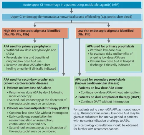
8 For patients using antiplatelet agents, ESGE recommends the management algorithm detailed in
●
"Fig. 2(strong recommendation, moderate quality evidence).9 ESGE recommends initiating high dose intravenous proton pump inhibitors (PPI), intravenous bolus followed by continuous infusion (80 mg then 8 mg/
hour), in patients presenting with acute UGIH awaiting upper endoscopy. However, PPI infusion should not delay the performance of early endoscopy (strong recommendation, high quality evidence).
10 ESGE does not recommend the use of tranexamic acid in patients with NVUGIH (strong recommendation, low quality evidence).
11 ESGE does not recommend the use of somatostatin, or its analogue octreotide, in patients with NVUGIH (strong recommendation, low quality evi- dence).
12 ESGE recommends intravenous erythromycin (single dose, 250 mg given 30–120 minutes prior to upper GI endoscopy) in patients with clinically se- vere or ongoing active UGIH. In selected patients, pre-endoscopic infusion of erythromycin significantly improves endoscopic visualization, reduces the need for second-look endoscopy, decreases the number of units of blood transfused, and reduces duration of hospital stay (strong recommendation, high quality evidence).
13 ESGE does not recommend the routine use of nasogastric or orogastric aspiration/lavage in patients presenting with acute UGIH (strong recommen- dation, moderate quality evidence).
14 In an effort to protect the patient's airway from potential aspiration of gastric contents, ESGE suggests endotracheal intubation prior to endoscopy in patients with ongoing active hematemesis, encephalopathy, or agitation (weak recommendation, low quality evidence).
15 ESGE recommends adopting the following definitions regarding the timing of upper GI endoscopy in acute overt UGIH relative to patient presentation:
very early < 12 hours, early≤24 hours, and delayed > 24 hours (strong recommendation, moderate quality evidence).
16 Following hemodynamic resuscitation, ESGE recommends early (≤24 hours) upper GI endoscopy. Very early ( < 12 hours) upper GI endoscopy may be considered in patients with high risk clinical features, namely: hemodynamic instability (tachycardia, hypotension) that persists despite ongoing attempts at volume resuscitation; in-hospital bloody emesis/nasogastric aspirate; or contraindication to the interruption of anticoagulation (strong recommenda- tion, moderate quality evidence).
17 ESGE recommends the availability of both an on-call GI endoscopist proficient in endoscopic hemostasis and on-call nursing staff with technical ex- pertise in the use of endoscopic devices to allow performance of endoscopy on a 24 /7 basis (strong recommendation, moderate quality evidence).
Endoscopic therapy (peptic ulcer bleeding)
18 ESGE recommends the Forrest (F) classification be used in all patients with peptic ulcer hemorrhage in order to differentiate low and high risk endo- scopic stigmata (strong recommendation, high quality evidence).
19 ESGE recommends that peptic ulcers with spurting or oozing bleeding (Forrest classification Ia and Ib respectively), or with a nonbleeding visible vessel (Forrest classification IIa) receive endoscopic hemostasis because these lesions are at high risk for persistent bleeding or rebleeding (strong recommen- dation, high quality evidence).
20 ESGE recommends that peptic ulcers with an adherent clot (Forrest classification IIb) be considered for endoscopic clot removal. Once the clot is re- moved, any identified underlying active bleeding (Forrest classification Ia or Ib) or nonbleeding visible vessel (Forrest classification IIa) should receive endoscopic hemostasis (weak recommendation, moderate quality evidence).
21 In patients with peptic ulcers having a flat pigmented spot (Forrest classification IIc) or clean base (Forrest classification III), ESGE does not recommend endoscopic hemostasis as these stigmata present a low risk of recurrent bleeding. In selected clinical settings, these patients may be discharged to home on standard PPI therapy, e. g., oral PPI once-daily (strong recommendation, moderate quality evidence).
22 ESGE does not recommend the routine use of Doppler ultrasound or magnification endoscopy in the evaluation of endoscopic stigmata of peptic ulcer bleeding (strong recommendation, low quality evidence).
23 For patients with actively bleeding ulcers (FIa, FIb), ESGE recommends combining epinephrine injection with a second hemostasis modality (contact thermal, mechanical therapy, or injection of a sclerosing agent). ESGE recommends that epinephrine injection therapy not be used as endoscopic mono- therapy (strong recommendation, high quality evidence).
Risk stratification
ESGE recommends the use of a validated risk stratification tool to stratify pa- tients into high and low risk groups. Risk stratification can aid clinical decision making regarding timing of endoscopy and hospital discharge (strong re- commendation, moderate quality evidence).
ESGE recommends the use of the Glasgow-Blatchford Score (GBS) for pre- endoscopy risk stratification. Outpatients determined to be at very low risk, based upon a GBS score of 0–1, do not require early endoscopy nor hospital
admission. Discharged patients should be informed of the risk of recurrent bleeding and be advised to maintain contact with the discharging hospital (strong recommendation, moderate quality evidence).
Risk stratification of patients presenting with acute UGIH can as- sist in identifying those who may require more urgent interven- tion and help triage patients to in-hospital vs. out-of-hospital management. A number of scoring tools have been created for predicting outcomes following acute UGIH, with the Glasgow- Blatchford Score (GBS) (
●
" Table 2) and Rockall score being the most widely evaluated and adopted [17–19]. However, no single scoring tool has been shown to excel at predicting all relevant Table 1 (Continuation)Initial patient evaluation and hemodynamic resuscitation
24 For patients with nonbleeding visible vessel (FIIa), ESGE recommends mechanical therapy, thermal therapy, or injection of a sclerosing agent as monotherapy or in combination with epinephrine injection. ESGE recommends that epinephrine injection therapy not be used as endoscopic monotherapy (strong recommendation, high quality evidence).
25 For patients with active NVUGIH bleeding not controlled by standard endoscopic hemostasis therapies, ESGE suggests the use of a topical hemostatic spray or over-the-scope clip as salvage endoscopic therapy (weak recommendation, low quality evidence).
Endoscopic therapy (other causes of NVUGIH)
26 For patients with acid-related causes of NVUGIH different from peptic ulcers (e. g., erosive esophagitis, gastritis, duodenitis), ESGE recommends treatment with high dose PPI. Endoscopic hemostasis is usually not required and selected patients may be discharged early (strong recommendation, low quality evidence).
27 ESGE recommends that patients with a Mallory–Weiss lesion that is actively bleeding receive endoscopic hemostasis. There is currently inadequate evidence to recommend a specific endoscopic hemostasis modality. Patients with a Mallory–Weiss lesion and no active bleeding can receive high dose PPI therapy alone (strong recommendation, moderate quality evidence).
28 ESGE recommends that a Dieulafoy lesion receive endoscopic hemostasis using thermal, mechanical (hemoclip or band ligation), or combination therapy (dilute epinephrine injection combined with contact thermal or mechanical therapy) (strong recommendation, moderate quality evidence).
Transcatheter angiographic embolization (TAE) or surgery should be considered if endoscopic treatment fails or is not technically feasible (strong recom- mendation, low quality evidence).
29 In patients bleeding from upper GI angioectasias, ESGE recommends endoscopic hemostasis therapy. However, there is currently inadequate evidence to recommend a specific endoscopic hemostasis modality (strong recommendation, low quality evidence).
30 In patients bleeding from upper GI neoplasia, ESGE recommends considering endoscopic hemostasis in order to avert urgent surgery and reduce blood transfusion requirements. However, no currently available endoscopic treatment appears to have long-term efficacy (weak recommendation, low quality evidence).
Post endoscopy/endoscopic hemostasis management
31 ESGE recommends PPI therapy for patients who receive endoscopic hemostasis and for patients with adherent clot not receiving endoscopic hemo- stasis. PPI therapy should be high dose and administered as an intravenous bolus followed by continuous infusion (80 mg then 8 mg /hour) for 72 hours post endoscopy (strong recommendation, high quality evidence).
32 ESGE suggests considering PPI therapy as intermittent intravenous bolus dosing (at least twice-daily) for 72 hours post endoscopy for patients who receive endoscopic hemostasis and for patients with adherent clot not receiving endoscopic hemostasis. If the patient’s condition permits, high dose oral PPI may also be an option in those able to tolerate oral medications (weak recommendation, moderate quality evidence).
33 In patients with clinical evidence of rebleeding following successful initial endoscopic hemostasis, ESGE recommends repeat upper endoscopy with hemostasis if indicated. In the case of failure of this second attempt at hemostasis, transcatheter angiographic embolization (TAE) or surgery should be considered (strong recommendation, high quality evidence).
34 ESGE does not recommend routine second-look endoscopy as part of the management of NVUGIH. However, second-look endoscopy may be consid- ered in selected patients at high risk for rebleeding (strong recommendation, high quality evidence).
35 In patients with NVUGIH secondary to peptic ulcer, ESGE recommends investigating for the presence ofHelicobacter pyloriin the acute setting with initiation of appropriate antibiotic therapy whenH. pyloriis detected. Re-testing forH. pylorishould be performed in those patients with a negative test in the acute setting. Documentation of successfulH. pylorieradication is recommended (strong recommendation, high quality evidence).
36 ESGE recommends restarting anticoagulant therapy following NVUGIH in patients with an indication for long-term anticoagulation. The timing for resumption of anticoagulation should be assessed on a patient by patient basis. Resuming warfarin between 7 and 15 days following the bleeding event appears safe and effective in preventing thromboembolic complications for most patients. Earlier resumption, within the first 7 days, may be indicated for patients at high thrombotic risk (strong recommendation, moderate quality evidence).
37 In patients receiving low dose aspirin for primary cardiovascular prophylaxis who develop peptic ulcer bleeding, ESGE recommends withholding as- pirin, re-evaluating the risks/benefits of ongoing aspirin use in consultation with a cardiologist, and resuming low dose aspirin following ulcer healing or earlier if clinically indicated (strong recommendation, low quality evidence).
38 In patients receiving low dose aspirin for secondary cardiovascular prophylaxis who develop peptic ulcer bleeding, ESGE recommends aspirin be re- sumed immediately following index endoscopy if the risk of rebleeding is low (e. g., FIIc, FIII). In patients with high risk peptic ulcer (FIa, FIb, FIIa, FIIb), early reintroduction of aspirin by day 3 after index endoscopy is recommended, provided that adequate hemostasis has been established (strong recommen- dation, moderate quality evidence).
39 In patients receiving dual antiplatelet therapy (DAPT) who develop peptic ulcer bleeding, ESGE recommends continuing low dose aspirin therapy. Early cardiology consultation should be obtained regarding the timing of resuming the second antiplatelet agent (strong recommendation, low quality evi- dence).
40 In patients requiring dual antiplatelet therapy (DAPT) and who have had NVUGIH, ESGE recommends the use of a PPI as co-therapy (strong recom- mendation, moderate quality evidence).
outcomes in acute UGIH (e. g., rebleeding, need for intervention, mortality) [19]. This is not surprising as the most validated risk scores were derived to assess a specific UGIH outcome: that for the Rockall score being mortality and for the GBS being the need for intervention [17, 18].
A recent systematic review evaluating the accuracy of the avail- able UGIH risk stratification tools demonstrated substantial het- erogeneity in predicted outcomes and highlighted that methodo- logical quality of the prediction scores was less than optimal [19].
Regarding the need for intervention, retrospective and prospec- tive studies have assessed the prognostic value of the GBS vs. the Rockall score. These studies showed that the GBS correctly iden- tified 98 % (95 %CI 89 %–100 %) of those patients who did not re- quire any subsequent intervention while 83 % (95 %CI 71 %–91 %) of those patients were identified using the Rockall score. Ran- domized controlled trials and observational studies consistently indicate that clinical, endoscopic, and social factors may identify patients who may be safely discharged for outpatient manage- ment [20–28]. The most frequent adverse event reported is re- bleeding ranging between 0.5 % and 4 %, with no deaths or hospi- tal readmissions for surgery reported. Moreover, studies consis- tently indicate that outpatient management of appropriately se- lected patients with acute UGIH reduces resource utilization [20, 21, 27]. Emergency department discharge without inpatient endoscopy (i. e., outpatient management) should be considered for patients if: systolic blood pressure≥110 mmHg, pulse < 100 beats/minute, hemoglobin≥13.0 g/dL for men or≥12.0 g/dL for women, blood urea nitrogen < 18.2 mg/dL, along with the absence of melena, syncope, hepatic disease, and cardiac failure [18]. (See Appendix e2, online-only.)
Pre-endoscopy management
Initial management of antithrombotic agents (anticoagulants and antiplatelet agents)
For patients taking vitamin K antagonists (VKAs), ESGE recommends with- holding the VKA and correcting coagulopathy while taking into account the patient’s cardiovascular risk in consultation with a cardiologist. In patients with hemodynamic instability, administration of vitamin K, supplemented with intravenous prothrombin complex concentrate (PCC) or fresh frozen plasma (FFP) if PCC is unavailable, is recommended (strong recommendation, low quality evidence).
If the clinical situation allows, ESGE suggests an international normalized ratio (INR) value < 2.5 before performing endoscopy with or without endoscopic hemostasis (weak recommendation, moderate quality evidence).
GI bleeding represents a serious complication of VKA therapy, with an incidence of 1 %–4 % per year [29, 30]. Discontinuation of anticoagulants and correction of coagulopathy before endos- copy is the“standard of practice”in patients with clinically sig- nificant GI bleeding [31–33]. Because data are limited, specific strategies to reverse VKAs in a patient with acute overt UGIH vary [34]. Practice guidelines recommend urgent reversal in all patients presenting with serious, life-threatening bleeding (i. e., hemodynamic instability or shock), either in the case of thera- peutic or supratherapeutic INR elevations [32, 35]. For patients who are not actively bleeding and are hemodynamically stable, intravenous vitamin K administration may be an option. When more urgent reversal is required, administration of prothrombin complex concentrates (PCCs) or fresh frozen plasma (FFP) is nec- essary, with concomitant intravenous administration of 5–10 mg vitamin K to prevent “rebound coagulopathy” once the trans- fused factors have been cleared. Prothrombin complex concen- trates contain clotting factors prepared from pooled and concen- trated human plasma and are preferred over FFP because of sev- eral advantages, including no need to check the patient’s blood group, less risk for volume overload because of smaller transfu- sion volume, faster onset of action, similar thrombotic risk pro- file, and minimal risk of infectious transmission, albeit at a higher cost [36–40]. A recent prospective, nonrandomized, comparative study of 40 warfarin users who presented with UGIH and an INR
> 2.1 reported that patients who received PCC had a near normal- ized INR at 2 hours following infusion (INR = 1.5) while those who received FFP had an INR of 2.4 at 6 hours following infusion [38].
No patient in the PCC group had active bleeding at endoscopy compared with 7 in the FFP group (0 vs. 35 %,P< 0.01). The risk of thrombosis following PCC administration approximates 1 %, and is similar to that reported with FFP [39, 40].
ESGE recommends temporarily withholding new direct oral anticoagulants (DOACs) in patients with suspected acute NVUGIH in coordination/consulta- tion with the local hematologist/cardiologist (strong recommendation, very low quality evidence).
As an alternative to heparin and VKAs, the new non-VKA oral an- ticoagulants (NOACs; also referred to as direct oral anticoagulants [DOACs]) are being rapidly adopted worldwide, primarily for thromboembolic prevention in patients with nonvalvular atrial fibrillation and for prophylaxis or treatment of venous throm- boembolism [41]. These pharmacological agents do however, present a risk of significant GI bleeding similar to or greater than that reported with warfarin [42, 43]. Moreover, DOACs differ in comparison with heparin and VKA. Specifically, in the absence of renal or hepatic failure, DOAC clearance and the subsequent Table 2 Glasgow-Blatchford Score (GBS).
Points Systolic blood pressure, mmHg
100–109 1
90–99 2
< 90 3
Blood urea nitrogen, mmol/L
6.5–7.9 2
8.0–9.9 3
10.0–24.9 4
≥25.0 6
Hemoglobin for men, g/dL
12.0–12.9 1
10.0–11.9 3
< 10.0 6
Hemoglobin for women, g/dL
10.0–11.9 1
< 10.0 6
Other risk variables
Pulse≥100 1
Melena 1
Syncope 2
Hepatic disease 2
Cardiac failure 2
TOTAL GBS __________________
GBS restricted for use only in nonhospitalized, ambulatory patients Risk variables measured at time of patient presentation GBS = 0–1 denotes“low-risk”
loss of anticoagulation effect is rapid and predictable (occurring gradually over 12–24 hours), routine laboratory tests are not sensitive for the quantitative assessment of their anticoagulant activity, and there is currently no specific reversal agent/antidote for emergency use with any DOAC, although potential agents are in development and may be commercially available in the next 1–2 years [44–46]. As there are no published clinical trials ad- dressing the management of GI bleeding in patients using DOAC, current recommendations are based on expert opinion or labora- tory end-points [47–49].
At the time of patient presentation with acute UGIH, DOACs should be temporarily withheld. Given their relatively short half-life, time is the most important antidote against DOACs.
Strategies to accelerate anticoagulation reversal are supported only by data collected from healthy human volunteers, animal models, and in vitro studies [50]. Based on those data, vitamin K or FFP have no place as reversal agents for DOACs. Prothrombin complex concentrates or activated PCC may be considered in pa- tients with severe or life-threatening bleeding, and hemodialysis can be used to reduce the blood concentration of dabigatran, but not that of rivaroxaban and apixaban which are more tightly bound to plasma proteins [48, 49, 51]. Additional data on the clin- ical effectiveness of these strategies in acutely bleeding patients are urgently needed.
For patients using antiplatelet agents, ESGE recommends the management algorithm detailed in
●
" Fig. 1(strong recommendation, moderate quality evidence).Antiplatelet agents include low dose aspirin and thienopyridines (e. g., clopidogrel, prasugrel, ticlopidine) that irreversibly inhibit platelet aggregation, ticagrelor a reversible P2Y12 receptor an- tagonist, and vorapaxar, a protease-activated receptor (PAR-1)
antagonist that inhibits thrombin. The minimum duration of an- tiplatelet agent discontinuation that allows for restoration of nor- mal platelet aggregation is 5–7 days [52].
Studies have shown that in patients taking low dose aspirin for secondary cardiovascular prophylaxis, all-cause mortality was lower if aspirin was not discontinued following peptic ulcer bleeding [53, 54]. In an RCT, 156 recipients of low dose aspirin for secondary prophylaxis who had peptic ulcer bleeding were randomized to receive continuous aspirin or placebo [53]. At 8- week follow up, all-cause mortality was lower in the patients randomized to aspirin compared with placebo (1.3 % vs. 12.9 %, 95 %CI 3.7 %–19.5 %; hazard ratio [HR] 0.20), with the difference being attributable to cardiovascular, cerebrovascular, or GI com- plications. The 30-day ulcer rebleeding rate was not significantly greater in the aspirin group. Patients who required dual antipla- telet therapy (DAPT) were excluded from this study. In a subse- quent retrospective analysis that included 118 low dose aspirin recipients who had been treated for peptic ulcer bleeding and fol- lowed-up for a median of 2 years, 47 (40 %) patients stopped as- pirin [54]. Patients who discontinued aspirin and those who con- tinued aspirin had similar mortality rates (31 %). However, in a subgroup analysis limited to patients with cardiovascular co- morbidities, those patients who discontinued aspirin had an al- most fourfold increase in the risk of death or acute cardiovascular event (P< 0.01) [54]. Randomized controlled trials have shown that neither aspirin nor clopidogrel use impede ulcer healing promoted by proton pump inhibitors (PPI) [55, 56].
Pharmacological therapy
ESGE recommends initiating high dose intravenous proton pump inhibitors (PPI), intravenous bolus followed by continuous infusion (80 mg then 8 mg/
hour), in patients presenting with acute UGIH awaiting upper endoscopy.
Acute upper GI hemorrhage in a patient using antiplatelet agent(s) (APA)
Upper GI endoscopy demonstrates a nonvariceal source of bleeding (e.g. peptic ulcer bleed)
High risk endoscopic stigmata identified (FIa, FIb, FIIa, FIIb)
Low risk endoscopic stigmata identified (FIIc, FIII)
APA used for primary prophylaxis
▪ Withhold low dose acetylsalicylic acid (ASA)
▪ Re-evaluate risks and benefits of ongoing low dose ASA use
▪ Resume low dose ASA after ulcer healing or earlier if clinically indicated
APA used for secondary prophylaxis (known cardiovascular disease) 1 Patients on low dose ASA alone ▪ Resume low dose ASA by day 3 following index endoscopy
▪ Second-look endoscopy at the discretion of the endoscopist may be considered 2 Patients on dual antiplatelet therapy (DAPT) ▪ Continue low dose ASA without interruption ▪ Early cardiology consultation for
recommendation on resumption/
continuation of second APA
▪ Second-look endoscopy at the discretion of the endoscopist may be considered
APA used for secondary prophylaxis (known cardiovascular disease) 1 Patients on low dose ASA alone
▪ Continue low dose ASA without interruption 2 Patients on dual antiplatelet therapy (DAPT) ▪ Continue DAPT without interruption For patients using a non-ASA APA as monotherapy (e.g., thienopyridine alone), low-dose ASA may be given as substitute for interval period in patients with no contraindication or allergy to ASA.
Early cardiology consultation should be obtained for further APA recommendations.
APA used for primary prophylaxis
▪ Withhold low dose ASA
▪ Re-evaluate risks and benefits of ongoing low dose ASA use
▪ Resume low dose ASA at hospital discharge if clinically indicated
Fig. 1 Algorithm for the management of patients with acute upper gastrointestinal hemorrhage who are using antiplatelet agent(s): European Society of Gastrointestinal Endoscopy (ESGE) Guideline.
However, PPI infusion should not delay the performance of early endoscopy (strong recommendation, high quality evidence).
A Cochrane meta-analysis of 6 RCTs (n = 2223 patients) showed that administering PPIs before endoscopy significantly decreases the incidence of high risk stigmata of hemorrhage at the time of index endoscopy (37.2 % vs. 46.5 %; OR 0.67, 95 %CI 0.54–0.84) and the need for endoscopic hemostasis (8.6 % vs. 11.7 %; OR 0.68, 95 %CI 0.50–0.93), but has no effect on rebleeding, need for surgery, or mortality [57].
Cost–effectiveness studies suggest that high dose PPI infusion prior to endoscopy for patients with UGIH is more effective and less costly than placebo [58, 59]. (SeeAppendix e3, online-only.)
ESGE does not recommend the use of tranexamic acid in patients with NVU- GIH (strong recommendation, low quality evidence).
Tranexamic acid reduces clot breakdown by inhibiting the fibri- nolytic action of plasmin. A recent RCT demonstrated that tra- nexamic acid significantly reduces bleeding-related and all-cause mortality in trauma patients with significant hemorrhage [60]. A Cochrane meta-analysis evaluating the use of tranexamic acid in 1654 UGIH patients showed a beneficial effect of tranexamic acid on mortality when compared with placebo (relative risk [RR]
0.61, 95 %CI 0.42–0.89), but not on other patient outcomes in- cluding bleeding, surgery, or transfusion requirements [61].
However, the beneficial effect on mortality did not persist in sub- group analysis. The studies included in this meta-analysis have important limitations that affect their generalizability including their methodological quality and the fact that the majority were conducted before the widespread use of therapeutic endoscopy and PPIs. To date, no controlled trial assessing the role of alterna- tive antifibrinolytic agents (e. g., aminocaproic acid, aprotinin) in patients with acute UGIH has been reported. (SeeAppendix e4, online-only.)
ESGE does not recommend the use of somatostatin, or its analogue octreo- tide, in patients with NVUGIH (strong recommendation, low quality evi- dence).
Somatostatin, and its analogue octreotide, inhibit both acid and pepsin secretion while also reducing gastroduodenal mucosal blood flow [62]. However, they are not routinely recommended in NVUGIH (e. g., peptic ulcer bleeding), either pre-endoscopy or as an adjunctive therapy post endoscopy, since published data show little or no benefit attributable to these pharmacological agents. (SeeAppendix e5, online-only.)
ESGE recommends intravenous erythromycin (single dose, 250 mg given 30– 120 minutes prior to upper GI endoscopy) in patients with clinically severe or ongoing active UGIH. In selected patients, pre-endoscopic infusion of ery- thromycin significantly improves endoscopic visualization, reduces the need for second-look endoscopy, decreases the number of units of blood trans- fused, and reduces duration of hospital stay (strong recommendation, high quality evidence).
It has been reported that in 3 % to 19 % of UGIH cases, no obvious cause of bleeding is identified [63, 64]. This may in part be related to the presence of blood and clots impairing endoscopic visuali- zation. There are four published meta-analyses evaluating the role of prokinetic agent infusion prior to upper GI endoscopy in patients presenting with acute UGIH [65–68]. The most recently published meta-analysis (n = 558 patients) showed that erythro- mycin infusion prior to endoscopy significantly improved gastric
mucosa visualization (OR 3.43, 95 %CI 1.81–6.50;P< 0.01), and decreased the need for second-look endoscopy (OR 0.47, 95 %CI 0.26−0.83,P= 0.01), RBC units transfused (weighted mean differ- ence−0.41, 95 %CI−0.82 to−0.01,P= 0.04), and duration of hospi- tal stay (weighted mean difference −1.51 days, 95 %CI −2.45 to
−0.56,P< 0.01) [68].
A single intravenous dose of erythromycin is safe and generally well tolerated, with no adverse events reported in the meta-ana- lyses. Studies that found a significant improvement in endoscopic visualization with pre-endoscopic erythromycin infusion includ- ed patients admitted to the intensive care unit because of UGIH with clinical evidence of active bleeding or hematemesis or blood seen on nasogastric lavage. These patients are most likely to ben- efit from erythromycin infusion prior to endoscopy. The dose of erythromycin most commonly used is 250 mg and is infused 30 to 120 minutes prior to upper GI endoscopy. A cost–effectiveness study found that pre-endoscopy erythromycin infusion in UGIH was cost-effective, primarily due to a reduction in the need for second-look endoscopies [69]. Contraindications to erythromy- cin administration include sensitivity to macrolide antibiotics and prolonged QT interval.
Metoclopramide has been less studied, it has been assigned a
“black box warning”by the United States Food and Drug Admin- istration because of the risk of neurologic side effects, and cau- tion should therefore be advised with the use of this prokinetic agent.
(SeeAppendix e6, online-only.)
Role of gastric lavage and prophylactic endotracheal intubation
ESGE does not recommend the routine use of nasogastric or orogastric as- piration/lavage in patients presenting with acute UGIH (strong recommenda- tion, moderate quality evidence).
A number of studies, including a meta-analysis, have evaluated the role of nasogastric aspiration/lavage in patients presenting with acute UGIH [70–73]. In distinguishing upper from lower GI bleeding, nasogastric aspiration has low sensitivity 44 % (95 %CI 39 %–48 %) yet high specificity 95 % (95 %CI 90 %–98 %). In identi- fying severe UGIH, its sensitivity and specificity are 77 % (95 %CI 57 %–90 %) and 76 % (95 %CI 32 %–95 %), respectively [70]. This meta-analysis also found that as compared to nasogastric aspira- tion/lavage, clinical signs and laboratory findings (e. g., hemody- namic shock and hemoglobin < 8 g/dL) had similar ability to iden- tify severe UGIH [70]. Others have reported that nasogastric as- piration/lavage failed to assist clinicians in correctly predicting the need for endoscopic hemostasis, did not improve visualiza- tion of the stomach at endoscopy, or improve clinically relevant outcomes such as rebleeding, need for second-look endoscopy, or blood transfusion requirements [71–73]. It also should be no- ted that nasogastric aspiration/lavage is a very uncomfortable procedure that is not well tolerated or desired by patients [74].
In an effort to protect the patient’s airway from potential aspiration of gastric contents, ESGE suggests endotracheal intubation prior to endoscopy in pa- tients with ongoing active hematemesis, encephalopathy, or agitation (weak recommendation, low quality evidence).
It has been hypothesized that pre-endoscopic endotracheal intu- bation may prevent cardiorespiratory adverse events in patients with acute UGIH. However, between those patients who were prophylactically intubated prior to upper GI endoscopy as com-
pared to those patients not intubated, published data show no significant difference in patient outcomes (e. g., pulmonary as- piration, in-hospital mortality) [75–77]. One study suggested that aspiration was actually more frequent in those patients who had undergone endotracheal intubation prior to upper GI endoscopy [75]. At this time, endotracheal intubation prior to upper GI endoscopy in patients with UGIH does not seem to make a difference in patient outcome but published data are lim- ited with small numbers of subjects and low methodological quality.
Timing of endoscopy
ESGE recommends adopting the following definitions regarding the timing of upper GI endoscopy in acute overt UGIH relative to patient presentation: very early < 12 hours, early≤24 hours, and delayed > 24 hours (strong recommen- dation, moderate quality evidence).
Following hemodynamic resuscitation, ESGE recommends early (≤24 hours) upper GI endoscopy. Very early ( < 12 hours) upper GI endoscopy may be considered in patients with high risk clinical features, namely: hemodynamic instability (tachycardia, hypotension) that persists despite ongoing attempts at volume resuscitation; in-hospital bloody emesis/nasogastric aspirate; or contraindication to the interruption of anticoagulation (strong recommenda- tion, moderate quality evidence).
ESGE recommends the availability of both an on-call GI endoscopist proficient in endoscopic hemostasis and on-call nursing staff with technical expertise in the use of endoscopic devices to allow performance of endoscopy on a 24 /7 basis (strong recommendation, moderate quality evidence).
Performance of upper GI endoscopy within 24 hours of patient presentation with suspected NVUGIH and no contraindication to endoscopy has been proposed as a key quality indicator in the management of upper GI bleeding [78]. In a large European ob- servational study that included 123 centers in 7 countries, there was wide variation in practice where anywhere from 70 % to 93 % of 2660 unselected patients with UGIH underwent upper endos- copy within 24 hours of hospital admission [79].
Two systematic reviews evaluating the timing of upper GI endos- copy demonstrated improved risk assessment and reduction in hospital length of stay if endoscopy was performed within 24 hours of patient presentation, yet the impact on need for surgery and in-hospital mortality was variable [80, 81]. More recently, a retrospective analysis of risk factors for mortality in more than 400 000 patients with NVUGIH found an increased mortality in patients who failed to receive upper endoscopy within 1 day of hospital admission (OR 1.32, 95 %CI 1.26–1.38) [82]. (SeeAppen- dix e7, online-only.)
With respect to very early upper GI endoscopy, an RCT that in- cluded 325 patients with peptic ulcer bleeding showed that up- per GI endoscopy performed within 12 hours of admission (as compared with 12–24 hours) resulted in a significant reduction in transfusion requirements in patients with bloody nasogastric lavage (P< 0.001). No such reduction was observed in patients with“coffee grounds”or clear lavage [83]. A retrospective analy- sis that included 934 UGIH patients showed that in the subset of patients having a GBS≥12 (n = 97, 10.4 %), the time lapse between presentation to endoscopy was the lone independent risk factor associated with all-cause in-hospital mortality [84]. In this study, a cutoff time of 13 hours in delay to endoscopy best discrimina- ted between patient survival and nonsurvival.
In patients who are hemodynamically stable and without serious co-morbidities, RCTs have shown that performing endoscopy
without hospital admission facilitates discharge in up to 46 % of patients and reduces costs/resource utilization [20, 85]. Dischar- ging low risk suspected NVUGIH patients (GBS = 0) directly from the emergency department without undergoing upper GI endos- copy has been proposed as a safe and cost-saving option in multi- ple studies in various clinical settings [18, 86–89]. Some investi- gators have suggested that using a GBS≤1 (see
●
" Table 2) could double the number of patients eligible for ambulatory manage- ment while maintaining safety [89].There are four published studies, one RCT and three prospective case series, that have evaluated the test characteristics and accu- racy parameters of video capsule endoscopy (VCE) in risk stratifi- cation of patients presenting with acute UGIH [90–93]. The over- all sensitivity, specificity, positive predictive value, and negative predictive value of VCE for detecting blood in the upper GI tract in patients suspected of acute UGIH are 75 %, 76 %, 67 %, and 82 % respectively. Because the data are limited, at this time there is no role for VCE in the emergency department setting in evaluating acute upper GIH. However, additional studies are needed to fur- ther assess VCE in this patient population since, for low to mod- erate risk UGIH patients, VCE may be a cost-effective modality if post-VCE low risk patients are discharged directly home from the emergency department and hospital admission is avoided [94, 95].
Endoscopic management Endoscopic diagnosis
ESGE recommends the Forrest (F) classification be used in all patients with peptic ulcer hemorrhage in order to differentiate low and high risk endoscopic stigmata (strong recommendation, high quality evidence).
ESGE recommends that peptic ulcers with spurting or oozing bleeding (Forr- est classification Ia and Ib, respectively) or with a nonbleeding visible vessel (Forrest classification IIa) receive endoscopic hemostasis because these le- sions are at high risk for persistent bleeding or rebleeding (strong recom- mendation, high quality evidence).
ESGE recommends that peptic ulcers with an adherent clot (Forrest classifica- tion IIb) be considered for endoscopic clot removal. Once the clot is removed, any identified underlying active bleeding (Forrest classification Ia or Ib) or nonbleeding visible vessel (Forrest classification IIa) should receive endo- scopic hemostasis (weak recommendation, moderate quality evidence).
In patients with peptic ulcers having a flat pigmented spot (Forrest classifica- tion IIc) or clean base (Forrest classification III), ESGE does not recommend endoscopic hemostasis as these stigmata present a low risk of recurrent bleeding. In selected clinical settings, these patients may be discharged to home on standard PPI therapy, e. g., oral PPI once-daily (strong recommen- dation, moderate quality evidence).
The Forrest (F) classification was developed more than 40 years ago in an attempt to standardize the characterization of peptic ulcers [96]. The Forrest classification is defined as follows: FIa spurting hemorrhage, FIb oozing hemorrhage, FIIa nonbleeding visible vessel, FIIb an adherent clot, FIIc flat pigmented spot, and FIII clean base ulcer [97–99]. This classification has been used in numerous studies that aimed to identify patients at risk of per- sistent ulcer bleeding, rebleeding and mortality. Most of these studies have shown that the presence of an ulcer endoscopically classified as FIa or FIb is an independent risk factor for persistent bleeding or rebleeding [100–107]. A potential limitation of the Forrest classification is that stigmata recognition and identifica-
tion, as well as interobserver agreement, may be less than opti- mal, although the data are conflicting [108, 109].
In addition to the Forrest classification, there are other endo- scopic features of peptic ulcers that can predict adverse outcomes and/or endoscopic treatment failure. These include large-size ul- cer (> 2 cm), large-size nonbleeding visible vessel, presence of blood in the gastric lumen, and ulcer location on the posterior duodenal wall or the proximal lesser curvature of the stomach [100, 101, 103, 105, 110, 111].
A meta-analysis of RCTs that evaluated endoscopic hemostasis vs.
no endoscopic hemostasis demonstrated that endoscopic hemo- stasis was effective in preventing persistent or recurrent bleeding in actively bleeding ulcers (FIa, FIb: RR 0.29, 95 %CI 0.20–0.43;
number needed to treat [NNT] 2, 95 %CI 2–2) as well as in ulcers with a nonbleeding visible vessel (FIIa: RR 0.49, 95 %CI 0.40– 0.59; NNT 5, 95 %CI 4–6) [112].
●
" Fig. 2presents an algorithm for the endoscopic management ofbleeding peptic ulcer, stratified by endoscopic stigmata.
With respect to the incremental benefit of acid suppression in addition to endoscopic hemostasis, an RCT and a subsequent meta-analysis found a clear advantage for endoscopic hemostasis combined with PPI therapy over PPI therapy alone in preventing
recurrent ulcer bleeding and need for surgery in patients with FIIa and FIIb ulcers [113, 114].
The indication for endoscopic treatment of FIIb ulcers (adherent clot) remains controversial because of conflicting data. In evalua- tion of the natural history of FIIb ulcers (that did not receive endoscopic hemostasis), it was found that 25 % of patients re- bled within 30 days of follow-up [115]. In patients with FIIb ul- cers, RCTs and a meta-analysis comparing medical therapy alone with endoscopic hemostasis demonstrated a significant advan- tage for endoscopic hemostasis in reducing ulcer rebleeding (8.2 % vs. 24.7 %,P< 0.01, yet there was no difference in need for surgery or mortality [116–118]. In contrast, in a separate RCT, Sung and colleagues reported no ulcer rebleeding in those pa- tients with adherent clots who received medical therapy alone;
however the numbers of such patients in the trial were quite limited (n = 24) [113]. Moreover, a meta-analysis restricted only to RCTs showed no benefit for endoscopic hemostasis in patients with an adherent clot (RR 0.31, 95 %CI 0.06–1.77) [112].
In patients with peptic ulcers having a flat pigmented spot (FIIc) or clean base (FIII), rebleeding is rare and therefore endoscopic hemostasis does not provide a significant advantage [97–99].
Performance of upper GI endoscopy1
▪ High dose intravenous PPI given as bolus + continuous infusion;
can consider intermittent intravenous bolus dosing (minimum twice-daily) for 72 hours5
▪ May start clear liquids soon after endoscopy
▪ Test for H. pylori, treat if positive
▪ Document H. pylori eradication
If endoscopic hemostasis performed:
▪ Dilute epinephrine injection circumferential to base of clot followed by clot removal using cold polyp snare guillotine technique
▪ If underlying high risk stigmata identified after clot removal, apply endoscopic hemostasis as described for FIa, FIb, FIIa stigmata High risk stigmata
FIa (active spurting) FIb (active oozing) FIIa (nonbleeding visible vessel)
FIIb (adherent clot) Low risk stigmata
FIIc (flat pigmented spot) FIII (clean base)
Perform endoscopic hemostasis Consider performing clot removal followed by endoscopic hemostasis of underlying
high risk stigmata2 OR
Medical management with high dose intravenous PPI
No endoscopic hemostasis required In select clinical settings, these patients may
have expedited hospital discharge
For FIa and FIb stigmata
Combination endoscopic therapy using dilute epinephrine injection plus a second hemostasis modality (contact thermal3, mechanical, or sclerosant4)
For FIIa stigmata
Contact thermal4, mechanical, or injection of a sclerosant can be used alone as monotherapy or in combination with dilute epinephrine injection
– Once daily oral PPI – Start regular diet
– Test for Helicobacter pylori, treat if positive – Document H. pylori eradication
If clinical evidence of ulcer rebleeding, repeat upper endoscopy with endoscopic hemostasis where indicated If hemostasis not achieved or recurrent rebleeding following second attempt at endoscopic hemostasis,
▪ Consider endoscopic salvage therapy with topical hemostatic spray/over-the-scope clip
▪ Or refer for transcatheter angiographic embolization (TAE) or surgery
Fig. 2 Algorithm for the endoscopic management of patients with nonvariceal upper gastrointestinal hemorrhage (NVUGIH) secondary to peptic ulcer, stra- tified by endoscopic stigmata: European Society of Gastrointestinal Endoscopy (ESGE) Guideline. GI, gastrointestinal; PPI, proton pump inhibitor.
1Use of a large single-channel or double-channel therapeutic upper GI endoscope is recommended.
2The benefit of endoscopic hemostasis may be greater in patients at higher risk for rebleeding, e. g., older age, co-morbidities, in-hospital UGIH.
3Large size 10-Fr probe recommended.
4Absolute alcohol, polidocanol, or ethanolamine injected in limited volumes.
5High dose oral PPI may be an option in those able to tolerate oral medications.