Muito obrigada por me aceitar como sua aluna desde o dia que soube que a Bibi iria para Portugal e eu ficaria sem tutor. E aos representantes masculinos do nosso grupo, Marcelo e Paulete, por estarem sempre dispostos a me ajudar e me apoiar também. Às minhas queridas colegas que estão do outro lado do mundo estudando e crescendo ainda mais nas pesquisas, Gabi e Déia, obrigada por sempre estarem dispostas a me ajudar e também me ensinar.
À família Torres de Ávila, por estar sempre ao meu lado, confortando-me e tranquilizando-me, preparando o jantar, o chá, tudo para me agradar e me fazer sentir melhor. Um grande obrigado aos meus amigos e familiares por de alguma forma me apoiarem e torcerem pelo meu sucesso.
1
INTRODUÇÃO
- Cafeína
- Classificação e histórico
- Principais fontes alimentares e consumo no mundo
- Metabolismo
- Mecanismo de ação
- Desenvolvimento do Sistema Nervoso Central – comparação
- Estudos epidemiológicos – cafeína na gestação
- Efeitos do consumo perinatal de cafeína em
- Vias de sinalização cruciais para o desenvolvimento e maturação do
- Glutamato
- Receptores ionotrópicos
- Objetivos específicos
Metabólitos da cafeína e principais diferenças entre humanos e roedores no metabolismo da cafeína (Adaptado de Porciúncula et al., 2013). No SNC, a cafeína é um potente estimulante, aumentando o estado de alerta, reduzindo o sono e a sensação de fadiga (Rogers et al., 2005). Muitos fatores são cruciais para este processo de desenvolvimento, incluindo neurotrofinas (Zweifel et al., 2005).
Além disso, o BDNF parece regular a expressão das subunidades do receptor NMDA nos neurônios do hipocampo (Caldeira et al., 2007). Este morfogênio é altamente expresso no cérebro de ratos desde o 12º dia embrionário até o 2º dia pós-natal (Petralia et al., 2011). No entanto, sua fosforilação e ativação ocorrem apenas no período pós-natal, a partir do 4º dia de vida (Kataoka et al., 2006).
A maioria dos receptores AMPA no SNC contém as subunidades GluA1 e GluA2 ou GluA2 e GluA3 (Lu et al., 2009).

48
The central mechanisms of action of caffeine in adults are well documented and reviewed elsewhere (Fredholm et al., 1999; Ribeiro and Sebastião, 2010). In another study, postnatal administration of caffeine also impaired spatial learning ability in adult Long Evans rats (Zimmermberg et al., 1991). Studies on the effects of caffeine on adenosine receptors in adults have been extensively reviewed elsewhere (Chen et al., 2010; Ribeiro and Sebastião, 2010).
Furthermore, saturation analysis in the cortex showed an increase in maximal A1 receptor density (Guillet and Kellogg 1991a; Marangos et al., 1984). Apart from the brainstem (Gaytan et al., 2006), the thalamus and cerebellum presented upregulation of adenosine A1 receptors upon exposure to caffeine in the early neonatal period (Etzel and Guillet, 1994). The effects of caffeine (1 g/l in the drinking water) intake during pregnancy and lactation were investigated in hippocampal activity and expression of NTPDases and 50nucleotidase from rat pups at PND 7, 15 and 21 (da Silva et al., 2012).
Thus, the arousal effects of caffeine were associated with increased cholinergic activity in the mammalian cerebral cortex, including the hippocampus (Carter et al., 1995). Elevated levels of 5-HT and its metabolites have also been described with a single administration of caffeine (Haleem et al., 1995). Recently, Silva and colleagues showed that caffeine (0.3 g/L) and KW 6002 (istradefylline, 2 mg/kg daily, selective A2AR antagonist) administered during pregnancy and lactation stimulated migration and insertion of GABA neurons in the hippocampal circuits in the offspring (PND 6) (Silva et al., 2013).
However, they did not find a dose response with increased caffeine intake (Schmidt et al., 2009). Caffeine intake was collected using the Caffeine Assessment Tool (CAT) previously reported (Boylan et al., 2008). There is no association between IUGR and caffeine intake during the first and seventh months of pregnancy (Grosso et al., 2001).

65
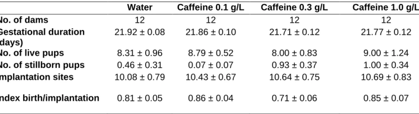
Regarding the effects of caffeine during brain development, epidemiological studies have linked caffeine intake to birth outcomes such as low birth weight, intrauterine growth retardation, and spontaneous abortion (Brent et al., 2011; Bakker et al., 2010; Giannellietal., 2003). . , the effects of caffeine consumed during fetal and even postnatal brain development have been poorly studied (for review, see Porciúncula et al., 2013; Temple, 2009). Caffeine enters all fetal tissues (Brackenetal., 2003) as it can easily cross biological membranes, including the site of the natal septum and the fetal brain (Arnaud, 1987). Some experimental studies on rodents have shown that caffeine administered during. Adult female Wistar rats were treated with caffeine in the drinking water (0.1, 0.3 and 1.0 g/L, corresponding to low, moderate and high intake, respectively) (Fredholmetal., 1999) during the active period (dark cycle) during the week, two weeks before delivery. Although 1 mg/mL of caffeine is considered high values Caffeine consumption resulted in plasma concentrations below 60Minadultrat (Costenlaetal., 2010; Duarteetal.,2009). At the beginning of the light cycle, all experimental groups received tap water. The presence of a vaginal plug was considered embryonic day 0 (E0). The same caffeine treatment was continued throughout pregnancy until embryonic days 18 or 20 (E18 and E20, respectively), when the mothers were sacrificed under anesthesia. .Fruits were weighed and the entire cortex and iliac pocampus were immediately excised at 4°C for Western blot analysis; or whole brains were immersed in 4% paraformaldehyde in 0.1 MPBS to fix brain tissue to determine the total number of neuronal and non-neuronal cells in the cortex and hippocampus. Litter size and number of implantations were also analyzed. Figure 1. Immunological analysis of BDNF (black circle) and its receptor, TrkB (black square) in the hippocampus at E18 (A) and E20 (B) and in the whole cortex at E18 (C) and E20 (D) in water-exposed fetuses or caffeine. Data are means ± S.E.M (n = 5–6 samples/group). )line of density units obtained from the protein/tubulin ratio.* P<0.05, significantly different from the control group (water). One-way ANOVA followed by Newman–Keuls multiple comparison test.
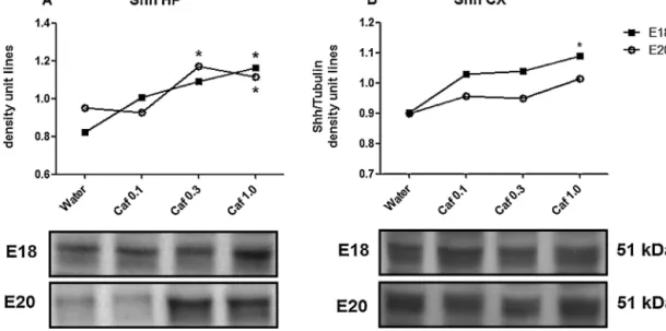
Fig. 2. Immunoblotting analysis of Shinde hippocampus (A) at E18 (black square) and E20 (white circle), and in whole cortex (B) at E18 (black square) and E20 (white circle) of fetuses exposed to water or caffeine. The data are ± S.E.M.(n=5–6 samples/group) of density unit lines obtained from the Shh/tubulin ratio. *P < 0.05, significantly different from control group (water). One-way ANOVA followed by Newman-Keul's multiple comparison test. The impact of prenatal caffeine intake at different doses was also evaluated in neuronal and non-neuronal cell counts. Caffeine 0.3 g/L increased the total number of cells and neuronal cells in the hippocampus of the fetus exposed to caffeine to E18 (Fig. 5A). The lowest dose of caffeine increased neuronal cells and total cell numbers in the cortex at E20 (Fig. 5D). We did not find any effect of caffeine treatment in the hippocampus at E20 or in the cortex at E18 (Fig. 5B and C, respectively). Consistent with previous studies, albeit with different dosing schedules, the absence of effect on the number of implantations and litter size was also observed in animals treated with caffeine (Everekliogluetal., 2003; Lorenzoetal., 2010). One of the expected effects of a high dose of caffeine is the decrease in body weight, which was confirmed at E20. reported for pregnant Wistar rats given caffeine.
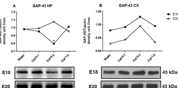
Fig.4.Immunoblotting analysis of SNAP-25 in the hippocampus (A) at E18 (black square) and E20 (white circle), and in the whole cortex (B) at E18 (black square) and E20 (white circle) from fetuses exposed to water or caffeine (±S.up.s. ) unit lines obtained from SNAP-25/ tubulin ratio. No statistical and differences were found. Fig.5.Number of NeuN-stained nuclei (estimated as neuronal cells) and Dapi-stained nuclei (estimated as non-neuronal cells) (permgtissue) in the hippocampus E18 (A) or E20(B), and in the whole cortex of E18(Cs) and E20(Cs) water to E18(Cs) water. P<0.05, significantly different from control group (water). One-way ANOVA followed by Newman–Keuls multiple comparison test. Unlike the hippocampus, higher caffeine increased cortical BDNFatE18 and decreased this protein from E20, without any effect on TrkB receptors. ration of telencephalon development, an event previously reported in in vitro studies (Marretetal., 1997; Sahir . etal. Shh modulates cell proliferation and the migration of progenitor cells in the adult brain (Charytoniuketal., 2002; Dave et al., 2011; Palmaetal. . ,2005;Traiffortetal.,2001).This morphogen highly expressed in rat brains from E12 to postnatal day2 (P2)(Petraliaetal.,2011), and recently its expression was identified in cortical pyramidal neurons.which caffeine relatively high concentrations an overexpression of Shh- cultured neurons and astrocytes (Sahir et al., 2004). Importantly, Shh is associated with induction of GABAergic interneuron differentiation in the encephalon at early embryonic stages (E11.5) (Madyen Kohtz, 2007). One of the consequences of increasing the morphogenic input during pregnancy can be abnormal distribution of neuronal input to BA. -birth impact The moderate caffeine intake (0.3 g/L) during pregnancy and lactation delayed GABA neuron migration in the hippocampus and superficial cortical layers on postnatal day 6 (PND6) (Silvaetal., 2013).
Bakker, R., Steegers, E.A., Obradov, A., Raat, H., Hofman, A., Jaddoe, V.W., 2010. Maternal intake of caffeine from coffee and tea, fetal growth and the risks of adverse birth outcomes outcomes: the Generation RS study. Am. J. Clin. Nutr. Sahir, N., Mas, C., Bourgeois, F., Simonneau, M., Evrard, P., Gressens, P., 2001. Caffeine-induced teleencephalic vesicle evaporation in early post-implantation mouse embryos involves cAMP-dependent protein kinase (PKA) inhibition. Cereb. Cortex11 ,343-349. Shikata,Y.,Okada,T.,Hashimoto,M.,Ellis,T.,Matsumaru,D.,Shiroishi,T.,Ogawa,M., Wainwright,B.,Motoyama,J.,2010.Ptch1-mediated dose-dependent action of Shh signaling regulates neural progenitor development in late stages of pregnancy. Dev Biol.

74
For the same reason, no effect of caffeine on exercise activity was observed. Adenosine is a neuromodulator in the central nervous system (CNS), which regulates the synaptic transmission of neurotransmitters such as dopamine and glutamate through mainly adenosine A1 and A2A metabotropic receptors (Cunha, 2001). BOLD) signal in the frontal cortex and nucleus accumbens of adult rats treated with MPH, but not in the hippocampus (Canese et al., 2009).
Since activation of adenosine A1 receptors initiates anxiolytic effects in rodents (Jain et al., 1995), this transient regulation of adenosine A1 receptors may be involved in the anxiolytic effects of MPH. Adenosine is a neuromodulator in the central nervous system (CNS), which mainly controls the synaptic transmission of neurotransmitters such as dopamine and glutamate via adenosine A1 and A2A metabotropic receptors (Cunha, 2001). As observed, acute administration of MPH 5 mg/kg resulted in an increase in the object recognition index.
Thus, MPH overdose of 50 mg/kg decreased the object recognition index in the test session. Furthermore, the role of A1 adenosine receptors has been implicated in the effects of MPH on recognition memory. In addition, dopamine signaling is critical for memory persistence, as demonstrated in a one-trial inhibitory avoidance task (Rossato et al., 2009).
6. Immunoblotting analysis of adenosine A1 receptor density in frontal cortex and hippocampus from adult rats.
123
DISCUSSÃO
Houve um aumento dos receptores A1 na ponte e no núcleo do tractus solitarius de ratos recém-nascidos de 6 dias de idade que receberam cafeína por gavagem (15 mg/kg) entre PND 2-6 (Gaytan et al., 2006). Em outro estudo, com maior dose de cafeína na água de beber (1,0 g/L) durante a gravidez, houve aumento do número de receptores A1 no cérebro materno e fetal, sendo este último associado ao aumento da afinidade pela cafeína (Lorenzo e outros, 2010). Em outro estudo, utilizando a mesma escala de administração de cafeína, mas em dose única (entre 11 e 20 dias de gestação, 120 mg/kg de cafeína por via intragástrica), houve redução no comprimento do fêmur de embriões de 20 dias e na a expressão de proteínas envolvidas na sinalização do fator de crescimento da insulina 1 (IGF-1) (Tan et al., 2012).
O tratamento com cafeína diminuiu a expressão de genes relacionados à adipogênese, como PPAR-γ, lipoproteína lipase, leptina e TNF-α de maneira dependente da dose ( Su et al., 2013 ). As neurotrofinas são fatores de crescimento com um papel crucial no desenvolvimento e maturação do cérebro (Cohen-Cory et al., 2010). Isso pode ser observado em um estudo recente em que o consumo moderado de cafeína (0,3 g/L) durante a gravidez e lactação levou a um atraso na migração de neurônios GABAérgicos no hipocampo e nas camadas corticais superficiais em ratos em todos os 6-estanho após o nascimento. -dia do nascimento (Silva et al., 2013).
133 O desenvolvimento motor e reflexo não foi afetado pela exposição perinatal à cafeína (0,3 g/L na água potável) quando avaliado pelos testes de reflexo de giro e aversão à queda (Silva et al., 2013). Nesse estudo, um atraso na migração de neurônios GABAérgicos do hipocampo foi observado em ratos de 6 dias de idade submetidos à cafeína do período (Silva et al., 2013). A subunidade GluN1 do receptor NMDA no hipocampo aumenta significativamente na primeira semana pós-natal, atingindo níveis semelhantes aos do animal adulto por volta dos 20 dias de idade (Ibaraki et al., 1999).
Em outro estudo, um aumento da suscetibilidade a convulsões foi observado em camundongos de 6 dias de idade que receberam cafeína durante a gestação e lactação (Silva et al., 2013). A ativação excessiva de receptores metabotrópicos de glutamato pode resultar em perda de resposta, conhecida como dessensibilização (De Blasi et al., 2001). O impacto desse aumento na idade adulta deve ser investigado, pois o tratamento crônico com cafeína na água de bebida durante a gestação e lactação levou a uma diminuição da hiperlocomoção induzida por um antagonista do receptor NMDA em filhotes adultos (da Silva et al., 2005).
CONCLUSÃO
PERSPECTIVAS
The role of adenosine A1 receptors was also investigated on the effects produced by MPH 50 mg/kg in the elevated plus maze. Thus, the role of adenosine A1 receptors was also investigated on behavioral changes of MPH in the elevated plus-maze. 2-The lack of involvement of adenosine A1 receptors on the MPH-induced behavioral changes in the elevated plus-maze.
MPH (5 and 50 mg/kg, i.p.) was administered 30 min before placing the mouse in the elevated plus maze. Mice subjected to the chronic treatment with MPH 5 mg/kg were also evaluated in the inhibitory avoidance task.

164