Für eine detaillierte Beschreibung der verschiedenen Formen von MCD verweisen wir auf die Arbeit von Barkovic et al.

Ictal SPECT in Focal Epilepsy
Survey on structural and functional imaging techniques to investigate localization-related
Other imaging modalities have provided useful data in epilepsy research, but these modalities have not yet been adequately investigated to support a definitional role.
The Role of PET in the Diagnosis of Epilepsies*
What are the abnormalities of MTLE-HS observed with these imaging modalities?
Atrophy-signal changes of the ipsilateral amygdala, temporal neocortex, temporal lobe white matter, fornix, mammillary body, insula, thalamus, or basal frontal cortex. For example, many proton MRS investigations use four voxels in a single thick imaging plane, typically as right and left mesial and lateral temporal lobe voxels when temporal lobe epilepsy (TLE) is to be imaged. Often, in unilateral MTLE-HS, interictal [Tc-99m]HMPAO or [Tc-99m]ECD retention is mildly or moderately reduced in the anterior temporal lobe unilaterally, but "false lateralization" of lobe hypoperfusion occurs temporal interictal. in perhaps 10% of patients.
When radioligand is injected during a complex partial seizure, the temporal lobe of ictal onset typically shows a large increase in radioligand retention, and false lateralization is quite rare when using co-registered ictal and interictal temporal lobe perfusion maps. False lateralization of temporal lobe hypometabolism on interictal FDG images is rare in MTLE-HS, unlike the more common occurrence of false lateralized temporal lobe hypoperfusion on interictal SPECT. Anterior temporal lobe hypometabolism ipsilateral to HS or bilateral but greater on the side of HS (detected by interictal FDG PET in 90-95% of cases in which HS is found in resected tissue) and .. extratemporal hypometabolism ipsilateral to predominant anterior temporal hypometabolism, usually involving the thalamus and often involving the basal ganglia, insula, inferior frontal cortex, and lateral parietal cortex. e) Interictal FMZ PET maps the density of cBZR, which thus maps the density of the major inhibitory site of the mammalian cortex, the GABAA receptor-chloride ionophore complex.
Thus, reductions in cBZRs are much more focal with the anterior mesial temporal lobe in MTLE-HS, than ictal hyperperfusion and interictal glucose hypo-.
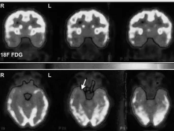
Examples of interictal and ictal PET studies in the context of presurgical evaluation
The results of this study were as follows [13]: preoperative MTLE-HS patients had significantly reduced regional glucose metabolic rates (rCMRglu) ipsilateral to the focus in the mesial temporal lobe. Postoperatively, rCMRglu values were decreased in the operated mesial temporal structures, as expected. In the first postoperative PET study (in the 3rd postoperative month), the ipsilateral lateral TL cortex showed significantly reduced glucose consumption compared to the control group.
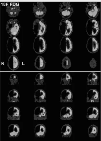
Ictal FDG PET study in a patient with a prolonged status-like seizure discharge in the left temporo-occipital cortex. During the prolonged seizure discharge in the left temporo-occipital cortex (see Ictal and the difference between Interictal and Ictal), the patient exhibited aphasic and visual symptoms and was anxious with insufficient response to external stimuli. Preoperative activation in the contralateral MTL during the ipsilateral learning task correlated positively with the postoperative outcome for ipsilateral memory.
Activity increases during the ipsilateral learning task are located in the ipsilateral right hippocampal area (non-verbal learning vs. control).
Tumor-associated epilepsies: Imaging gliomas with PET (see [2] )
In this study, postoperative memory deficits were absent if the contralateral MTL had taken over memory functions from the pathological ipsilateral MTL. Comparisons of the intra-arterial amobarbital tests with functional neuroimaging of memory have yielded a high agreement between the two methods. The importance of the number and placement of the electrodes is addressed along with recent developments in multichannel EEG recordings.
It is also described how the choice of time period for source localizations of the epileptic activity can affect the source localization results. Localizing the different modules of these – normal or pathological – networks is the main objective of functional neuroimaging studies, for example with PET or functional MRI [2]. However, these models require some a priori assumptions to be made about the generation of the EEG and MEG signals.
This article focuses on EEG recordings, although most of the aspects discussed here also apply to MEG.
Electric Source Imaging of Epileptic Foci
It is particularly easy to implement in realistic head models based on the patient's individual MRI. Shown below are the original surface data reconstructions corresponding to intracranial spikes in the 4 subdural regions. The results illustrate the importance of a sufficient number of recording electrodes for the accuracy of source localization results.
The disadvantage of the realistic head models described above is their complexity and the computational load required for the calculations. By transforming the MRI into a sphere, the inverse problem can be computed analytically (using a spherical model), but the solutions are computed directly for this (albeit slightly deformed) MRI. When the EEG source is integrated with MRI, the resolution is limited to the gray matter of the individual patient.
The position of the electrodes as well as the mounts can be progressively adjusted.
Peroperative Corticography (ECoG)
Intraoperative electrocortigraphy (ECoG) has been used as an adjunct to the surgical management of medically refractory partial epilepsy to identify the location and boundaries of the epileptic region at the time of resection. In this sense, preoperative electrocorticography (ECoG) may be a potential aid in guiding the surgical resection of both the lesion and the epileptogenic zone. Penfield and Jasper [1] pioneered this technique in the 1930s, recording interictal spikes to determine the extent of resection in epilepsy surgery.
Despite the widespread use of the technique, corticography does not help predict the outcome of surgery [2. The definition of the epileptic foci almost always relies on semiology, but localization can be difficult due to the greater variability of semiology compared to temporal lobe epilepsy. The outcome was favorable in 75% of patients whose cortical tissue showing continuous spikes could be included in the resection.
It can also provide information on the spike activity of the resection margin and provide the opportunity to tailor the surgical strategy.
Nouvelle technique dans l’évaluation préchirurgicale de l’épilepsie : IRM fonctionnelle triggée par l’EEG
Parmi ces nouvelles méthodes, l'IRM fonctionnelle déclenchée par l'EEG est une technique d'évaluation non invasive qui permet de localiser l'activité des foyers intercritiques chez les patients épileptiques. Ces dernières années, les techniques d'évaluation pré-chirurgicale de l'épilepsie ont connu un essor important. Le signal MR résultant est connu sous le nom d'"effet BOLD (dépendant du niveau d'oxygénation du sang)" [2].
Il a été observé que l'enregistrement de l'effet BOLD permettait une bonne localisation du foyer critique chez les patients ayant présenté des crises partielles sans mouvement lors de l'examen IRMf [3]. En effet, plusieurs études ont prouvé que l'utilisation d'un matériel adapté permet de réaliser des enregistrements EEG dans l'aimant IRM dans les limites de la sécurité pour le patient [8, 9]. De plus, un certain nombre de patients ont ensuite été évalués avec des électrodes implantées, qui ont pu confirmer les résultats de l'IRMf[6,.
L'activation de l'IRMf déclenchée par l'EEG se trouve dans le cortex calcarien ; les crises étaient caractérisées par une hémianopsie droite intermittente.

Sibylle - Ried - Preis
Localisation non invasive du foyer épileptique à l'aide de l'IRM fonctionnelle déclenchée par l'EEG et de la tomographie électromagnétique. Dans ce contexte, la spectroscopie protonique (1H-SRM) est particulièrement intéressante car elle renseigne sur le taux de certains métabolites comme le N-acétylaspartate (marqueur neural), le pic de ne ou encore l'acide lactique, signe d'une activité neuronale anormale. Le rôle de la spectroscopie dans l'évaluation de l'épilepsie va certainement s'accroître à l'avenir avec l'émergence de techniques de quantification fiables, qui donnent accès à d'autres composés comme la glutamine et le glutamate, et le développement de techniques d'imagerie spectroscopique.
Il est possible d'utiliser une impulsion radiofréquence pour exciter cette aimantation et la détecter à l'aide d'une antenne de réception. La spectroscopie protonique (1H-SRM) est basée sur le fait que chaque atome 1H, lorsqu'il est intégré dans une molécule, a une fréquence de résonance légèrement différente de celle de l'atome isolé. Ce petit décalage (quelques dizaines de Hz), appelé "décalage chimique", reflète la faible variation du champ magnétique local due à l'environnement électronique de la molécule.
La spectroscopie protonique, contrairement aux autres noyaux, nécessite la suppression des signaux eau et graisse dont l'intensité est 1000 à 10000 fois supérieure à l'intensité des métabolites à étudier.
Rôle de la spectroscopie du proton dans le bilan préchirurgical de l’épilepsie*
La suppression du signal gras est difficile à réaliser et nécessite des techniques d'acquisition de signal spécifiques (techniques d'édition). Le principal avantage du proton SRM est sa sensibilité, qui permet une meilleure localisation spatiale, et la relative simplicité de sa mise en œuvre. Les principales raisons sont un signal faible, la présence de pics de distorsion dus à des imperfections techniques (homogénéité du champ magnétique, courants de Foucault, mauvaise suppression du signal de l'eau), le chevauchement des résonances voisines, la distorsion par rapport à la ligne de base.
De plus, une diminution relative du NAA par rapport à la choline et/ou au Cre a été démontrée par de nombreux groupes, démontrant la sensibilité du 1H-SRM pour localiser le foyer épileptique. Ces résultats préliminaires suggèrent que la combinaison de l'EEG-IRMf et de la spectroscopie permet une meilleure caractérisation des foyers extratemporaux. La spectroscopie protonique s'est révélée être un complément précieux à l'imagerie anatomique et fonctionnelle.
Les spectres montrent une déficience distincte, avec des niveaux proches de la normale dans le lobe temporal médial (rangée du bas), une diminution marquée du niveau de NAA dans le pôle antérieur du lobe temporal droit (rangée du milieu), ainsi que dans le droit. foyer fronto-orbitaire (rangée du haut), ce qui suggère des dommages neuronaux importants.

Liga-Mitteilungen
Gemeinsame Jahrestagung der Deutschen, Österreichischen
Neue Geschäftsführerin
Ausschreibung – Forschungsförderung
Une expédition splendide
Sport treiben macht glücklich
Fachtagung für Neurophysiologie und angrenzende Gebiete
Informationen: AAN Member Services, 1080 Montreal Avenue, St. www.aan.com oder http://am.aan.com Recklinghausen, Deutschland Klinische Neurophysiologie: Evozierte Potenziale – Neurovegetative Funktionsdiagnostik. Gemeinsame Jahrestagung der Deutschen Gesellschaft für Neurotraumatologie und Klinische Neuropsychologie e.V. DGNKN) und der Deutschen Gesellschaft für Neurologische Rehabilitation e.V. Tagung der Schweizerischen Gesellschaft für Neurologie (SNG) zusammen mit der Jahrestagung der Schweizerischen Gesellschaft für Intensivmedizin Informationen: www.swissneuro.ch.
Kongresskalender
Gemeinsame Jahrestagung der Deutschen, Öster- reichischen und Schweizer Sektionen der Internationa-
Impressum