Many thanks to all members of the Laboratory of Cellular Neurophysiology of the Institute of Physiology of the Czech Academy of Sciences for their contribution to the implementation of the research mentioned in this thesis and for the very friendly, hospitable and pleasant environment they created. Combining electrophysiology, molecular biology and computational modeling, we identified the PE-S and EPA-But binding sites on the transmembrane domain of the GluN1/GluN2B receptor.
Ionotropic glutamate receptors
NMDARs represent the slow component of EPSCs due to their slow transient and desensitization kinetics and relatively weak desensitization. At the same time, excessive NMDAR-mediated Ca2+ influx can cause neuronal damage or death and has been implicated in the pathogenesis of many neurodegenerative disorders (Hansen et al., 2021; Traynelis et al., 2010).
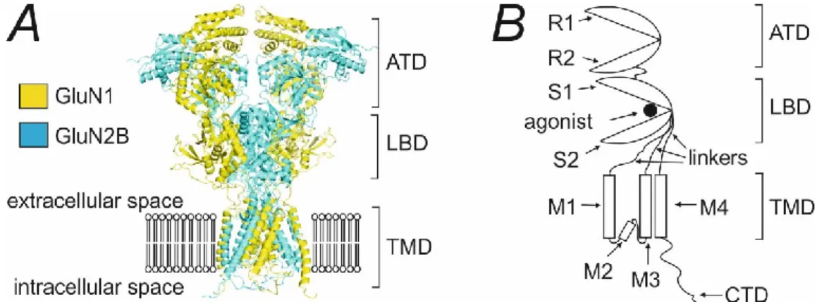
Structure of the NMDA receptor
Subunit composition of NMDA receptor
GluN2B and GluN2D subunits are ubiquitously expressed in the embryonic CNS, whereas GluN2A and GluN2C subunits are absent. The expression of GluN2B and GluN2D remains high in the neonatal brain, but then decreases significantly.
NMDA receptor activation
Kinetic scheme for NMDA receptor activation
On a simplified level, the activation of the conventional NMDAR can be described by the kinetic scheme (Figure 1.2) (Lester & Jahr, 1992). In this scheme, the glutamate binding is characterized by the on-rate constant kon and the glutamate release is characterized by the off-rate constant koff.
Pharmacology of NMDA receptor
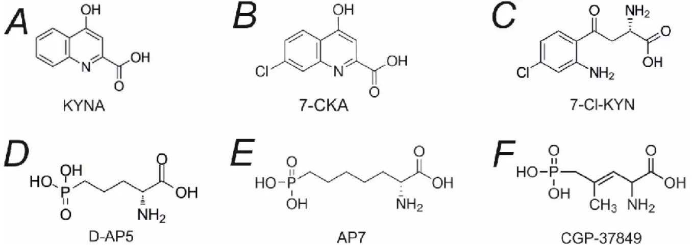
- Competitive antagonists
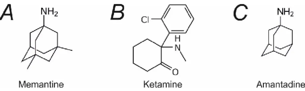
- Open channel blockers
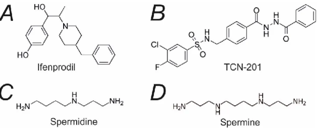
- Allosteric modulators
- Neuroactive steroids
Moreover, ifenprodil can inhibit triheteromeric GluN1/GluN2A/GluN2B receptors by 30% and GluN1/GluN2B/GluN2D receptors by 70% (Hansen et al., 2021). In contrast, cholesterol enrichment enhances the NMDAR responses by increasing the Po of the receptors (Korinek et al., 2015).
Role of NMDA receptors in the pathophysiology of neuropsychiatric disorders
- NMDA receptor and excitotoxicity
- Affective disorders
- Epilepsy
- Schizophrenia
- Intellectual disability and developmental delay
- Autism-spectrum disorders
Numerous studies have reported increased KYNA levels in the CNS of schizophrenia patients (Plitman et al., 2017). Nuclear magnetic resonance spectroscopy studies revealed reduced glutamate levels in the prefrontal cortex of schizophrenia patients ( Marsman et al., 2013 ).
Functional and pharmacological properties of disease-associated de novo mutations
Identification of the site of action for pregnenolone sulfate at the NMDAR
Structure requirements for potentiating neuroactive steroids
Materials and chemicals
- Lysogeny Broth medium (LB medium)
- Agar plates
- Phosphate-buffered saline (PBS)
- Trypsin-EDTA solution
- Culture medium
- Intracellular solution (ICS)
- Mg 2+ - free extracellular solution (ECS)
- Mg 2+ - containing extracellular solution (Mg 2+ -ECS)
- cDNA constructs
To avoid glycine contamination, ultrapure chemicals and HPLC water were used to prepare ECS for the experiments analyzing the glycine dose-response effect. ECS was sterilized by sterile filtration with a 0.2 μm Porafil membrane filter (Macherey-Nagel) and stored at 4ºC.
Site-directed mutagenesis
Enhanced green fluorescent protein (eGFP) in the pQBI 25 expression vector (Tacara Bio Inc., Tokyo, Japan). Selected colonies were picked, transferred to culture tubes LB medium with ampicillin (100 mg/l) and incubated overnight on a shaker at 37ºC and 200 rpm.
Cell culture and transfection
The cells were transferred to culture tubes with 200 μl of LB medium and incubated on a shaker with 200 rpm rotation at 37°C for 1 hour. The resulting culture was spread on agar plates and incubated overnight at 37°C to grow bacterial colonies.
Cholesterol depletion
After that, the cells were washed with PBS, detached from the surface of the well with trypsin-EDTA solution, aspirated by pipetting into the pre-warmed culture medium and plated on 24 mm round coverslips (Glaswarenfabrik Karl Hecht, Sondheim vor der Rhön, Germany) coated with poly-L-lysine (1 mg/ml, Serva, Heidelberg, Germany).
Immunofluorescence microscopy
Electrophysiology
Data analysis
- Electrophysiological recordings analysis
- Steroid effect assessment
- Agonist dose-response analysis
- Current density analysis
- Open probability analysis
- Comparative analysis of MK-801 blocking rate
- Statistical analysis
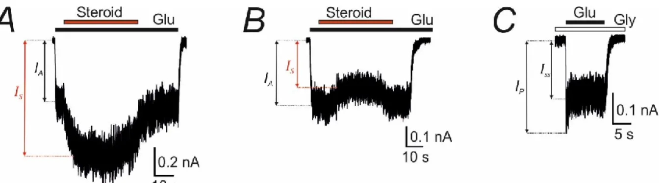
43 The glutamate dose-response assay was done in the presence of 30 μM glycine and the glycine dose-response assay was done in the presence of 1 mM glutamate. -B) Representative recordings demonstrate the effect of potentiating (A) and inhibitory (B) steroids on GluN1/GluN2B receptor responses to 1 μM glutamate in the presence of 30 μM glycine. Representative recording demonstrates the response of GluN1/GluN2B receptors induced by 1 mM glutamate in the presence of 30 μM glycine.
In the second step, the kinetic constants 𝑘𝑟 and 𝑘𝑑 were fixed to values determined in the first step. The NMDAR responses to 1 mM glutamate were inhibited by the application of MK-801 (1 μM) in the continuous presence of glutamate.
Functional and pharmacological properties of disease-associated de novo mutations
Disease-associated mutations affect the current density and surface expression
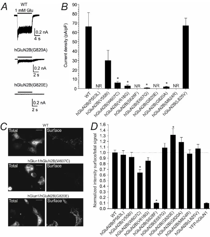
HEK293T cells were transfected with eGFP, hGluN1 subunit, and either WT or mutated hGluN2B subunit. The values of current densities mediated by hGluN1/hGluN2B(V558I) and hGluN1/hGluN2B(L825V) receptors were similar to those mediated by WT receptors Figure 4.2B. COS-7 cells were transfected with DNA vectors encoding YFP-tagged hGluN1 subunit and either WT or mutated hGluN2B subunit.
The analysis of acquired immunofluorescence microscopy images indicated that the surface expression of mutated hGluN1/hGluN2B(W607C; S628F) receptors was significantly lower than that of WT receptors (Figure 4.2C-D). The bar graph shows mean current densities ± SEM recorded in HEK293T cells expressing NMDARs with WT and mutated hGluN2B subunits (n = 5–23).
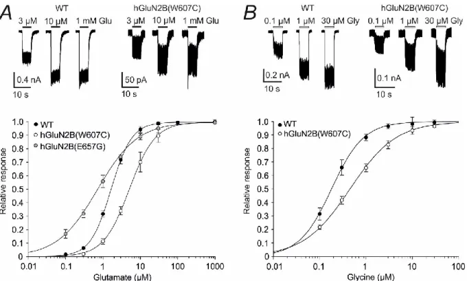
Mutations in the TMD alter NMDAR agonist affinity
At the bottom, glutamate dose–response curves recorded from the HEK293T cells expressing WT, hGluN1/hGluN2B(W607C), and hGluN1/hGluN2B(E657G) receptors. The experiments were performed in the continuous presence of 30 μM glycine; the duration of glutamate application is indicated by filled bars. At the top, representative current responses to 0.1, 1 and 30 μM glycine are recorded from the HEK 293T cells expressing the WT and hGluN1/hGluN2B(W607C) receptors.
The experiments were performed in the continuous presence of 1 mM glutamate; duration of glycine application is indicated by open bars. 52 Statistical analysis was performed on logEC50 andlogHill using one-way ANOVA and Tukey posthoc Dunnett's method; h is the Hill coefficient; n indicates the number of cells in the group; * < 0.05.
Disease-related mutations affect receptor desensitization and open probability 52
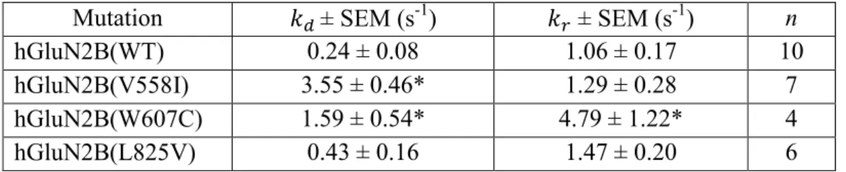
𝑘𝑑 values in hGluN1/hGluN2B(L825V) receptors were similar to those in WT receptors (Table 4.3). The 𝑘𝑟 value determined in hGluN1/hGluN2B(W607C) receptors was 4.5 times higher compared to that determined in WT receptors. No statistically significant difference was found between the 𝑘𝑑 values determined in WT receptors and the mutant hGluN1/hGluN2B(V558I) and hGluN1/hGluN2B(L825V) receptors (Table 4.3).
Summary of desensitization (𝑘𝑑) and resensitization (𝑘𝑟) kinetic constants determined in WT and mutant receptors. The results showed that the Po of hGluN1/hGluN2B(V558I), hGluN1/hGluN2B(W607C), hGluN1/hGluN2B(V618G), and hGluN1/hGluN2B(L825V) receptors was significantly lower than that of WT receptors ( Figure 4D ).
Identification of the site of action for pregnenolone sulfate at the NMDAR
- The transmembrane domain of NMDAR is crucial for PE-S potentiation
- PE-S and sterols act at distinct sites at the NMDAR
- Amino-acid residues at the TMD are essential for PE-S potentiation
- Identification of PE-S binding interface at the NMDAR
- In silico modelling of PE-S binding
Representative recordings of the current responses obtained from HEK293T cells transfected with GluN1/GluN2B receptors. The duration of glutamate and PE-S application is indicated by filled and open bars, respectively. To determine the PE-S binding site, we analyzed the effect of mutations in the TMD of the GluN1 and GluN2B subunits on PE-S potentiation.
The positive modulatory effect of PE-S is affected by mutations in the M1 and M4 helices of the GluN1 and GluN2B subunits. First, PE-S was docked into the TMD of an unliganded GluN1/GluN2B receptor model.

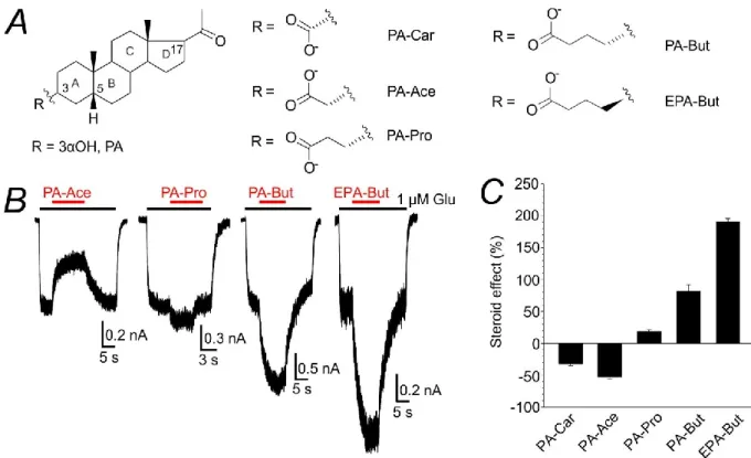
Structure requirements for potentiating neuroactive steroids
- ω5β-pregnan-3β-yl derivatives of carboxylic acids potentiate NMDAR
- EPA-But potentiates NMDAR responses in a disuse-dependent manner
- EPA-But site of action at the NMDAR is different from that for PE-S
- Mutations in the TMD affect the EPA-But potentiation
- In silico modelling of EPA-But binding at NMDAR
- EPA-But and PE-S effects on NMDARs with disease-related mutations
In addition, the potentiating effect of EPA-But is affected by the timing of steroid and glutamate administration. We hypothesized that we would observe the occlusion of the effects of EPA-But and PE-S in the case of the same site of action for both steroids (model 1) or the sum of their effects in the case of different sites of action (model 2). -C) Bar graphs show the degree of potentiation by EPA-But (15 μM) of glutamate-evoked responses in GluN1/GluN2B receptors with a mutated GluN1 (B) or GluN2B (C) subunit.
80 Dashed lines represent the average potentiation induced by EPA-But and PE-S in WT receptors. The effects of EPA-But (15 μM) and PE-S (100 μM) on WT and mutant receptors were evaluated after co-administration with glutamate (1 μM).
Functional and pharmacological properties of disease-associated de novo mutations
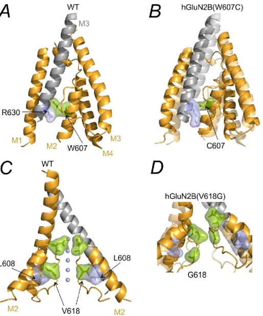
MD simulation shows that the hGluN2B(P553L) mutation leads to the elimination of the pre-M1 linker resulting in the formation of a nearly continuous M1 helix (Figure 5.1B). Helical organization of the TMD in WT (light colors) and GluN1/hGluN2B(P553L) receptors (deep colors). The hGluN2B(V618G) mutation disrupts the interaction between hGluN2B(V618) and hGluN2B(R630) thereby altering the orientation of the M2 helix.
The arrangement of the TMD in WT (light colors) and hGluN1/hGluN2B(S628F) receptors (deep colors). MD indicates that the hGluN2B(S628F) mutation leads to an increase in the distance between the M1 and M4. C) The arrangement of the TMD in WT (light colors) and hGluN1/hGluN2B(E657G) receptors (deep colors).
Identification of the site of action for pregnenolone sulfate at the NMDAR
95 interface is subjected to structural rearrangements that lead to a reduction in PE-S binding affinity, in line with the disuse-dependent action of PE-S ( Horak et al., 2004 ). This mechanism sheds light on the disuse-dependent potentiating effect of PE-S (Horak et al., 2004). Thus, the potentiating and inhibitory effects of PE-S differ in their mechanisms: whereas PE-S potentiation is non-use-dependent, PE-S inhibition is use-dependent ( Horak et al., 2004 ).
The results of a recent alanine-scanning study indicated that the GluN1 M4 helix is a potential negative allosteric modulatory site for PE-S (Langer et al., 2021). In addition, the previously identified binding site for inhibitory steroids at the extracellular vestibule of the ion channel is also likely to be a negative allosteric modulatory site for PE-S ( Vyklicky et al., 2015 ).
Structure requirements for potentiating neuroactive steroids
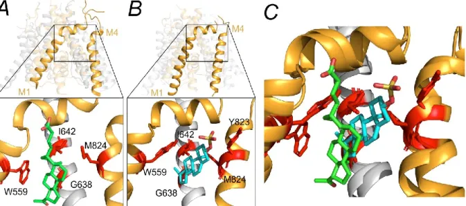
The EPA-But molecule intercalates between the residue GluN2B(W559) of the M1 helix and the residue GluN2B(M824) of the M4 helix (Figure 5.5A). It is necessary to mention that the experimental and in silico characterization of the EPA-But binding site is challenging due to several technical difficulties. The divergence in the EPA-But and PE-S binding sites leads to a significant difference in the effects of the steroids in seratin disease-related receptor variants.
Ribbon structure shows the arrangement of M1-M4 helices in the model of the GluN1/GluN2B receptor in the open state (Černý et al., 2019). Mutagenesis experiments suggested that the binding site of GNE-9278 is located in the extracellular region of the pre-M1/M1 helices of the GluN1 subunit ( Wang et al., 2017 ).
Functional and pharmacological properties of disease-associated de novo mutations
Identification of the site of action for pregnenolone sulfate at the NMDAR
Upon binding to the receptor, PE-S facilitates rotation of the GluN2B M4 helix promoting a transition to the open channel conformation. In addition, PE-S strengthens the interaction between GluN1(I642) and GluN2B(W559) residues, stabilizing the GluN1 M3 helix of the pore lining in the open position.
Structure requirements for potentiating neuroactive steroids
Funkční a farmakologické vlastnosti NMDAR s de novo mutacemi v podjednotce
Identifikace místa působení pregnenolon sulfátu na NMDAR
Strukturní požadavky na strukturu neuroaktivních steroidů s potencionálním
Genetic inactivation of the NMDA receptor NR2A subunit has anxiolytic and antidepressant-like effects in mice. Regulation of the NMDA receptor by its cytoplasmic domains: (How) the dog's tail wags. The bioactive protein-ligand conformation of GluN2C-selective positive allosteric modulators bound to the NMDA receptor.
Molecular determinants of agonist discrimination at NMDA receptor subunits: Analysis of the glutamate binding site on the NR2B subunit. Control of long-term synaptic potentiation and learning by alternative splicing of the NMDA receptor subunit GluN1. The role of NMDA receptor and neuroligin rare variants in synaptic dysfunction underlying neurodevelopmental disorders.
Blockade of NMDA receptor channels by endogenous neurosteroids: Implications for agonist-induced vestibular conformational states of the channel.