Henrique França Diniz Oliveira
NANOAGREGADOS BASEADOS EM CICLODEXTRINAS EM ASSOCIAÇÃO COM A TETRACICLINA: CARACTERIZAÇÃO FÍSICO-QUÍMICA E
AVALIAÇÃO ANTIMICROBIANA.
NANOASSEMBLIES BASED ON CYCLODEXTRIN AS CARRIER FOR
TETRACYCLINE: PHYSICOCHEMICAL CHARACTERIZATION AND
ANTIMICROBIAL EVALUATION.
Belo Horizonte
Henrique França Diniz Oliveira
NANOAGREGADOS BASEADOS EM CICLODEXTRINAS EM ASSOCIAÇÃO COM A TETRACICLINA: CARACTERIZAÇÃO FISICOMQUÍMICA E AVALIAÇÃO
ANTIMICROBIANA.
Dissertação apresentada à banca examinadora como requisito parcial para a obtenção do grau de Mestre em Odontologia. Área de Concentração: Materiais Odontológicos. Faculdade de Odontologia. Universidade Federal de Minas Gerais.
Orientadora: Profª. Drª. Maria Esperanza Cortés. Co-orientador: Prof. Dr. Rubén Dario Sinisterra Departamento de Química, ICEX-UFMG.
Oliveira, Henrique França Diniz
O48n Nanoagregados baseados em Ciclodextrinas em associação com a 2007 Tetraciclina: caracterização físico-química e avaliação antimicrobiana /
T Henrique França Diniz Oliveira, 2007. 80 fls.: il.
Orientadora: Maria Esperanza Cortés Co-orientador: Rubén Dario Sinisterra
Dissertação (Mestrado) – Universidade Federal de Minas Gerais, Faculdade de Odontologia.
1. Tetraciclina - Análise - Teses. 2. Ciclodextrinas - Análise - Teses I. Cortés Segura, Maria Esperanza. II. Sinisterra Millán, Rubén Dario. III. Universidade Federal de Minas Gerais. Faculdade de Odontologia. IV. Título
AGRADECIMENTOS
Gostaria de agradecer a todas as pessoas que de várias formas ajudaram na realização deste trabalho:
Aos meus pais, Sálvio Diniz Oliveira e Anamélia França Oliveira, pela dedicação e sacrifício. Para a realização do sonho dos seus filhos são capazes de mover montanhas, carregar o peso do mundo nas costas, passar por dificuldades financeiras. O amor infindável por eles demonstrado foi a maior motivação para a conclusão de mais uma etapa de minha vida.
Aos Professores Dra. Maria Esperanza Cortés e Dr. Rubén Dario Sinisterra, pela orientação, ajuda e paciência da iniciação científica até hoje. Além de grandes pesquisadores e profissionais, os considero hoje como amigos e referências na minha vida.
Ao Prof. Dr. João Maurício Lima de Figueiredo Mota pelas fundamentais contribuições a minha formação durante esse curso.
À colega Karina Imaculada Rosa Teixeira pela fundamental colaboração, dedicação, esforço pessoal além da grande amizade, nos momentos de tristeza e também de alegria.
Um agradecimento especial aos meus companheiros e hoje amigos do Laboratório de Encapsulamento Molecular e Biomateriais, DQ, ICEx – UFMG (Laboratório 290): Ângelo Marcio Denadai e Frederico Barros de Sousa, grandes professores e colaboradores.
Também do Departamento de Química agradeço aos colegas Michele Fabiane de Oliveira, Daniela Cristina dos Santos, Washington Xavier de Paula, Marina Christiane de Souza, Anayive Perez Rebolledo, Eliete Marçal Guimarães Raso, Pedro Pires Goulart Guimarães, Sonja Ellen Lobo e Ivana Silva Lula, pela paciência, fundamental ajuda e orientações.
AGRADECIMENTOS ESPECIAIS
Ao meu amor, Julia Mourão Braga (Jujuba), pelos momentos que passamos juntos, pela compreensão na minha ausência, pelo amor, amizade e carinho.
Aos meus irmãos, Chris juntamente com a Lu e Claudinha, pelo grande companheirismo, amizade e afeto.
À minha Vovó Dedeje pelo carinho, amor, orgulho e alegria, juntamente com minha Vovó Fia que, desde o início grande incentivadora da minha profissão, hoje deve estar dividindo esta alegria conosco, esteja onde estiver.
À Nice pelo grande carinho e amor, minha segunda mãe.
Aos meus tios: Dú e Ana Lúcia, Nélio e Mirian, Lulu e Vail, Aninha pelo grande incentivo e carinho.
Aos meus padrinhos: TiAna pelo suporte e apoio incondicional, e Paulinho que juntamente com a Lúcia foram grandes aliados nessa conquista.
A todos os meus colegas de mestrado, em especial à Juliene, Carolina e Luciano; e a todos os outros colegas.
Gostaria de agradecer também aos meus colegas Ashley Weiner, Ash Jayagopal, Chrissy Marasco, Sanjeet Rangarajan, Ananya Majumder e Elisabeth Vargis pelo apoio, paciência e acolhimento na Vanderbilt University.
Ao Fernando e à Isa pelo apoio incondicional e amizade.
LISTA DE ABREVIATURAS E SIGLAS
δ Chemical Shift
β-CD β-cyclodextrin
∆β-CDH° Partial molar enthalpy of β-CD
1:1 β-cyclodextrin:tetracycline 1:1 molar ratio
2:1 β-cyclodextrin:tetracycline 2:1 molar ratio
3:1 β-cyclodextrin:tetracycline 3:1 molar ratio
4:1 β-cyclodextrin:tetracycline 4:1 molar ratio
A.a.,A. Actinomycemcomitans Actinobacillus actinomycetemcomitans
ATCC American Type Culture Colection
BHI Brain Heart Infusion
CD Cyclodextrin CDs Cyclodextrins
CFU Colony Forming Units
D2O Heavy water
DLS Dynamic Light Scattering
DSC Differential Scanning Calorimetry
FTIR Fourier Transform Infrared
ITC Isothermal Titration Calorimetry
MIC Minimal Inhibitory Concentration
NMR Nuclear Magnetic Resonance
P.g., P.gingivalis Porphyromonas gingivalis
PLGA DL-co-polymers of lactic and glycolic acid PM 1:1; 2:1; 3:1 e 4:1 Physical mixtures in 1:1; 2:1; 3:1 and 4:1 β
-cyclodextrin:tetracycline molar ratio
PVA Polyvinyl Alcohol
SEM Scanning Electron Microscopy
TC Tetracycline
TG Thermogravimetric Analysis
UV Ultra Violet
XRD X-ray diffraction
SUMÁRIO
1. RESUMO 9
2. ABSTRACT 10
3. APRESENTAÇÃO 11
4. NANOASSEMBLIES BASED ON CYCLODEXTRIN AS CARRIER FOR TETRACYCLINE: PHYSICOCHEMICAL CHARACTERIZATION AND ANTIMICROBIAL EVALUATION 14
5. COMENTÁRIOS FINAIS 60
6. REFERÊNCIAS 62
RESUMO
A auto agregação molecular tem se mostrado como um método eficiente para produzir
estruturas medindo alguns nanômetros em tamanho. Esses compostos moleculares
sintetizados a partir da associação entre β-ciclodextrina:tetraciclina foram caracterizados por
espalhamento de luz dinâmico, calorimetria isotérmica de titulação, difração de raios-X,
espectroscopia em infravermelho, termogravimetria, calorimetria exploratória diferencial e
ressonância nuclear magnética. Além disto, esses compostos supramoleculares foram testados
para a determinação de sua atividade antimicrobiana contra A. actinomycetemcomitans e P.
gingivalis em solução e associados à nanoesferas poliméricas. Utilizando-se das técnicas de
caracterização, a formação do composto de inclusão entre TC e β-CD na razão molar de 1:1
foi evidenciada e, ao se aumentar a concentração de β-CD, ocorreu uma auto agregação
supramolecular espontaneamente, resultando em compostos de maior razão molar de β
-CD:TC. Estes complexos apresentaram propriedades fisico-químicas diferentes entre si e
diferentes da β-CD e TC puras, tamanho nanométrico, além de potenciarem a atividade
antimicrobiana do fármaco. Os compostos de razão molar 2:1 β-CD:TC mostraram uma
atividade antimicrobiana significativamente maior em solução (p<0.05). Dentre os outros
compostos, 4:1 foi mais eficiente contra P. gingivalis (zona de inibição = 41.67±1.4mm, MIC
0.25µg/mL, p<0.05). As nano esferas poliméricas, preparadas utilizando-se os nanoagregados,
mostraram liberação controlada do fármaco por 10 dias, em uma concentração superior à
concentração inibitória mínima das bactérias testadas.
Palavras chave: Tetraciclina, Ciclodextrinas, Nanoagregados, Nanoesferas, Liberação
ABSTRACT
Molecular self-assembling has been shown to be an efficient method to produce structures of
few hundreds of nanometers in size. The supramolecular compounds made from β
-cyclodextrin:tetracycline in aqueous solution were evaluated. The physicochemical
interactions between β-cyclodextrin:tetracycline were characterized by dynamic light
scattering (DLS), isothermal titration calorimetry (ITC), X-ray diffraction (XD), infrared
spectroscopy (FTIR), thermogravimetric analyses (TG), differential scanning calorimetry
(DSC), and nuclear magnetic resonance (NMR). The supra-molecular interaction with A.
actinomycetemcomitans and P. gingivalis in solution and in association with polymeric
nanospheres were determined. Using the characterization techniques, it was demonstrated that
the formation of inclusion complex takes place at a 1:1 β-CD:TC molar ratio and, increasing
β-CD concentration, supramolecular spontaneous aggregation occurred. The resulting
complexes showed different physicochemical properties, nanometric size and improved
antimicrobial activity. The 2:1 β-CD:TC showed significantly higher antimicrobial activity in
nsolution (p<0.05). Among the other compounds, 4:1 was the most effective against P.
gingivalis (inhibition zone = 41.67±1.4mm, MIC 0.25µg/mL, p<0.05). Polymeric
nanospheres were then manufactured using these nanoassemblies. The nanospheres showed
controlled TC release for 10 days, and concentrations above minimum inhibitory
concentrations of tested bacteria.
APRESENTAÇÃO
Recentemente, estudos reportam o uso de nano agregados utilizando-se polissacarídeos
cíclicos para a alteração das características fisico-químicas de substâncias. Esses poderiam
solubilizar fármacos pouco hidrossolúveis em água, através de interações em seus
microdomínios hidrofóbicos. A auto organização molecular tem se mostrado um fenômeno
útil para a produção de estruturas com o tamanho de poucos nanômetros. Devido às
propriedades especiais apresentadas pelas nanopartículas, que incluem propriedades químicas
e biológicas entre outras, elas exibem uma performance superior em relação aos materiais
tradicionais [1]
Estruturas supramoleculares auto organizadas podem também ser alcançadas utilizando-se as
ciclodextrinas. Estas são moléculas cíclicas que atuam como hospedeiras, apresentando
características anfifílicas. Apesar de a molécula como um todo ser hidrosolúvel, o interior da
sua cavidade é relativamente apolar, criando um microambiente hidrofóbico. Sua parte
exterior é predominantemente hidrofílica. Estas características são responsáveis pela
solubilidade destas moléculas em meio aquoso e ainda por apresentar a capacidade de
interagir com substâncias hidrofóbicas em solução, mudando suas propriedades
farmacológicas no caso de medicamentos, incluindo o aumento de sua biodisponibilidade
[2-5].
Compostos supramoleculares, nos quais as ciclodextrinas se associam à fármacos para a
formação de compostos de inclusão, estão sendo desenvolvidos com sucesso. Os compostos
anfifílico das ciclodextrinas e a capacidade desta molécula para formar ligações de hidrogênio
e de Van der Waals, sugerem que, aumentando-se a concentração de β-ciclodextrinas no
sistema, novos complexos de razões molares diferentes poderiam ser formados. Esta nova
conformação espacial poderia conferir diferentes propriedades ao fármaco incluído. Os
processos de auto organização podem ser pré-determinados para que as condições
experimentais se tornem favoráveis para a formação e a estabilização desse novo composto
[6-8].
Estudos prévios mostraram vantagens ao se usar o composto de inclusão β
-ciclodextrina:tetraciclina (1:1) em solução em relação à tetraciclina pura, resultando em
melhores propriedades antimicrobianas e menores concentrações, in vitro. Esse composto de
inclusão mostrou ainda um padrão de liberação de tetraciclina de maneira mais lenta e gradual
em comparação ao fámaco puro [9]. Além disso, as ciclodextrinas podem agir como
conservantes em formulações, protegendo o fármaco da degradação, aumentando seu tempo
de estoque. Esta propriadade foi também descrita para as formulações contendo
hidroxipropil-β-ciclodextrina:tetraciclina. Os autores sugerem que esta ciclodextrina poderia proteger
alguns radicais mais sensíveis presentes na molécula do agente farmacológico [10].
Como a tetraciclina possui um amplo espectro de ação antimicrobiano além de propriedades
antiinflamatórias, e, vem sendo testada ultimamente em sistemas de liberação controlada de
fármacos [11-16] com diversas aplicações tais como na odontologia [17-25], com diferentes
veículos [11-12; 26-34], foi levantada a hipótese que a utilização do fármaco em novas
conformações espaciais associado às ciclodextrinas poderia levar a novas propriedades e
formulações. A formação dos nano agregados poderia ser vantajosa mudando as
Assim, o objetivo deste trabalho foi desenvolver uma nova abordagem para o
desenvolvimento dos compostos entre β-ciclodextrina e tetraciclina, combinando as vantagens
das ciclodextrinas em uma técnica de nano organização. Então, a caracterização
fisico-química dos compostos foi realizada, assim como a medida de suas atividades
antimicrobianas através de suas interações biológicas com microrganismos anaeróbicos em
solução e em associação a nanoesferas poliméricas. Estas foram ainda testadas em relação a
NANOASSEMBLIES BASED ON CYCLODEXTRIN AS CARRIER FOR TETRACYCLINE: PHYSICOCHEMICAL CHARACTERIZATION AND ANTIMICROBIAL EVALUATION.
Henrique França Diniz1, Karina Imaculada Rosa Teixeira1, Frederico Barros de Sousa2, Ângelo Marcio Leite Denadai2, Rubén Dario Sinisterra2, Maria Esperanza Cortés1*
1
Departamento de Odontologia Restauradora da Faculdade de Odontologia da Universidade Federal de Minas Gerais, Brasil
3
Departamento de Química do Instituto de Ciências Exatas da Universidade Federal de Minas Gerais
* Author for Correspondence Dr. Maria Esperanza Cortés
Departamento de Odontologia Restauradora
Faculdade de Odontologia da Universidade Federal de Minas Gerais Avenida Antônio Carlos, 6627 Pampulha
CEP 31270-901
Abstract:
Molecular self-assembling has been shown to be an efficient method to produce structures of
few hundreds of nanometers in size. The supramolecular compounds made from β
-cyclodextrin:tetracycline in aqueous solution were evaluated. The physicochemical
interactions between β-cyclodextrin:tetracycline were characterized by dynamic light
scattering (DLS), isothermal titration calorimetry (ITC), X-ray diffraction (XD), infrared
spectroscopy (FTIR), thermogravimetric analyses (TG), differential scanning calorimetry
(DSC), and nuclear magnetic resonance (NMR). The supra-molecular interaction with A.
actinomycetemcomitans and P. gingivalis in solution and in association with polymeric
nanospheres were determined. Using the characterization techniques, it was demonstrated that
the formation of inclusion complex takes place at a 1:1 β-CD:TC molar ratio and, increasing
β-CD concentration, supramolecular spontaneous aggregation occurred. The resulting
complexes showed different physicochemical properties, nanometric size and improved
antimicrobial activity. The 2:1 β-CD:TC showed significantly higher antimicrobial activity in
nsolution (p<0.05). Among the other compounds, 4:1 was the most effective against P.
gingivalis (inhibition zone = 41.67±1.4mm, MIC 0.25µg/mL, p<0.05). Polymeric
nanospheres were then manufactured using these nanoassemblies. The nanospheres showed
controlled TC release for 10 days, and concentrations above minimum inhibitory
concentrations of tested bacteria.
1. Introduction
In recent years many studies reported that the physicochemical and biochemical stability of
drugs were altered by the formation of nanoagregates using cyclic polysaccharides. Those
were able to solubilize, in water, poorly hydrophilic drugs entrapped into their hydrophobic
microdomains. Molecular self-assembling has been shown to be a useful property to produce
structures at a few hundreds nanometers in size. Because nanoparticles exhibit new chemical
and biological properties, their performance are usually improved comparing to traditional
materials [1].
The supramolecular self-assembled species may also be achieved by using cyclodextrin
(CDs). Indeed, cyclodextrins are host cyclic carbohydrates with amphiphilic characteristics.
Although the entire molecule is water soluble, the interior part is relatively apolar, creating a
hydrophobic microenvironment, being the outer part of the molecule more hydrophilic. These
properties are responsible for their aqueous solubility and ability to interact with hydrophobic
and hydrophilic substances in solution, changing drug pharmacological properties, including
improving their bioavailability [2-5].
The supramolecular assemblies, in which CDs were associated to drug molecules in order to
form inclusion compounds, are being successfully developed. Fixed stoichiometries (1:1) are
often assumed for β-cyclodextrin (β-CD) inclusion compounds. However, CDs amphiphilic
behavior makes possible the formation of different types of chemical interactions such as Van
der Waals and hydrogen bondings, and suggests that by increasing β-CD molecules ratio in
the system, new complexes could be formed. Thus, spontaneous molecular self-assemblies
with different β-CD ratios are possible. The new spatial conformation could change
be pre-determinate to make the favorable conditions for new compound organization and
stabilization [6-8].
Previous studies showed advantages of using 1:1 β-CD:Tetracycline (TC) inclusion
compounds solutions resulting in improved in vitro antimicrobial activity when compared to
pure tetracycline. This inclusion compound showed also sustained release of the drug when
compared to pure tetracycline [9]. Moreover, CD cyclic molecules protected the antibiotic
from degradation, increasing the formulation shelf life. This property was also described for
hydroxypropyl-β-cyclodextrin:TC (HP-β-CD) pharmaceuticals formulation. The authors
suggested that this cyclodextrin could protect some especially labile radicals of the TC
molecule [10].
Since TC is a drug with wide antibiotic spectrum and anti-inflammatory activity and is being
tested in controlled release devices [11-16] with several applications, for example in dentistry
[17-25], with different vehicles [11-12; 26-34], it is hypothesized that new spatial drug
conformations using CDs could lead to new formulations for tetracycline, with new biological
properties. The nanoassemblies could be advantageous changing drug properties, in this case,
enhancing antimicrobial action.
Hence, the aim of this work was to develop a new approach for TC inclusion compound
combining advantages of the CD properties and nanoassembly strategy. Thus, the
physicochemical interactions between β-CD:TC were characterized and the biological
supra-molecular interactions with anaerobic microorganism in solution and in association with
polymeric nanospheres were determined. Polymeric nanospheres were also tested for TC
2. Materials and Methods
2.1 Materials
β-cyclodextrin (β-CD) was purchased from Cerestar® (USA), tetracycline hydrochloride (TC)
(Merck, Darmstadt, Germany), DL-co-polymers of lactic and glycolic acid 50:50 (PLGA)
were supplied by Birmingham Polymers, Inc., USA (MW 60000 g/mol-1). The Brain Heart
Infusion (BHI) and Blood Agar from Biobrás S.A. (Minas Gerais, Brazil); Actinobacillus
actinomycetemcomitans (Y4-FDC) from Universidade Federal de Rio de Janeiro (Rio de
Janeiro, Brazil) and Porphyromonas gingivalis strains were purchased from ATCC (ATCC
49417). All other chemicals were reagent grade and used without further purification.
Solid state inclusion compounds of TC were prepared by freeze-drying method from β
-cyclodextrin and tetracycline aqueous solutions in 1:1 [35]. The different molar ratios (2:1;
3:1 and 4:1) were also prepared using the same methods described before for 1:1 inclusion
compounds. The physical mixtures were prepared using a mortar and a pestle at the same
molar ratios and used as comparison groups.
2.2 The cyclodextrin:tetracycline complexes characterization and antimicrobial activity
The compounds and free agents were characterized by the following physicochemical
methods: dynamic light scattering (DLS), isothermal titration calorimetry (ITC), X-ray
diffraction (XD), infrared spectroscopy (FTIR), thermogravimetric analysis (TG), differential
scanning calorimetry (DSC), and nuclear magnetic resonance (NMR).
In order to further analyze the possible interactions between β-CD and TC, as well as size
distribution of these complexes as a function of concentration in aqueous solution, a titration
measurements and hydrodynamic diameter was determined by Dynamic Light Scattering
(DLS). The solutions were prepared by dissolution of the substances in milli-Q water at 25°C.
A TC solution was prepared at a concentration of 0.5 x 10⁻³ mol/L and a 12.3 x 10⁻³ mol/L β
-CD solution was used for titration [8]. The β-CD solution was added to the 1.5mL cuvette
containing TC solutions in 10µL increments increasing the β-CD concentration in steps of
8.14 x 10⁻⁷ mol/L. At each increment, the cuvette was positioned in the Zetasizer chamber and
hydrodynamic diameter was recorded. A total of 25 (250µL) increments were added for each
TC solution.
The hydrodynamic diameters of the complexes were measured using the Zetasizer Nano ZS
simultaneously to the titration experiment. The average sizes and standard deviations were
recorded.
The isothermal titration calorimetry was accomplished in duplicate in a Microcal
Microcalorimeter VP-ITC. The solutions were prepared by dissolution of the chemicals in
milli-Q water. Titration was accomplished by injecting solutions of 5µL of β-CD (12.3 x 10⁻³
mol/L) into TC solution (0.5 x 10⁻³ mol/L) after the electrical and chemical calibration of the
calorimeter. The initial 1 µL injection was discarded from each dataset in order to eliminate
the effect of titrant diffusion across the syringe tip during the pre-equilibration process. The
initial cell volume was 1.6mL. The concentration correction as well as the integration of the
heat flow peaks involved in partial molar enthalpy of β-CD were made with software
Microcal Origin 5.0 for ITC [8].
The X-ray powder diffraction patterns of the samples were recorded on a Rigaku X-ray
analyzed with 2θ angles between 5 e 40o C. The voltages, current, and time per step were
30Kv, 5 mA, and 1 min, respectively.
FTIR spectra of the pure TC, β-CD, and their complexes at different β-CD:TC ratios 1:1, 2:1,
3:1, 4:1 and their respective physical mixtures (PM 1:1, PM 2:1, PM 3:1 and PM 4:1) were
recorded in a Perkin Elmer spectrometer model Spectrum GX (Perkin Elmer, Boston, MA,
USA) at 4000 – 400cm-1 in KBr pellets at a resolution of 4cm-1 using 32 scans per sample.
The Thermogravimetric analysis (TG) curves were recorded on a Shimadzu TGA-50H at a
scan rate of 10°C/min, among 25-750°C, under nitrogen atmosphere by using a platinum
crucible. The samples mass were 2mg each. The Differential Scanning Calorimetric curves
(DSC) were recorded on a Shimadzu DSC-50. The samples mass were 3.5mg in an aluminum
pan with lid using a heating rate of 10°C/min, under nitrogen atmosphere.
NMR spectra were recorded at 27ºC on a Bruker DRX 400 – AVANCE spectrometer
operating at 400MHz, equipped with a 5mm inverse probe with z-gradient coil. 1H NMR
experiment was achieved using the WATERGATE technique for suppression the residual
water signal. NOESY spectra were acquired using a standard experiment from the
spectrometer library with a mixing time of 600ms. All NMR experiments were used to
confirm the assignments of the protons signals of the TC molecule and its inclusion
compound.
The β-CD:TC system at the 1:1, 2:1, 3:1 and 4:1 molar ratio were dissolved in D2O
(Cambridge Isotope Laboratories, Inc – 99.9% of isotopic purity) to confirm the interactions
The minimal inhibitory concentration (MIC) was determined using broth dilution
susceptibility test against reference strains of A. actinomycemcomitans (Y4-FDC) and P.
gingivalis (ATCC 49417). Serial two fold dilutions of antimicrobial agents were prepared in
Brain Heart Infusion agar (BHI) supplemented with 0.5% yeast extract, menadione and
haemin. After 24 hours of incubation, 100µL of bacterial strain cultures with density adjusted
to 108/mL colony-forming units (CFU) were inoculated. Serial fresh solutions of 1:1; 2:1; 3:1;
4:1 β-CD:TC compounds were used. TC and β-CD alone, plain broth or inoculate with
bacteria were used as control groups. The TC concentrations ranged from 128 to 0.25µg/mL.
The cultures were placed at 37°C in an incubator for 24h in anaerobic conditions (75% N2;
25% CO2). Tubes absorbance were then measured and compared to the controls by
spectrophotometry and analysed by ANOVA.
Moreover, based on the MIC found in the previous test (Table 1) agar diffusion susceptibility
test was carried out to determine the antimicrobial activity. Solutions were prepared with the
same groups listed above at different drug concentrations (1, 10, 30 µg/mL). Pure TC solution
was prepared as positive control groups, and cyclodextrin solutions and plane blank discs as
negative control groups were used. The plates with blood agar were swabbed with the
microorganisms A.a. and P.g). Blank discs (6 mm diameter) were immersed in the solutions
and placed on the culture media. The plates were then incubated for 48h at 37°C under
anaerobic conditions. Afterward, the inhibition zone diameters were measured. The results
2.2 Nanospheres preparation, release study and antimicrobial activity
The TC, β-CD and the β-CD:TC compounds (1:1; 2:1; 3:1 and 4:1) loaded in the polymer
nanospheres were prepared by a solvent evaporation process by water-oil-water emulsion. An
aqueous phase containing 10mg of TC, β-CD or the β-CD:TC compounds (1:1, 2:1, 3:1 and
4:1) in 100µL of water was injected into an organic phase solution containing 100mg of the
polymer dissolved in 1mL of dichloromethane. The mixture was immediately ultra-sonicated
for 2 minutes. This first emulsion was added drop wise inside 50mL of a 1% polyvinyl
alcohol (PVA) solution under constant homogenization for 2 minutes. The emulsion was left
under magnetic stirring for 2 hours for solvent evaporation. Following, the particles
suspension was concentrated to the desired final volume by sedimentation using centrifuge
under a speed of 13000rpm for 20 minutes and removal of the supernatants. This procedure
was repeated three times and then, freeze dried for 48 hours. Control nanospheres were
prepared without drug.
Drug loading in the nanospheres was determined using 10mg samples. They were dissolved in
50mL of 3M NaOH solution and filtered through a 0.2µm filter. The aqueous solutions
containing extracted drug were assayed using UV-visible spectrophotometry in the
365-370nm region. The calibration curve was prepared diluting TC in 3mol/L NaOH at known
concentrations. The experimental drug loading efficiency was calculated.
The nanospheres size and morphology were determined by scanning electron microscopy
(SEM). No chemical fixation or freezing methods were used in the preparation of samples for
SEM. Samples were placed on round brass stubs and sputter coated under an argon
atmosphere with gold. Samples were examined using a Jeol JFM 840A scanning electron
group in 2mL milli-Q water inside a plastic transparent cuvette. The cuvette was placed in the
Zetasizer Nano-ZS (Malvern) and measured by DLS. The measurements were performed
using a low intensity Helium-Neon laser (4mW), at 25°C. The particles hydrodynamic
diameters were obtained from analysis of resultant size histogram.
TC release profiles were obtained by soaking the test samples resulting from the inclusion
compound nanoencapsulation process, in 1mL of distilled water with constant stirring of
70rpm at 37°C. In spite of different weight percentage of TC, the drug was normalized at
1mg/mL. The tubes were placed in a greenhouse incubator at a shaking speed of 70rpm at
37°C. At pre-determined intervals, the samples were centrifuged at 5000rpm for 5 minutes
and the entire media was removed from the tubes and replaced with fresh distilled water. The
concentration of released drug in media was evaluated by UV spectrophotometry at
365-370nm wavelengths using a HP 8452A-diode array UV-visible spectrophotometer.
Nanospheres antimicrobial activity was assessed by agar diffusion susceptibility test
according to the NCCLS standards [36]. In addition, antimicrobial activity was determined
from the media of the release experiment to asses nanospheres released drug for all tested
groups at the same period test. Blank disks were immersed into the released aliquots and
placed previously inoculated plates using the same bacteria and culture medium as described
above. As a control groups were used TC alone or drug empty nanospheres. Then the plates
3. Results and Discussion
3.1 The cyclodextrin:tetracycline complexes characterization and antimicrobial activity
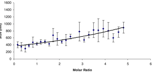
The mean hydrodynamic diameter of the β-CD:TC compounds measured by the dynamic
light scattering is shown in Figure 1. The titration of β-CD solution in TC solution showed an
increase of this parameter as a function β-CD concentration. The experiment was designed so
as to include all the fixed molar ratios used in the physicochemical characterizations. The
average hydrodynamic diameter of the associations was of 433.3±41.9nm, 492.3±46.1nm,
542.3±57.5nm and 861.0±236.8nm for the 1:1, 2:1, 3:1 and 4:1, respectively. The size of the
complex increased upon β-CD concentration increment. An increase of dispersity could be
due to a non-geometric shape of these growing aggregates [37]. TC seems to have seeding
properties among β-CD molecules, inducing these self-assembling phenomena [8].
0 200 400 600 800 1000 1200 1400 1600
0 1 2 3 4 5 6
Molar Ratio Siz e ( n m)
Figure 1. Size measurements obtained by DLS titration of a 0.5 x 10⁻³ solution of TC in a 12.3
x 10-3mol/L β-CD solution.
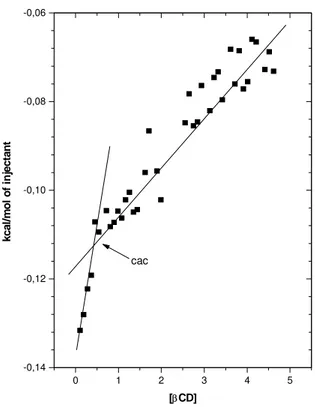
ITC experiments were also performed using the same conditions as those of the DLS
experiment. The curves showed, by linear regression, two domains with the increase of β-CD
modulus, attributed to host-guest interactions, which may be attributed to the formation of
hydrogen bonds between TC aromatic hydrogens with β-CD cavity, releasing highly energetic
water molecules. This suggested the formation of an inclusion compound (Figure 2). The
second domain was also exothermic but showed that as β-CD concentration increased the ∆β
-CDH° modulus became smaller. This reduction could be related to the complexation and
desolvation of outer water molecules of the high stoichiometry complexes, suggesting the
formation of nano assemblies [8].
0 1 2 3 4 5
-0,14 -0,12 -0,10 -0,08 -0,06 cac
[βCD]
kc al /m ol of i n ject ant
Figure 2. ITC experiments expansion to β-CD 12.3 x 10⁻³ mol/L in TC 0.5 x 10⁻³ mol/L.
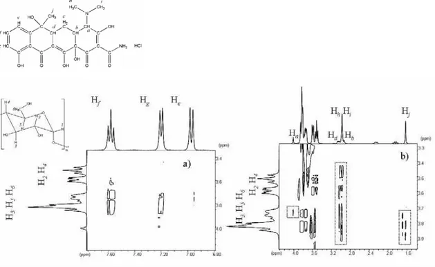
The NMR spectrum of pure TC and the inclusion compound as in the 1:1 molar ratio are
presented in Figure 3a and 3b respectively. Some modifications in the profile of the spectrum
of pure TC are observed in the presence of β-CD. These modifications in the chemical shits
are caused by the change in the electronic density upon the inclusion of the TC in the β-CD
Figure 3. 1H NMR (400MHz) in D2O at 25 ºC of: a) pure TC and b) IC at 1:1 TC β-CD molar
ratio.
To confirm which region of TC molecule was included into the β-CD cavity, NOESY
experiments were conducted. The 1:1 molar ratio NOESY experiment showed a cross peak
correlation, at short distance (less than 5 Å) between the aromatic protons of the TC, He (
δ
6.98), Hf (
δ
7.59) and Hg (δ
7.21), with the cavity protons of β-CD, H3, H5 and H6 (region atδ
In addition, the correlation between the protons Hb, Hd (
δ
3.01), Hh and Hi (δ
3.98) of TCwere determined with all β-CD protons. Also the Hj(
δ
1.63) of TC were correlated to protonsH3, H5 and H6 from the β-CD, Figure 4b.
Figure 4. Expansion of the NMR NOESY contour map (400MHz, mixing time 600ms) in
D2O of IC at molar ratio of 1:1, a) to aromatic region and b) to other correlations between TC
and β-CD.
These results suggested that multiple associations were present in this system. The
correlations between the outer part of the molecule hydrogens (H2 and H4) and TC
non-aromatic protons, besides not being less than 5Å interactions, could be interpreted as
proximity between these molecules. No change in the chemical shifts at the higher molar
ratios 2:1, 3:1 and 4:1 were observed when these are compared to the 1:1 β-CD:TC system
and similar cross peak correlations between the aromatic region of TC with internal protons of
In order to further characterize the supramolecular complexes, solid state physicochemical
analysis were also performed.
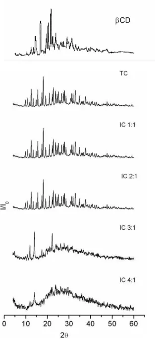
X-ray free substances and mechanical mixtures spectra showed high crystallinity (Figure 5)
[38; 39], a pattern that changed to an amorphous profile as long as the β-CD concentration
also increased, relatively. This could suggest new molecular organization phenomenon upon
Figure 5. X-ray powder diffraction of the β-CD:TC compounds (in the labels TC represents
tetracycline and IC represents β-CD:TC respectively molar ratio of 1:1; 2:1; 3:1 and 4:1).
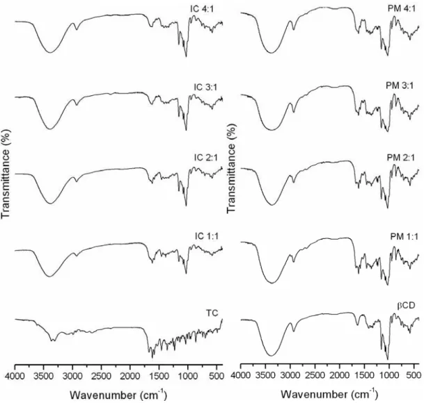
A host:guest interaction is also suggested by infrared analysis. The infrared spectra of β
-cyclodextrin, tetracycline, physical mixture and the supramolecular compounds are shown in
Figure 6. The FTIR spectrum of pure TC is presented, and the most important vibrations
modes are: ν (C-N) at 3365 cm-1, ν (C=O) and ν (C=C) of aromatic ring between 1674 to
1580 cm-1, at 1460 – 1310 cm-1 related to δ (OH), δ (C-C) and ν (C-C) and finally the bands
at 1250 – 1200 cm-1 corresponding to δ (N-H) and ν (C-N) [40].
A sharpening of the ν (O-H) at 3300 cm-1 and ν (C-O-C) at 1070 cm-1 modes are observed in
the spectra of the inclusion compounds at 1:1 to 4:1 molar ratio, when compared to the
spectrum of pure β-CD. These modifications may be attributed to the reduction in the
hydrogen bond forming upon the inclusion of the TC as result of the loss of intra cavity water
molecules [41]
In addition, a strong modification in the vibration mode relating to the ν (C=O) and ν (C=C)
of aromatic ring between 1674 to 1580 cm-1 are observed with the gradual increase of the β
-CD concentration in the inclusion compound. Modifications in the bands below 900 cm-1 are
also observed in all the inclusion compounds. These results suggested the formation of a new
species in solid state when the freeze-drying method is used to prepare the inclusion
Figure 6. FTIR spectra at 4000 – 400cm-1 of pure TC, β-CD:TC (IC) 1:1, 2:1, 3:1, 4:1, pure β
-CD, and physical mixtures (PM) 1:1, 2:1, 3:1 and 4:1.
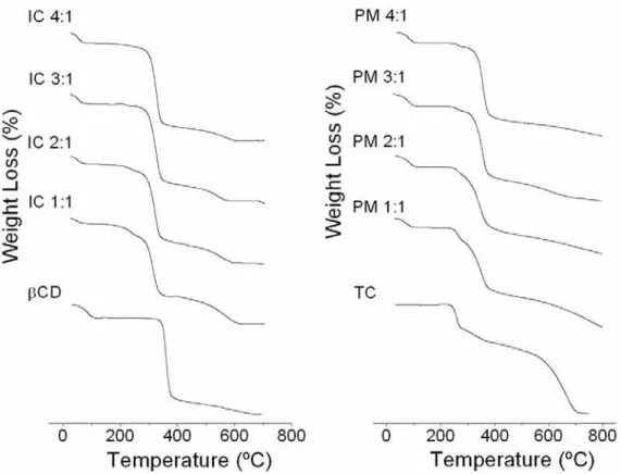
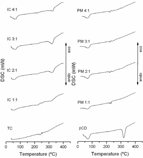
The TG and DSC curves are presented in Figure 7 and 8. Tetracycline hydrochloride showed
great thermal stability between 25 and 200°C and mass losses in two stages, between 200 and
738°C, as shown in its TG and DSC curves. The first event occurred at 287°C, a fast process
of 23.81% mass loss; and the second stage (287-737°C) showed 76.57% mass loss, due to the
thermal decomposition with the formation of carbonaceous products. TC DSC curve showed
an endothermic event at 225°C and exothermic event at 235°C. This result suggested fusion
oxidation, corresponding to the first mass loss showed at the TG curve. The exothermic
events after this point were due to the pirolysis of the carbonaceous products [38].
β-cyclodextrin showed mass losses in two main phases between 25 and 700°C. The first, from
27.2 to 111.56°C, corresponded mainly to water loss (12%) from β-CD cavity. The second
phase (111.56 to 682.94°C) represented the substance degradation and mass loss of 88%. The
respective DSC curves showed two endothermic events at the same temperature ranges and an
endothermic event at 320°C [39].
In the case of 1:1 inclusion compound, the first event in TG curve showed mass loss with less
intensity (4.5% at 25-96.5°C) comparing to β-CD courve. Therefore, it suggested that water
and TC occupied β-CDs’ cavities. This event could be confirmed with less pronounced
endothermic peak when compared to β-CD alone DSC curve at the same temperature range;
and lower β-CD and TC stability between 96.5-282.9°C. This mass loss (12.3%)
corresponded an endothermic event in the DSC curve, which mainly represented TC fusion.
The third event showed 55% mass loss (283-405°C), and an endothermic peak at the same
region of β-CD alone, representing the final thermal decomposition.
In relation to 2:1 compound TG curve, it showed water mass loss in the first stage of
degradation in the range of 25-107°C (6.9%). The endothermic event showed in DSC curve
confirms this finding. The water mass loss was more pronounced in this case when compare
with 1:1 inclusion compound due to β-CD excess in the system. The second event at
107-280°C had lower intensity (7%) and corresponded to probably TC degradation. The DSC
curves showed an endothermic event with lower intensity than in TC alone and 1:1 IC curves
in the 280-424.6°C range and was higher than in 1:1 endothermic event registered in DSC
curves that could be related to excess of β-CD in the system.
In the case of the 3:1 compound, TG curve showed mass loss of 8.2% at the first stage of
degradation (25-102°C), higher than in 2:1 compound group, due to the increased β-CD
concentration in the system. The endothermic peak in the DSC was more pronounced,
corroborating that interpretation. The following stage of degradation showed in 1:1 and 2:1
groups was not seen in this group. This fact can be related to a higher protection of TC
molecule inside the β-CD aggregate, providing higher thermal stability to the drug. In
consequence, the second event of thermal degradation in this group showed an increased mass
loss of 74% (103-423°C). A higher endothermic event was observed in the same temperature
range.
In the case of 4:1 compound, TG curves showed mass loss of 9.1% at the first event of
degradation (25-102°C). This curve resembled to the β-CD TG curve due to the excess of this
molecule in the system. The next stages of degradation showed mass loss of 78%
(103-430°C) and 13% (430-748°C). The 4:1 DSC curve was similar to the 3:1 compound showing
the equivalent endothermic events described above.
The β-CD:TC continuous thermal decomposition process was different from TC and/or β-CD
alone. It could be noticed that with the increase of β-CD molar ratio, the thermal
decomposition profile of the compounds became similar to β-CD alone profile. Then, thermal
instability of TC and β-CD occurred after forming the inclusion compounds, evidencing the
β-CD degradation event near 100°C, TC fusion and thermal decomposition. The second β-CD
endothermic event near 320°C was not observed [35; 38; 39].
Figure 7. TG of tetracycline (TC), β-cyclodextrin (βCD) and the β-CD:TC compounds at
different molar ratios (IC 1:1; IC 2:1; IC 3:1 and IC 4:1) and their physical mixtures (PM 1:1;
Figure 8. DSC curves of tetracycline (TC), β-cyclodextrin (βCD) and the β-CD:TC
compounds at different molar ratios (IC 1:1; IC 2:1; IC 3:1 and IC 4:1) and their physical
mixtures (PM 1:1; PM 2:1; PM 3:1 and PM 4:1).
The physicochemical characterization strongly suggested important structural changes in the
molecules characteristics, revealing the formation of an inclusion compound and, with the
gradual increase of β-CD amount in the system, these compounds could self assemble as
supramolecular complexes. This interpretation differs from other studies that suggested that
the interactions take place with the outer part of the β-CD molecule which is more
hydrophilic, and also contradicts the affirmative that TC molecule seems to be too large to fit
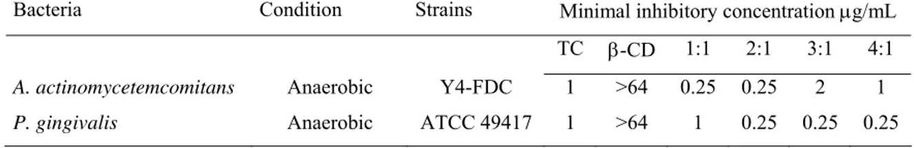
The structural changes that happened upon molecules interaction reflected in TC
antimicrobial properties. The antimicrobial tests showed that 2:1 β-CD:TC compound
exhibited significant antimicrobial activity against A. actinomycetemcomitans with MIC of
0.25µg/mL of TC concentration(1:1; 2:1 groups), followed by 1µg/mL (4:1 and TC control
group) and 2µg/mL (3:1) (Table 1). In spite of TC concentrations used in the test solutions
being normalized with the same TC concentration, the results suggested different interactions
between this bacteria strain and the different β-CD:TC compounds in solution. A.
actinomycetemcomitans is frequently resistant to TC showing MIC values between
0.8-1.2µg/mL [43; 44]. These findings have also to be confirmed against rough-type
microorganisms [45]. The MIC values found for P. gingivalis were 0.25µg/mL (2:1; 3:1 and
4:1 groups), followed by 1µg/mL (1:1 and TC control groups). Other studies showed MIC100
values for TC of 1-4µg/mL against the same bacteria, isolated from humans or reference
strains [46; 47]. β-CD showed no activity against the tested bacteria. Thus, nanoassemblies
antimicrobial properties evidenced that MIC concentrations could decrease four times and
show the same inhibition as non-complexed drug.
Table 1. Minimal inhibitory concentrations values (MIC in µg/mL) of the studied compounds
against tested reference bacteria strains in vitro.
Bacteria Condition Strains Minimal inhibitory concentration µg/mL
TC β-CD 1:1 2:1 3:1 4:1
A. actinomycetemcomitans Anaerobic Y4-FDC 1 >64 0.25 0.25 2 1
P. gingivalis Anaerobic ATCC 49417 1 >64 1 0.25 0.25 0.25
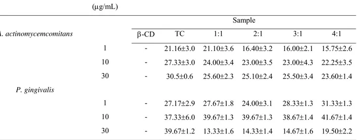
Agar diffusion susceptibility test revealed that all compounds successfully inhibited growth of
A. actinomycetemcomitans, disclosing that the inclusion process did not affected negatively
were found against this strain. β-CD:TC compounds and TC alone showed comparable
growth inhibition of P. gingivalis, at the concentration of 30µg/mL. The exception was the
1:1 compound that demonstrated significantly lower inhibition (p<0.05). For the
concentration of 10µg/mL, all groups showed similar inhibition. 1µg/mL of TC showed
inhibition in all tested groups, being 4:1 with larger inhibition zone when compared to 2:1
group (p<0.05).
Table 2. Inhibition of growth of β-CD:TC compounds (1:1, 2:1, 3:1 and 4:1) and against A.
actinomycemcomitans and P.gingivalis.
Bacteria Tetracycline
Concentration
(µg/mL)
Mean inhibition zone diameter (mm ± S. D.)
Sample
A. actinomycemcomitans β-CD TC 1:1 2:1 3:1 4:1
1 - 21.16±3.0 21.10±3.6 16.40±3.2 16.00±2.1 15.75±2.6
10 - 27.33±3.0 24.00±3.4 23.00±3.5 23.00±4.3 22.25±3.5
30 - 30.5±0.6 25.60±2.3 25.10±2.4 25.50±3.4 23.60±1.4
P. gingivalis
1 - 27.17±2.9 27.67±1.8 24.00±3.1 28.33±1.3 31.33±1.3
10 - 37.33±6.0 39.67±1.3 39.67±1.3 38.67±1.4 41.67±1.4
30 - 39.67±1.2 13.33±1.6 14.33±1.4 14.67±1.6 19.50±2.2
3.2 Nanospheres preparation, release study and antimicrobial activity
The TC encapsulation efficiency of nanospheres prepared with PLGA by double emulsion
process showed values of 46.80% w/w (1:1), 16.73% (2:1 group), 19.07% (3:1), 25.63% (TC
alone) and 42.90% (4:1) (Table 2). Thus, different proportions of β-CD influenced the TC
encapsulation efficiency. The encapsulation efficiency obtained in this study was higher than
double emulsion [49; 50]. The SEM images disclosed nanospheres morphology and also a
smooth surface (Figure 9).
Figure 9. SEM images from the PLGA nanospheres in association with TC; β-CD:TC 1:1;
2:1; 3:1; 4:1.
Nanospheres size distribution is shown in Table 3 and also reflected the influence of β-CD in
the encapsulation process. Nanospheres measuring 285 to 396nm were observed when the
cyclic molecule was present in the process comparing to 958nm with TC alone group.
T
TCC 11::11 ``22::11
3
Table 3. Characteristics of PLGA/TC particles prepared in the presence of β-CD with respect
to drug encapsulation efficiency and size distribuition.
Size distribution Group TC Loading (%)
(%) (nm) Pdi
TC 25.63 82.9
8.60 8.40 958 5400 309 0.5
1:1 46.80 100 307 1.0
2:1 16.73 100 396 0.7
3:1 19.07 82.90
17.10
285
5240
0.7
4:1 42.90 100 360 0.6
Tetracycline release kinetics from the tested compounds in association with PLGA for 10
days is shown in Figure 10. All the groups were prepared with the same TC initial amount. β
-CD influenced drug release from nanospheres with the 2:1 group nearer to a zero order
release. This sample showed a burst effect of 12.74±1.4% (78.9µg/mL) of the total released
drug. The other groups showed a significantly higher 24h release relative to the total amount
of released drug, including TC (23.84±1.0% - 105.3µg/mL), 1:1 (29.39±11.3 - 46.2µg/mL),
3:1 (28.08±0.7 - 52.7µg/mL) and 4:1 (22.23±0.2% - 64.2µg/mL) (p<0.01). These results
showed more constant release kinetics than Actisite®, the commercially available TC local
release device [51]. PLGA degradation is mainly by bulk erosion and this could explain the
drug delivery for the subsequent days. β-CD showed to have great influence in TC release
0 2 4 6 8 10 0 100 200 300 400 500 600 700
Concentrat
ion (
µg/mL)
Time (Days)
TC 1:1 2:1 3:1 4:1Figure 10. In vitro cumulative TC release from PLGA nanospheres in water at 37°C.
The in vitro release study therefore revealed that the concentration of the drug released at each
time point throughout the 10 days test period was above 4µg/mL. This amount of the drug
released was enough to provide appropriate TC levels that correspond to MIC values in vivo
[43-47]. This fact associated to the initial high release and prolonged subsequent release was
favorable to increase antibiotic potency
Agar diffusion susceptibility test revealed that the TC concentrations released from
nanospheres produced satisfactory growth inhibition. The groups showed efficient
antimicrobial activity during 10 days of release. 2:1 group revealed higher inhibition
compared to the other groups in days, which correlated with the release findings (p<0.05).
These results suggest that the drug can be released in a controlled fashion and exert efficiently
5. Conclusion
The physicochemical characterization and drug release data obtained in this study confirms
the potential of this system for optimizing antimicrobial activity.
The interactions between β-CD and TC suggested by X-ray diffraction, thermal analysis, IR
were strongly confirmed by ITC and DLS. NMR analysis also disclosed short distance
interactions between these two molecules in different molar ratios and, TC seems to act as a
seeding molecule capable of inducing self-assembly phenomena with β-CD. The
supramolecular conformation in stoichiometries of 2:1, 3:1 and 4:1 increased four times TC
efficiency against P. gingivalis. 1:1 and 2:1 complexes were also more efficient against A.
actinomycetemcomitans.
The water-oil-water emulsion technique used was efficient to produce PLGA nanospheres
containing β-CD:TC compounds. The cyclodextrin influenced the encapsulation efficiency,
size distribution and also increased the antimicrobial effectiveness of the antibiotic.
The nanoparticles showed a controlled release profile mainly with the 2:1 molar ratio,
releasing TC over minimum inhibitory concentration of A. actinomycetemcomitans and P.
Acknowledgments
The authors thank CNPq - Brazil (Conselho Nacional de Desenvolvimento Científico e
Tecnológico) and FAPEMIG – Minas Gerais, Brazil (Fundação de Amparo à Pesquisa do
Estado de Minas Gerais) for the financial support and scholarship, and to Vanderbilt
6. References
[1] L.X. Kong, Z. Peng, S.D. Li, P.M. Bartold, Nanotechnology and its role in management
of periodontal diseases, Periodontol. 2000. 40 (2006) 184-196.
[2] I.W. Saenger, Cyclodextrin Inclusion Compounds in Research and Industry. Angew.
Chem. Int. Ed. Engl. 19(5) (1980) 344 – 362.
[3] J. Szejtli, The cyclodextrins and their applications in biotechnology. Carbohydr. polym.
12(4) (1990) 375.
[4] A. Korolkovas, Inclusão molecular e ciclodextrinas: propriedades e aplicações
terapêuticas. Enlace Farmalab. 2(2) (1991) 6-15.
[5] K. Uekama, Design and evaluation of cyclodextrin-based drug formulation. Chem.
Pharm. Bull. (Tokyo). 52(8) (2004) 900-915.
[6] S. Zhang, D.M. Marini, W. Hwang, S. Santoso, Design of nanostructured biological
materials through self-assembly of peptides and proteins. Curr. Opin. Chem. Biol. 6(6)
(2002) 865–871.
[7] S. Zhang, Fabrication of biomaterials through molecular self-assembly. Nat. Biotechnol.
21(10) (2003) 1171-1178.
[8] A.M.L. Denadai, M.M. Santoro, L.H. Da Silva, A.T. Viana, R.A.S. Santos, R.D.
Sinisterra, Self-assembly Characterization of the β-Cyclodextrin and
Hydrochlorothiazide System: NMR, Phase Solubility, ITC and QELS. J. Incl. Phenom.
Macro. (55) (2006) 41–49.
[9] A.L. Pataro, C.F. Franco, V.R. Santos, M.E. Cortes, R.D. Sinisterra, Surface effects and
desorption of tetracycline supramolecular complex on bovine dentine. Biomaterials.
24(6) (2003) 1075-1080.
[10] J.M. Moreno-Cerezo, M. Córdoba-Díaz, D. Córdoba-Díaz, M. Córdoba-Borrego, A
stability study of tetracycline and tetracycline cyclodextrins in tablets using a new
HPLC method. J. Pharm. Biomed. Anal. (26) (2001) 417–426.
[11] I.C. Yue, J. Poff, M.E. Cortés, R.D. Sinisterra, C.B. Faris, P. Hildgen, R. Langer, V.P.
Shastri, A novel polymeric chlorhexidine delivery device for the treatment of
[12] R.A. Seymour, P.A. Heasman, Pharmacological control of periodontal disease. II.
Antimicrobial agents. J. Dent. 23(1) (1995) 5-14.
[13] A. Marzo, L. Dal Bo, Chromatography as an analytical tool for selected antibiotic
classes: a reappraisal addressed to pharmacokinetic applications. J. Chromatogr. A.
(812) (1998) 17–34.
[14] E.L. Carter, Antibiotics in cutaneous medicine: an update. Semin. Cutan. Med. Surg. 22
(3) (2003) 196-211.
[15] A. Mombelli, Antibióticos em Terapia Periodontal. In: Lindhe J. Tratado de Periodontia
e Implantologia Oral, 3ed, Guanabara-Koogan, Rio de Janeiro, 1999, p. 350-363.
[16] C.B. Walker, K. Karpinia, P. Baehni, Chemotherapeutics: antibiotics and other
antimicrobials. Periodontol. 2000. (36) (2004) 146–165.
[17] S.L. Morrison, C.M. Cobb, G.M. Kazakos, W.J. Killoy. Root surface characteristic
associated with subgingival placement of monolithic tetracycline-impregnated fibers. J.
Periodontol. 63 (2) (1992) 137-43.
[18] L.R. Friesen, K.B. Williams, L.S. Krause, W.J. Killoy, Controlled local delivery of
tetracycline with polymer strips in the treatment of periodontitis. J. Periodontol. 73 (1)
(2002) 13-9.
[19] R.J. Oringer, K.F. Al-Shammari, W.A. Aldredge, V.J. Iacono, R.M. Eber, H.L. Wang,
B. Berwald, R. Nejat, W.V. Giannobile, Effect of locally delivered minocycline
microspheres on markers of bone resorption. J. Periodontol. 73(8) (2002) 835-42.
[20] G.E. Salvi, A. Mombelli, L. Mayfield, A. Rutar, J. Suvan, S. Garret, NP. Lang. Local
antimicrobial therapy after initial periodontal treatment. A randomized clinical trial
comparing three biodegradable polymers. J. Clin. Periodontol. (29) (2002) 540-550.
[21] L. Silverstein, N. Bissada, M. Manouchehr-Pour, H. Greenwell. Clinical and
microbioloogical effects of local tetracycline irrigation on periodontitis. J. Periodontol.
59(5) (1988) 301-5.
[22] T.A. Eckles, R.A. Reinhardt, J.K. Dyer, G.J. Tussing, W.M. Szydlowski, L.M. DuBous.
Intracrevicular application of tetracycline in white petrolatum for the treatment of
periodontal disease. J. Clin. Periodontol. 17(7 pt 1) (1990) 454-62.
[23] G.I. Maze, R.A. Reinhardt, R.K. Agarwal, J.K. Dyer, D.H. Robinson, L.M. Du Bois,
G.J. Tussing, C.R. Maze. Response to intracrevicular controlled delivery of 25%
tetracycline from poly(lactide/glycolide) film strips in SPT patients. J. Clin. Periodontol.
[24] T.F. Flemmig, S. Weinacht, S. Rüdiger, M. Rumetsch, A. Jung, B. Klaiber, Adjunctive
controlled topical application of tetracycline HCL in the treatment of localized
persistent or recurrent periodontitis. Effects on clinical parameters and elastase-α
1-proteinase inhibitor in gingival crevicular fluid. J. Clin. Periodontol. (23) (1996)
914-921.
[25] N. Babay, Attachment of human gingival fibroblasts to periodontally involved root
surface following scaling and/or etching procedures: a scanning electron microscopy
study, Braz. Dent. J. 12(1) (2000) 17-21.
[26] E. Esposito, V. Carotta, A. Scabbia, L. Trombelli, P. D’Antona, E. Menegatti, C.
Nastruzzi, Comparative analysis of tetracycline dental gels: poloxamer- and
monoglyceride-based formulations. Int. J. Pharm. (142) (1996) 9-23.
[27] D.S. Vienneau, C.G. Kindberg, Development and validation of a sensitive method for
tetracycline in gingival crevicular fluid by HPLC using fluorescence detection. J.
Pharm. Biomed. Anal. 16(1) (1997) 111-117.
[28] D. Sendil, I. Gursel, D.L. Wise, V. Hasýrcý, Antibiotic release from biodegradable
PHBV microparticles. J. Control. Release. 59(2) (1999) 207-217.
[29] D.S. Jones, A.D. Woolfson, A.F. Brown, W.A. Coulter, C. McClelland, C.R. Irwin,
Design, characterization and preliminary clinical evaluation of a novel mucoadesive
topical formulation containing tetracycline for treatment of periodontal disease. J.
Control. Release. (67) (2000) 357-368.
[30] K. Schwach-Abdellaoui, A. Monti, J. Barr, J. Heller, R. Gurny, Optimization of a novel
bioerodible device based on auto-catalyzed poly(ortho esters) for controlled delivery of
tetracycline to periodontal pocket. Biomaterials. 22(11) (2001) 1659-1666.
[31] L.E. Bromberg, D.K. Buxton, P.M. Friden, Novel periodontal drug delivery system for
treatment of periodontitis. J. Control. Release. (71) (2001) 251-259.
[32] J. Heller, J. Barr, S.Y. Ng, H.R. Shen, K. Schwach-Abdellaoui, R. Gurny, Vivien- N.
Castioni, P.J. Loup, P. Baehni, A. Mombelli, Development and applications of
injectable poly(ortho esters) for pain control and periodontal treatment. Biomaterials.
(23) (2002) 4397-4404.
[33] H.M. Kelly, P.B. Deasy, E. Ziaka, N. Claffey, Formulation and preliminary in vivo dog
studies of a novel drug delivery system for the treatment of periodontitis. Int. J. Pharm.
[34] Z.R. Domingues, M.E. Cortés, T.A. Gomes, H.F. Diniz, C.S. Freitas, J.B. Gomes,
A.M.C. Faria, R.D. Sinisterra, Bioactive glass as a drug delivery system of tetracycline
and tetracycline associated with β-cyclodextrin. Biomaterials. (25) (2004) 327–333.
[35] M.E. Cortés, R.D. Sinisterra, M.J. Avila-Campos, N. Tortamano, R.G. Rocha, The
chlorhexidine: -cyclodextrin inclusion compound: preparation, characterization and
microbiological evaluation. J. Incl. Phenom. Macro. (40) (2001) 297–302.
[36] NCCLS-National Committee for Clinical Laboratory Standards (2000): Methods for
dilution antimicrobial susceptibility tests for bacteria that grow aerobically. Wayne,
Pennsylvania: National Committee for Clinical Laboratory Standards. NCCLS
Document M7-A5.
[37] R.C. Petter, J.S. Salek, C.T. Sikorski, G. Kumaravel, F.T. Lin, Cooperative binding by
aggregated mono-6-(alkylamino)-β-cyclodextrins. J. Am. Chem. Soc. 112(10) (1990)
3860-3868.
[38] N.S. Fernandes, M.A.S. Carvalho Filho, R.A. Mendes, M. Ionashiro, Thermal
Decomposition of Some Chemotherapic Substances. J. Braz. Chem. Soc. 10(6) (1999)
459-462.
[39] G. Bettinetti, Cs. Novák, M. Sorrenti, Thermal and structural characterization of
commercial α-, β-, and -cyclodextrins. J. Therm. Anal. Cal. (68) (2002) 517-529.
[40] C.F. Leypold, M. Reiher, G. Brehm, M.O. Schmitt, S. Schneider, P. Matousek, M.
Towrie, Tetracycline and derivatives – assignment of IR and Raman spectra via DFT
calculations. Phys. Chem. Chem. Phys. (5) (1149) 2003-1157.
[41] E. Lamcharfi, G. Kunesch, C. Meyer, B. Robert, Investigation of cyclodextrin inclusion
compounds using FT-IR and Raman spectroscopy. Spectrochim. (51) (1995)
1861-1870.
[42] M. Plätzer, M.A. Schwarz, R.H.H. Neubert, Determination of formation constants of
cyclodextrin inclusion complexes using affinity capillary electrophoresis. J. Micro. Sep.
(11) (1999) 215-222.
[43] M.J. Pavicic, A.J. van Winkelhoff, J. Graaff, In vitro susceptibilities of Actinobacillus
actinomycetemcomitans to a number of antimicrobial combinations. Antimicrob.
Agents. Chemother. 36(12) (1992) 2634-2638.
[44] M.J. Avila-Campos, M.A.R. Carvalho, F. Zelante, Distribuition of biotypes and
antimicrobial susceptibility of Actinobacillus actinomycetemcomitans. Oral. Microbiol.
[45] N. Takahashi, K. Ishihara, T. Kato, K. Okuda, Susceptibility of Actinobacillus
actinomycetemcomitans to six antibiotics decreases as biofilm matures. J. Antimicrob.
Chemoth. 59(1) (2007) 59-65.
[46] F.A. Santos, E.M.A. Bastos, P.H. Rodrigues, M. Uzeda, M.A.R. Carvalho, L.M. Farias,
E.S.A. Moreira, Susceptibility of Prevotella intermedia/ Prevotella nigrescens (and
Porphyromonas gingivalis) to propolis (Bee Glue) and other antimicrobial agents.
Anaerobe. 8(1) (2002) 9-15.
[47] M.T. Andres, W.O. Chung, M.C. Roberts, J.F. Fierro, Antimicrobial susceptibilities of
Porphyromonas gingivalis, Prevotella intermedia, and Prevotella nigrescens spp.
isolated in Spain. Antimicrob. Agents. Chemother. 42(11) (1998) 3022-3.
[48] B. Bittner, K. Mäder, C. Kroll, H-H. Borchert, T. Kissela, Tetracycline-HCl-loaded
poly(DL-lactide-co-glycolide) microspheres prepared by a spray drying technique:
influence of g-irradiation on radical formation and polymer degradation. J. Control.
Release. (59) (1999) 23–32.
[49] H. Kim, D.J. Burgess, Effect of drug stability on the analysis of release data from
controlled release microspheres. J. Microencapsul. 19(5) (2002) 631-640.
[50] E. Esposito, R. Cortesi, F. Cervellati, E. Menegatti, C. Nastruzzi, Biodegradable
microparticles for sustained delivery of tetracycline to the periodontal pocket:
formulatory and drug release studies. J. Microencapsul. 14(2) (1997) 175-87.
[51] E.R. Kenawy, G.L. Bowlin, K. Mansfield, J. Layman, D.G. Simpson, E.H. Sanders,
G.E. Wnek, Release of tetracycline hydrochloride from electrospun
Table 1. Minimal inhibitory concentrations values (MIC in µg/mL) of the studied compounds
against tested reference bacteria strains in vitro.
Bacteria Condition Strains Minimal inhibitory concentration µg/mL
TC β-CD 1:1 2:1 3:1 4:1
A. actinomycetemcomitans anaerobic Y4-FDC 1 >64 0.25 0.25 2 1
P. gingivalis anaerobic ATCC 49417 1 >64 1 0.25 0.25 0.25
Table 2. Inhibition of growth of β-CD:TC compounds (1:1, 2:1, 3:1 and 4:1) and against A.
actinomycemcomitans and P.gingivalis.
Bacteria Tetracycline
Concentration
(µg/mL)
Mean inhibition zone diameter (mm ± S. D.)
Sample
A. actinomycemcomitans β-CD TC 1:1 2:1 3:1 4:1
1 - 21.16±3.0 21.10±3.6 16.40±3.2 16.00±2.1 15.75±2.6
10 - 27.33±3.0 24.00±3.4 23.00±3.5 23.00±4.3 22.25±3.5
30 - 30.5±0.6 25.60±2.3 25.10±2.4 25.50±3.4 23.60±1.4
P. gingivalis
1 - 27.17±2.9 27.67±1.8 24.00±3.1 28.33±1.3 31.33±1.3
10 - 37.33±6.0 39.67±1.3 39.67±1.3 38.67±1.4 41.67±1.4
Table 3. Characteristics of PLGA/TC particles prepared in the presence of β-CD with
respect to drug encapsulation efficiency and size distribuition.
Size distribution Group TC Loading (%)
(%) (nm) Pdi
TC 25.63 82.9
8.60
8.40
958
5400
309
0.5
1:1 46.80 100 307 1.0
2:1 16.73 100 396 0.7
3:1 19.07 82.90
17.10
285
5240
0.7
Figure Legends
Figure 1. Size measurements obtained by DLS titration of a 0.5 x 10⁻³ solution of TC in a 12.3 x 10⁻³mol/L β-CD solution.
Figure 2. ITC experiments expansion to β-CD 12.3 x 10⁻³ mol/L in TC 0.5 x 10⁻³ mol/L.
Figure 3. 1H NMR (400MHz) in D2O at 25 ºC of: a) pure TC and b) IC at 1:1 TC β-CD molar
ratio.
Figure 4. Expansion of the NMR NOESY contour map (400MHz, mixing time 600ms) in D2O of IC at molar ratio of 1:1, a) to aromatic region and b) to other correlations between TC
and β-CD.
Figure 5. X-ray powder diffraction of the β-CD:TC compounds (in the labels TC represents tetracycline and β-CD:TC represents respectively molar ratio of IC 1:1; IC 2:1; IC 3:1 and IC 4:1). PM 1:1; PM 2:1; PM 3:1 and PM 4:1 represents the physical mixtures.
Figure 6. Infrared spectra of tetracycline (TC), β-cyclodextrin (βCD), the β-CD:TC compounds at different molar ratios (IC 1:1; IC 2:1; IC 3:1 and IC 4:1) and their physical mixtures (PM 1:1; PM 2:1; PM 3:1 and PM 4:1).
Figure 7. TG of tetracycline (TC), β-cyclodextrin (βCD) and the β-CD:TC compounds at different molar ratios (IC 1:1; IC 2:1; IC 3:1 and IC 4:1) and their physical mixtures (PM 1:1; PM 2:1; PM 3:1 and PM 4:1).
Figure 8. DSC curves of tetracycline (TC), β-cyclodextrin (βCD) and the β-CD:TC compounds at different molar ratios (IC 1:1; IC 2:1; IC 3:1 and IC 4:1) and their mechanical mixtures (PM 1:1; PM 2:1; PM 3:1 and PM 4:1).
Figure 9. SEM images from the PLGA microspheres in association with TC; β-CD:TC 1:1; 2:1; 3:1; 4:1.
Figure 1
0 200 400 600 800 1000 1200 1400 1600
0 1 2 3 4 5 6
Molar Ratio
Siz
e
(
n
Figure 2
0 1 2 3 4 5
-0,14 -0,12 -0,10 -0,08 -0,06
cac
[βCD]
kc
al
/m
ol
of
i
n
ject
Figura 4
Figure 5
Figure 9
T
TCC 11::11 ``22::11
3
Figure 10
0 2 4 6 8 10
0 100 200 300 400 500 600 700
Conc
ent
rat
ion (
µ
g/
mL)
Time (Days)
COMENTÁRIOS FINAIS
O presente trabalho teve como intenção a fabricação e a caracterização de compostos
supramoleculares entre β-ciclodextrinas e tetraciclinas. A elucidação das interações entre as
duas moléculas é de fundamental importância para o futuro desenvolvimento de formulações
e produtos que as contenham.
A caracterização físico-química revelou interação, mudanças estruturais e conformacionais
entre as duas moléculas, relacionadas ao aumento da concentração de β-ciclodextrina no
sistema. Na medida em que a razão molar de β-ciclodextrina cresceu no sistema, foi
observado um aumento de tamanho das partículas formadas pelos compostos em solução,
devido a interações dentro e fora da cavidade, além de mudanças na cristalinidade e em
relação às suas propriedades de estabilidade térmica.
Essas interações tiveram impacto na atividade antimicrobiana desses compostos. Foi
mostrados que esses nano agregados, quando em solução, apresentaram maior eficácia
antimicrobiana contra as bactérias testadas em determinadas concentrações. Estas novas
propriedades antimicrobianas abrem novos questionamentos à cerca de como esses nano
agregados interagem com as bactérias, porque nestes experimentos, todos os compostos
apresentavam a mesma concentração de tetraciclina, diferindo apenas na razão molar de β
-ciclodextrina. Possivelmente as β-ciclodextrinas poderiam modular a liberação do fármaco de
uma maneira mais controlada por um período mais prolongado, diferindo da ação da
tetraciclina pura e melhorando suas propriedades [33; 38]. Outra possibilidade seria uma
diferente interação dos nanoagregados com a parede celular bacteriana o que poderia explicar
A técnica de nano encapsulamento no polímero PLGA mostrou-se eficaz para a produção de
nano esferas de tetraciclina. A presença da β-ciclodextrina mostrou influenciar o percentual
de encapsulamento de tetraciclina no polímero além de ter marcante influência no tamanho
das nano esferas.
As nano esferas poliméricas exibiram uma liberação controlada por cerca de 10 dias. Foi
observado que a liberação obtida das nano esferas sintetizadas a partir do composto na razão
molar de 2:1 β-ciclodextrina:tetraciclina esteve perto de uma liberação de ordem zero. As
concentrações da droga obtidas a partir da concentração inicial de 1mg/mL mostraram-se
acima das concentrações inibitórias mínimas para as bactérias testadas A.
actinomycetemcomitans e P. gingivalis.
Assim, foi demonstrado que é possível a formação de nano agregados com tetraciclina e β
-ciclodextrina e que estes podem ser utilizados para a formulação de nano esferas de PLGA