catenin-dependent reporter gene activity. Although the mechanism through which the compounds activate b-catenin signaling has yet to be determined, several key regulators of Wnt/b-catenin signaling, including glycogen synthase kinase 3 and Frizzled receptors, were excluded as the molecular target. The compounds displayed remarkable selectivity, as they only inducedb-catenin signaling in a human osteosarcoma U2OS cell line and not in a variety of other cell lines examined. Our data indicate that differences in cellular Wnt/b-catenin signaling machinery can be exploited to identify cell type-specific activators of Wnt/b-catenin signaling.
Citation:Verkaar F, van der Stelt M, Blankesteijn WM, van der Doelen AA, Zaman GJR (2011) Discovery of Novel Small Molecule Activators ofb-Catenin Signaling. PLoS ONE 6(4): e19185. doi:10.1371/journal.pone.0019185
Editor:Cara Gottardi, Northwestern University Feinberg School of Medicine, United States of America
ReceivedDecember 10, 2010;AcceptedMarch 22, 2011;PublishedApril 29, 2011
Copyright:ß2011 Verkaar et al. This is an open-access article distributed under the terms of the Creative Commons Attribution License, which permits unrestricted use, distribution, and reproduction in any medium, provided the original author and source are credited.
Funding:The work was supported by the Dutch government BSIK grant 03033 (to FV and WMB) (http://www.senternovem.nl/bsik) and carried out at Merck Research Laboratories in Oss, The Netherlands. The funders had no role in study design, data collection and analysis, decision to publish, or preparation of the manuscript.
Competing Interests:GZ, MS and AD are employees of Merck Sharp & Dohme. FV is an employee of the University of Maastricht. All work was carried out at Merck Research Laboratories in Oss, The Netherlands. The authors confirm that this does not alter their adherence to all the PLoS ONE policies on sharing data and materials, as detailed online in the guide for authors.
* E-mail: [email protected]
Introduction
Wnt/b-catenin signaling orchestrates embryogenesis and adult stem cell maintenance in mammals [1]. It is initiated when Wnt ligands bind to seven transmembrane receptors of the Frizzled family and to representatives of the single-pass low-density lipoprotein receptor-related protein family (LRP5 or -6) [2,3,4]. Wnt, Frizzled and LRP5/6 form a ternary complex that initiates a cascade of molecular interactions that ultimately leads to the cytoplasmic stabilization of the transcriptional modulator b -catenin. b-Catenin subsequently enters the nucleus where it interacts with T cell factor/Lymphoid enhancer factor (TCF/LEF) and influences the transcription ofb-catenin-dependent genes [5]. In non-stimulated cells,b-catenin protein stability is compromised by the glycogen synthase kinase 3 (GSK3)-mediated phosphory-lation of b-catenin on several conserved N-terminal residues. These phosphorylations serve as cues for proteasomal degradation ofb-catenin. As a result, quiescent cells typically contain low levels of cytoplasmic and nuclearb-catenin.
Aberrations in Wnt/b-catenin signaling activity are associated with several malignancies [6]. For example, mutations that lead to increasedb-catenin stability are observed in the large majority of colon cancers [6], and drug discovery efforts have mainly focused on identifying inhibitors for the b-catenin pathway [7]. The potential medical application of activators of b-catenin signaling has been largely overlooked, despite evidence that reduced
b-catenin signaling underlies neurodegenerative disorders and aberrations in bone formation [8,9]. We have previously described a cell-based assay that measures the nuclear translocation of b -catenin using enzyme fragment complementation (EFC) [10]. In this assay, complementation occurs between a peptide fragment of
b-galactosidase (called a-peptide) that is genetically fused to b -catenin and a nuclear-resident complementary enzyme fragment (termed Da-Nuc). By applying this assay to screening of a low molecular weight compound library, we have identified novel activators ofb-catenin signaling.
Results
To investigate whether the cannabinoid CB2 receptor was involved in the activation of Wnt/b-catenin signaling, reference cannabinoid CB1/2 agonists CP-55940, WIN55,212-2, and several structural analogs of Cpd1 and Cpd2 were tested in the assay. However, none of these potent cannabinoid CB2 agonists exerted any significant effect (Figure 1A), indicating that the effect was compound specific and activation of the cannabinoid CB2 receptors was not the mechanism through which Cpd1 and Cpd2 increasedb–galactosidase activity in the assay.
At a 10mM concentration, both Cpd1 and Cpd2 induced transcription of a transiently transfected SuperTOPflash reporter gene in U2OS-EFC cells (Figure 1C). We observed micromolar potencies for both compounds (data not shown), similar to those observed in the EFC assay (Figure 1A). As controls, we treated
these cells with 12 nM recombinant mouse Wnt-3a (rmWnt-3a) and 30 mM lithium chloride (LiCl) (Figure 1C). Wnt-3a is a naturally occurring Wnt ligand that activates Wnt/b-catenin signaling in several contexts [12,13]. LiCl and MG132 (see below) inhibit GSK3 and the proteasome, respectively. Treating cells with these agents causes b-catenin stabilization and subsequent activation of Wnt/b-catenin signaling [10]. Cpd1 and Cpd2 did not induce transcription of a mutant luciferase reporter gene construct containing b-catenin-responsive elements that do not bind TCF (SuperFOPflash; Figure 1C), demonstrating selectivity of the response. We subsequently studied the cytoplasmic accumulation ofb-catenin in U2OS-EFC cells following a three hour treatment with rmWnt-3a, LiCl, sotrastaurin and MG132. As predicted, these treatments resulted in an increase in Figure 1. Cpd1 and Cpd2 activate Wnt/b-catenin signaling in U2OS-EFC cells.(A) U2OS-EFC cells were treated with vehicle, 12 nM rmWnt-3a or increasing concentrations of Cpd1, Cpd2 and CP-55940 for 3 hrs, followed by measurement ofb-galactosidase activity. (B) Structures of Cpd1 and Cpd2. (C) U2OS-EFC cells transiently transfected with SuperTOPflash (white bars) or SuperFOPflash (black bars) reporter gene construct were incubated with vehicle, 12 nM rmWnt-3a, 30 mM lithium chloride (LiCl) or 10mM Cpd1, Cpd2 or CP-55940 for 5 hrs before measurement of luciferase activity. (D) U2OS-EFC cells were treated with vehicle, 12 nM rmWnt-3a, 30 mM LiCl, 20mM MG132 or 10mM sotrastaurin, Cpd1, Cpd2 or CP-55940 for 3 hrs. Cytoplasmic protein fractions were isolated and subjected to western blotting forb-catenin andb-actin. (E) U2OS-EFC cells were treated with vehicle, 12 nM rmWnt-3a or 10mM Cpd1 for 8 hrs before analysis of axin2 mRNA levels relative to the expression of the housekeeping gene TATA box-binding protein (TAFII) by means of qPCR. Asterisks (*) represent statistically significant differences (P,0.05).
desensitization by incubating U2OS-EFC cells with increasing concentrations of rhDkk-1 in the presence of 10 nM rmWnt-3a. As can be seen in Figure 2A, rhDkk-1 dose-dependently decreased
b-catenin signaling induced by rmWnt-3a. However, 2mg/ml
rhDkk-1 did not reduceb-galactosidase activity induced by Cpd1 or Cpd2, whereas it reduced rmWnt-3a-induced signaling to background levels (Figure 2B). Furthermore, Cpd1 and Cpd2 did not induce phosphorylation of LRP6 at Ser1490 (Figure 2C), an early event during Wnt/b-catenin pathway activation [18,19]. This suggests that both compounds interact with Wnt/b-catenin signaling at a level between LRP5/6 receptor activation and cytoplasmic accumulation ofb-catenin.
Although biochemical profiling indicated that Cpd1 or Cpd2 did not act through inhibition of GSK3 or any other protein kinase, the possibility that the compounds inhibit GSK3 activity by
stabilization and downstream of LRP5/6 phosphorylation (Figure S2).
696 structural analogs of Cpd1 and Cpd2 were selected from the Merck research Laboratories compound library and tested in theb-catenin EFC assay. 18 compounds were found to increaseb -galactosidase activity at 10mM and two compounds (Cpd4 and 5
in Figure S3) were even more efficacious than Cpd1. There was no apparent correlation between biophysical parameters, such as solubility or lipophylicity, and activity in theb-catenin EFC assay (data not shown), indicating that the activity in the assay was through interaction with a specific target. We have attempted to identify the target by profiling Cpd1 against 80 GPCR-, transporter- and ion channel targets (performed at Cerep, Paris, France) and 21 human phosphatases (performed at Millipore), but these screens did not reveal any specific interactions that could
Figure 2. Cpd1 and Cpd2 do not activate Frizzleds.(A) U2OS-EFC cells were treated with vehicle or 10 nM rmWnt-3a in the absence or presence of increasing concentrations of rhDkk-1 for 3 hrs, followed by measurement ofb-galactosidase activity. (B) U2OS-EFC cells were stimulated with 10 nM rmWnt-3a, 30 mM LiCl, 20mM MG132 or 10mM sotrastaurin, Cpd1 and Cpd2 in the presence of absence of 48 nM rhDkk-1 for 3 hrs, after whichb-galactosidase activities were measured. (C) U2OS-EFC cells were treated with vehicle, 12 nM rmWnt-3a, 30 mM LiCl or 10mM Cpd1, Cpd2, Win55,512-2 and CP-55940 for 1 hr, followed by cell lysis and western blotting forb-actin, total LRP6 and LRP6 phosphorylated at Ser1490. Asterisks (*) represent statistically significant differences (P,0.05).
account for the activating effect of Cpd1 onb-catenin signaling in our cell-based assays.
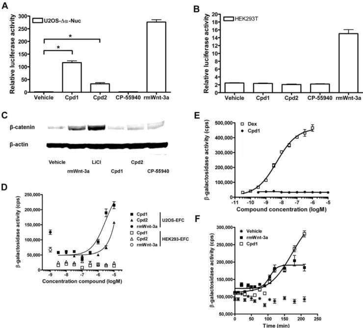
We extended our analysis of Cpd1/2 to different cell lines by transient transfection of superTOPflash reporter gene construct, followed by treatment with compound. As expected, U2OS-Da -Nuc cells responded to Cpd1 and Cpd2 with a marked increase in reporter gene activity (Figure 4A). In contrast, the superTOPflash reporter was not activated in human embryonic kidney (HEK293T) cells, cervical cancer (HeLa) cells or Chinese hamster ovary (CHO-K1) cells after Cpd1/2 treatment (Figure 4B, Figure S4A,B). Consistent with these findings, cytoplasmic b-catenin levels of HEK293T cells were not increased by treatment with 10mM Cpd1 or Cpd2 (Figure 4C). Notably, Cpd1 and Cpd2 did not activate reporter gene activity in another human osteosarcoma cell line (Saos2), or in a rat osteosarcoma (UMR106) cell line (Figure S4C,D). Most strikingly, the compounds were inactive in a U2OS cell batch directly obtained from the American Type Culture Collection (ATCC; Figure S4E). We confirmed that the U2OS-EFC cells shared a common ancestry with the U2OS cells from ATCC by microsatellite analysis, although the analysis indicated that genetic drift had occurred (performed at Biore-liance, Glasgow, UK; data not shown).
Despite the absence ofb-catenin activation in any cell line other than U2OS-EFC cells and U2OS-Da-Nuc cells, our data do not suggest that activation of b-galactosidase activity by Cpd1 and Cpd2 is an assay artifact. Firstly, other parameters of Wnt/b -catenin signaling, such asb-catenin-dependent reporter genes and Western blotting of cytoplasmic b-catenin levels also imply activation of Wnt/b-catenin signaling in these cells (Figure 1C– E, Figure 4A). As an additional control experiment, we transfected U2OS-Da-Nuc cells with luciferase reporter genes sensitive to cAMP (CREB), Ca2+ (NFAT) and glucocorticoid hormone receptor (GR) signaling. None of these reporter genes was activated by Cpd1, whereas SuperTOPflash reporter gene activity was dose-dependently induced by Cpd1 treatment (Figure S5A– D). Furthermore, HEK293 cells engineered to complement b -galactosidase in response to rmWnt-3a (HEK293-EFC cells) did not activateb-catenin signaling when treated with Cpd1 or Cpd2 (Figure 4D). In addition, Cpd1 did not induce b-galactosidase activity in a cellular EFC assay for the recruitment of the scaffolding protein b-arrestin2 to human parathyroid hormone receptor 1 (Figure S6). Furthermore, Cpd1 did not induce the nuclear translocation of human GR in U2OS cells (U2OS-GR)
genetically engineered to complementb-galactosidase in response to GR agonists [10], such as dexamethasone (Dex; Figure 4E). Of note, U2OS-GR cells are derivatives of the U2OS-Da-Nuc cell line. Lastly, time-course EFC experiments revealed that the onset of signal generation in U2OS-EFC cells stimulated with 10mM
Cpd1 is similar to that generated in U2OS-EFC cells treated with 500 ng/ml rmWnt-3a (Figure 4F), although b-galactosidase activity in response to rmWnt-3a seems to reach a plateau earlier. A signal generation profile similar to that seen for Cpd1 was observed when U2OS-EFC cells were treated with 30 mM LiCl (Figure S7). All these experiments indicate that Cpd1/2’s ability to activate Wnt/b-catenin is a highly specific, cell-type dependent process.
Discussion
Activation of the Wnt/b-catenin pathway might provide a new therapeutic opportunity to treat neurodegenerative disorders and aberrations in bone formation. Stimulation of the Wnt/b-catenin pathway by compounds only in specific tissues is expected to generate a better side-effect profile. We have identified small molecule activators of Wnt/b-catenin signaling in a U2OS cell line that did not activate this pathway in various other cell types from different histogenic origin. The molecular target through which the compounds activate b-catenin signaling has yet to be determined, although several key regulators ofb-catenin signaling, including GSK3 and Frizzled receptors, were excluded.
Integrative approaches coupling protein interaction maps to siRNA screening data have suggested that the components that constitute the Wnt/b-catenin signaling machinery in a given cell type are highly variable [21]. Our data confirm that small molecule-mediated cell-type specific activation of Wnt/b-catenin signaling can be achieved. However, elucidation of the molecular target is essential to fully appreciate this finding, and is desirable before these compounds are considered as a starting point for drug discovery. A possible strategy for target identification is biotin-labeling, followed by affinity capture of binding partners in cell lysates. However, such approaches are generally more successful with compounds that bind to their target with high affinity, while screening of several hundreds of analogs did not reveal compounds with potencies lower than 1mM.
In conclusion, we have identified small molecule compounds that activate Wnt/b-catenin signaling in a highly cell-type specific Figure 3. Cpd1 and Cpd2 do not cause activation of Akt/PKB signaling in U2OS-EFC cells.U2OS-EFC cells were stimulated with vehicle, 100 nM insulin, 100 ng/ml platelet-derived growth factor (PDGF), 12 nM rmWnt-3a, 30 mM LiCl or 10mM Cpd1, Cpd2 and CP-55940 for 30 min, followed by cell lysis. Akt/protein kinase B (PKB) and glycogen synthase kinase 3b(GSK3b) phosphorylations at Ser473 and Ser9, respectively, were analyzed by western blotting.
manner. Our data hold promise for the development of tissue-specificb-catenin signaling activators.
Materials and Methods
Cell lines
HEK293T, U2OS, CHO-K1, LM-TK and HeLa cells were obtained from the American Type Culture Collection (ATCC) and cultured in DMEM F12 containing 10% fetal bovine serum
(FBS; Cambrex, Verviers, Belgium), 100 U/ml penicillin and 100mg/ml streptomycin (Invitrogen, Breda, The Netherlands). U2OS-Da-Nuc cells (DiscoveRx; Hannover, Germany) were maintained in the same medium, supplemented with 150mg/ml
hygromycin (Invitrogen). U2OS-EFC, U2OS-GR and HEK293-EFC cells, the generation of which is described elsewhere [10], were cultured in DMEM F12 supplemented with 10% FBS, 100 U/ml penicillin, 100mg/ml streptomycin, 150mg/ml hygro-mycin and 500mg/ml geneticin (Invitrogen).
Figure 4. Cpd1 and Cpd2 do not activate Wnt/b-catenin signaling in HEK293 cells.(A,B) U2OS-Da-Nuc cells (parental to U2OS-EFC cells) (A) and HEK293T cells (B) transiently transfected with SuperTOPflash reporter gene construct were stimulated with 12 nM rmWnt-3a or 10mM Cpd1, Cpd2 and CP-55940 for 5 hrs followed by measurement of luciferase activity. (C) HEK293T cells were stimulated with vehicle, 12 nM rmWnt-3a, 30 mM LiCl or 10mM Cpd1, Cpd2 and CP-55940 for 3 hrs, followed by isolation of cytoplasmic protein fractions and probing ofb-catenin andb-actin using western blotting. (D) U2OS-EFC cells and HEK293 cells genetically engineered to complementb-galactosidase upon nuclear translocation ofb -catenin (HEK293-EFC cells) were stimulated with increasing concentrations of Cpd1 and Cpd2 or 12 nM rmWnt-3a for 3 hrs, followed by measurement ofb-galactosidase activity. (E) U2OS-Da-Nuc cells stably transfected with a vector coding fora-peptide-tagged human glucocorticoid receptor (U2OS-GR cells) were treated with increasing doses of dexamethasone (Dex; a GR agonist) or Cpd1 for 3 hrs before measurement ofb -galactosidase activity. (F) U2OS-EFC cells were stimulated with 12 nM rmWnt-3a or 10mM Cpd1 for several time points before measurement ofb -galactosidase activity.
LMW compounds and recombinant proteins
Synthetic organic low-molecular weight (LMW) compounds were selected from the Merck Research Laboratories compound collection (Oss, The Netherlands) based on previously determined activity on various G protein-coupled receptor (GPCR) or protein kinase targets. Recombinant mouse Wnt-3a (rmWnt-3a), recom-binant human Wnt inhibitory factor-1 (WIF1) and recomrecom-binant human Dickkopf-1 (rhDkk-1) were purchased from R&D Systems (Abingdon, U.K.). Platelet-derived growth factor (PDGF) was obtained from PeproTech (Rocky Hill, NJ).
b-Galactosidase fragment complementation assays
The b-catenin EFC assay was performed as described previously [10].
Reporter gene assays
Luciferase assays on transiently transfected cells were performed as described before [10]. The SuperTOPflash and pTA-SuperFOPflash reporter gene constructs were provided by Prof. dr. R.T Moon (University of Washington, WA, USA). p21xCRE-luc was obtained from the VU University (Amsterdam, The Netherlands). pTA-NFAT-luc was purchased from Clontech. pMMTV-luc and pNGV1-GR were provided by Hans van der Maaden (Merck Research Laboratories). For MMTV-luciferase reporter gene experiments, pMMTV-luc and pNGV1-GR were co-transfected in a 5:1 molar ratio.
Western blotting
Isolation of cytoplasmic fractions and total cell lysates, followed by western blotting, was described previously [10]. The following antibodies were used at the indicated dilutions: mouse anti-b -catenin: 1:2000 (BD Transduction Laboratories; Lexington, KY), mouse anti-b-actin: 1:5000 (Abcam; Cambridge, UK), rabbit-anti-Akt: 1:1000, rabbit anti-GSK3b: 1:1000, rabbit anti-phospho-Ser473 Akt: 1:1000, rabbit anti-phospho-Ser9 GSK3b: 1:1000, rabbit anti-LRP6: 1:1000 and rabbit anti-phospho-Ser1490 LRP6: 1:1000 (Cell Signaling Technology; Danvers, MA), anti-mouse-HRP conjugate: 1:2000 and anti-rabbit-anti-mouse-HRP conjugate: 1:1000 (Promega; Leiden, The Netherlands).
qPCR analysis
Isolation of total mRNA and subsequent analysis of transcript levels by qPCR was performed as described previously [22].
Data analysis
The concentration at which half-maximal activation of the cellular response was reached (EC50values) for rhDkk1 and rmWnt-3a dose response curves was calculated from the expected molecular mass of rhDkk-1 and rmWnt-3a (both 41 kDa) using Graphpad Prism 4.0 software. All data are represented as averages6standard error in the mean (SEM). Statistical significance of observed differences was determined using Student’s t-test and indicated in the figures with asterisks (*). P,0.05 was regarded as statistically significant.
Supporting Information
Figure S1 Cpd1 induces transcription of theb -catenin-responsive gene axin2 in U2OS-Da-Nuc cells. U2OS-Da -Nuc cells were treated with vehicle, 12 nM rmWnt-3a or 10mM
Cpd1 for 8 hrs before analysis of axin2 mRNA levels relative to the expression of the housekeeping gene TATA box-binding protein (TAFII) by means of qPCR. Asterisk represent statistically significant differences (p,0.05).
(TIF)
Figure S2 Schematic representation of the mode of action of Cpd1 and Cpd2. A model of Wnt/b-catenin signaling, in which activation of Frizzled receptors by Wnt-3a leads to subsequent phosphorylation of LRP5/6, inhibition of GSK3, b-catenin accumulation, b-catenin nuclear translocation and b-catenin-dependent gene transcription. Cpd1 and Cpd2 activate Wnt/b-catenin signaling at the level of b-catenin accumulation.
(TIF)
Figure S3 Structural analogs of Cpd1 and Cpd2 activate Wnt/b-catenin signaling in U2OS-EFC cells. U2OS-EFC cells were treated for 3 hrs with 10 nM rmWnt-3a or 10mM of
Cpd1-5, before measurement ofb-galactosidase activity. (TIF)
Figure S4 Cpd1 and Cpd2 do not activate Wnt/b-catenin signaling in several commonly used cell lines from different origins.HeLa (A), CHO-k1 (B), Saos-2 (C), UMR106 (D) and U2OS cells derived from ATCC (E) were transiently transfected with TOPflash reporter gene construct and stimulated with 10mM Cpd1, Cpd2 or CP55940 or 12 nM rmWnt-3a for 5 hrs prior to measurement of luciferase activity.
(TIF)
Figure S5 Cpd1 activates SuperTOPflash, but not CREB-, NFAT- and GR-dependent reporter gene activity in U2OS-Da-Nuc cells. U2OS-Da-Nuc cells were transiently transfected with vectors encoding luciferase under the transcrip-tional control of (A) b-catenin (SuperTOPflash), (B) CREB (21xCRE-luc), (C) NFAT/Ca2+ (NFAT-luc) and (D) GR (MMTV-luc). Cells were stimulated with ascending concentrations of Cpd1 and reference agonists: (A) 10 nM rmWnt-3a, (B) 10mM
isoproterenol, an agonist for endogenously expressed Gs-coupled
b2 adrenergic receptors, (C) a combination of 100 nM phorbol 12-myristate 13-acetate (PMA) and 100 nM thapsigargin (Thaps), which activate protein kinase C and Ca2+-signaling, respectively, and (D) 1mM of the GR agonist dexamethasone (Dex).
(TIF)
Figure S6 Cpd1 does not activateb-galactosidase activ-ity in a CHO cell line genetically engineered to couple recruitment of the scaffolding proteinb-arrestin2 to the human parathyroid hormone receptor 1 (hPTH1R).
CHO-PTH1R cells were stimulated with increasing concentra-tions of Cpd1 or with 100 nM human PTH1-34 for 90 min, followed by measurement ofb-galactosidase activity.
(TIF)
Figure S7 The increase in b-galactosidase activity in U2OS-EFC cells following treatment with Cpd1 has a profile that is similar to that observed for U2OS-EFC cells treated with LiCl. U2OS-EFC cells were treated with 10mM Cpd1 or 30 mM LiCl for several time periods before
measurement ofb-galactosidase activity. (TIF)
Acknowledgments
Godsave S, et al. (1996) XTcf-3 transcription factor mediates beta-catenin-induced axis formation in Xenopus embryos. Cell 86: 391–399.
6. Clevers H (2006) Wnt/beta-catenin signaling in development and disease. Cell 127: 469–480.
7. Barker N, Clevers H (2006) Mining the Wnt pathway for cancer therapeutics. Nat Rev Drug Discov 5: 997–1014.
8. Hoeppner LH, Secreto FJ, Westendorf JJ (2009) Wnt signaling as a therapeutic target for bone diseases. Expert Opin Ther Targets 13: 485–496.
9. Terstappen GC, Gaviraghi G, Caricasole A (2006) The Wnt signaling pathway as a target for the treatment of neurodegenerative disorders. IDrugs 9: 35–38. 10. Verkaar F, Blankesteijn WM, Smits JF, Zaman GJ (2010) Beta-galactosidase
enzyme fragment complementation for the measurement of Wnt/beta-catenin signaling. FASEB J 24: 1205–1217.
11. Van der Stelt M, Cals JM, Klomp JP (2010) 1-(4-(pyridin-2-yl)benzyl)imidazo-lidine-2,4-dione derivatives. WO/2010/063666/A1.
12. Shimizu H, Julius MA, Giarre M, Zheng Z, Brown AM, et al. (1997) Transformation by Wnt family proteins correlates with regulation of beta-catenin. Cell Growth Differ 8: 1349–1358.
664–667.
17. Mao B, Wu W, Li Y, Hoppe D, Stannek P, et al. (2001) LDL-receptor-related protein 6 is a receptor for Dickkopf proteins. Nature 411: 321–325. 18. Tamai K, Zeng X, Liu C, Zhang X, Harada Y, et al. (2004) A mechanism for
Wnt coreceptor activation. Mol Cell 13: 149–156.
19. Davidson G, Wu W, Shen J, Bilic J, Fenger U, et al. (2005) Casein kinase 1 gamma couples Wnt receptor activation to cytoplasmic signal transduction. Nature 438: 867–872.
20. Cross DA, Alessi DR, Cohen P, Andjelkovich M, Hemmings BA (1995) Inhibition of glycogen synthase kinase-3 by insulin mediated by protein kinase B. Nature 378: 785–789.
21. Major MB, Roberts BS, Berndt JD, Marine S, Anastas J, et al. (2008) New regulators of Wnt/beta-catenin signaling revealed by integrative molecular screening. Sci Signal 1: ra12.