Osteoblast-Specific Transcription Factor Osterix (Osx) in
Osteoblasts
Dafu Chen1, Yang Li2, Zhiyu Zhou3, Yonggang Xing1, Yu Zhong3, Xuenong Zou3, Wei Tian1*, Chi Zhang2*
1Laboratory of Bone Tissue Engineering, Beijing Research Institute of Traumatology and Orthopaedics, Beijing JiShuiTan Hospital, Beijing, China,2Bone Research Laboratory, Texas Scottish Rite Hospital for Children, University of Texas Southwestern Medical Center, Dallas, Texas, United States of America,3Department of Spine, The First Affiliated Hospital of Sun Yat-sen University, Guangzhou, China
Abstract
Osterix (Osx) is an osteoblast-specific transcription factor required for osteoblast differentiation. Inhibition of Wnt pathway by Osx highlights the potential for feedback control mechanisms involved in bone formation. Hypoxia-inducible factor-1a
(HIF-1a) is a master regulator of hypoxia. HIF-1ahas been reported to couple angiogenesis to osteogenesis. Our recent study has demonstrated that Osx and HIF-1acooperatively regulate VEGF expression in osteoblasts. Effects of hypoxia/HIF-1aon osteoblast proliferation and related mechanisms are not well understood. In this study, osteoblast growth under hypoxia was examined. We observed that osteoblast growth was inhibited under hypoxia. To explore possible mechanisms for hypoxia/HIF-1ato inhibit osteoblast proliferation, we tested the effect of hypoxia/HIF-1aon Wnt pathway. Quantitative RT-PCR results revealed that Wnt target genes such as cyclin D1 and c-Myc were downregulated under hypoxia while HIF-1a
was upregulated. Treatment of desferrioxamine, a HIF-1aactivator, led to further downregulation of expressions of cyclin D1 and c-Myc in osteoblasts. On the contrary, the inhibition of HIF-1aby siRNA in osteoblasts led to the expression increase of cyclin D1 and c-Myc. These data suggest that HIF-1ainhibits Wnt pathway in osteoblasts. To examine the effect of HIF-1a
on Wnt pathway, HIF-1awas cotransfected withb-catenin along with Topflash reporter in transient transfection assay. Our results showed that HIF-1ainhibitedb-catenin-induced Topflash reporter activity. Interestingly, a synergistic interplay was observed between Osx and HIF-1ain the inhibition ofb-catenin-induced Topflash expression. Our findings indicate that Osx and HIF-1acooperatively inhibit Wnt pathway. This study revealed additional new information of the cooperation between HIF-1aand Osx in osteoblasts.
Citation:Chen D, Li Y, Zhou Z, Xing Y, Zhong Y, et al. (2012) Synergistic Inhibition of Wnt Pathway by HIF-1aand Osteoblast-Specific Transcription Factor Osterix (Osx) in Osteoblasts. PLoS ONE 7(12): e52948. doi:10.1371/journal.pone.0052948
Editor:Rajeev Samant, University of Alabama at Birmingham, United States of America
ReceivedOctober 3, 2012;AcceptedNovember 22, 2012;PublishedDecember 27, 2012
Copyright:ß2012 Chen et al. This is an open-access article distributed under the terms of the Creative Commons Attribution License, which permits unrestricted use, distribution, and reproduction in any medium, provided the original author and source are credited.
Funding:Bone Research Laboratory is supported by a Research Grant from the Arthritis Foundation (http://www.arthritis.org). This work is also supported by the National Basic Research Program of China (973 Program, No. 2012CB619105) and is in part supported by the National Natural Science Foundation of China (Grant Number 81171682). The funders had no role in study design, data collection and analysis, decision to publish, or preparation of the manuscript.
Competing Interests:The authors have declared that no competing interests exist. * E-mail: Chi5.Zhang@utsouthwestern.edu (CZ); tianweijst@yahoo.com.cn (WT)
Introduction
Bone formation includes two distinct processes: endochondral ossification which requires a cartilage intermediate and intra-membranous ossification which forms directly from mesenchymal condensations without cartilage template. Bone formation is a highly regulated process involving the differentiation of mesen-chymal stem cells to osteoblasts. Osteoblast differentiation from mesenchymal stem cells is controlled by various transcription factors and signaling proteins, including Indian Hedgehog, Runx2, Osterix (Osx), and Wnt pathway [1]. Indian Hedgehog is indispensible for endochondral ossification and the initial activa-tion of Runx2 [2]. Runx2 is required for both endochondral and membranous ossification and needed for mesenchymal cell differentiation into preosteoblasts [3]. Osx, downstream of Runx2, is specifically expressed in osteoblasts and of low amount in prehypertrophic chondrocytes [4]. Osx was first discovered as a bone morphogenetic protein 2 (BMP-2) inducible gene in mesenchymal stem cells.Osxknockout mice lack bone formation,
while cartilage is normal. Osx is required for osteoblast differentiation and bone formation.
formation and bone mass of heterozygous Dkk1 mutant mice increase with an increased number of osteoblasts [13]. In contrast, the overexpression ofDkk1in osteoblasts leads to severe osteopenia with decreased osteoblast numbers [14]. Thus, Wnt/b-catenin signaling stimulates osteoblast proliferation. It has been reported that Osx inhibits osteoblast proliferation while it induces osteoblast differentiation [15]. The discovery that Osx inhibits the Wnt pathway highlights the potential for novel feedback control mechanisms involved in bone formation [15].
Replacing the avascular cartilage template with highly vascu-larized bone is the key step of endochondral ossification. During endochondral bone formation, chondrocytes model the growth plate at the long bone distal ends and become hypertrophic and hypoxic. Growth plate chondrocytes go through well-ordered and regulated phases of cell proliferation, differentiation, and apoptosis [16,17]. Differentiation is followed by hypertrophic chondrocyte death, blood vessel invasion, and replacement of the cartilage matrix with a trabecular bone matrix. Angiogenesis and osteo-genesis are coupled spatially and temporally in bone formation [18]. Blood vessel invasion from the metaphyseal region into the avascular cartilage coincides with bone formation on the cartilaginous template. The processes of endochondral bone formation and fracture repair are dependent on the blood vessel invasion [19]. Vascular endothelial growth factor (VEGF) is involved in both angiogenesis and osteogenesis. The nature of the cellular and molecular mechanisms for the transition of cartilage replacement with bone remains poorly understood. One of the driving forces is hypoxia. Hypoxia-inducible factor-1a(HIF-1a) is a master regulator of cellular response to hypoxia. For endochon-dral ossification, HIF-1aupregulates VEGF, and causes enhanced bone modeling [20]. Our studies have provided the first evidence that Osx directly targets VEGF expression, involving direct binding of Osx to sequence specific, GC-rich promoter elements to activate the VEGF expression in osteoblasts [21]. The observations indicate that Osx positively regulates VEGF expres-sion while inducing osteoblast differentiation, suggesting a potential role for Osx in coordinating osteogenesis and angiogen-esis. Our recent observations have demonstrated that Osx and HIF-1a cooperatively regulate VEGF expression in osteoblasts [22]. It has been speculated that the hypoxia in the chondrocytes imposes energetic limitations on the cells as they evolve from a proliferative to a terminally differentiated state [23]. However, effects of hypoxia/HIF-1aon osteoblast proliferation and related mechanisms are not well understood.
In this study, we explored the role of hypoxia/HIF-1a in osteoblast proliferation. We found that osteoblast growth was inhibited under hypoxia and that HIF-1ainhibited Wnt pathway. Interestingly, Osx and HIF-1a cooperatively inhibited Wnt pathway.
Methods
Plasmid constructs and Cell cultures
pEX-Osx plasmid and Topflash reporter was subcloned and used as previously described [15]. PIP2N-HIF-1a plasmid was used as previously described [22]. MC3T3 cells (ATCC) were cultured in Alpha Minimum Essential Medium with ribonucleo-sides, deoxyribonucleoribonucleo-sides, 2 mM L-glutamine and 1 mM sodium pyruvate (GIBCO) and supplemented with 10% FBS and penicillin plus streptomycin. HEK293 cells (ATCC) were grown in Dulbecco’s Modified Eagle Medium (GIBCO) supple-mented with 10% FBS and 100 units/ml penicillin and 100 ug/ml streptomycin. Cells were cultured in 95% air/5% CO2humidified incubator. Cells were trypsinized and plated before transfection.
Hypoxia experiment and osteoblast proliferation assay MC3T3 osteoblastic cells were maintained in Alpha Minimum Essential Medium containing 10% FBS, and cultured in normoxic (21%O2) or hypoxia (1%O2) condition incubator with 5%CO2 and the balanced N2. For osteoblast proliferation assay, MC3T3 were plated in 6-well plates at cell density of 26105cells/well, and cultured under hypoxia for different time points from 4 hr to 72 hr before harvest and cell counting. All endpoints measured in hypoxia cells were compared with those in cells kept under normoxic condition. Desferrioxamine was purchased from Sigma (D9533-1G).
RNA isolation and Real-time RT-PCR
Total RNA was isolated from MC3T3 osteoblasts with TRIzol reagent (Invitrogen) followed by RNeasy mini kit (Qiagen) as previously described [24]. TaqMan One-Step RT-PCR Master Mix reagent (Applied Biosystems) was used for quantitative RT-PCR. Reaction volume is 50 ul per well on 96-well plates. Analysis was performed with ABI PRISM 7500 sequence detection system (Applied Biosystems). Primers were ordered from Applied Biosys-tems. Transcript levels were normalized to heat shock protein 90 (HSP90) levels. All reactions were done in duplicate and all experiments were repeated at least three times. The relative mRNA expression levels were calculated according to the comparative CT(DDCT) method as described by the manufacturer (User Bulletin #2, Applied Biosystems). Target quantity is normalized to endogenous control and relative to a calibrator, and is calculated using formula: Target amount = 22DDC
T.
Protein purification and Western blot
Protein was isolated by acetone precipitation from the cell lysates as previously described [25]. The protein pellet was dissolved in 1% SDS buffer, warmed for 15 min at 55uC, and centrifuged for 5 min at 14000 rpm. Protein concentrations in the supernatant were determined using a BCA Protein Assay Kit (Pierce). Proteins were separated on 10% SDS-PAGE gels and transferred to a PVDF membrane followed by Western blot analysis. Briefly, 3% milk in TBS containing 0.1% Tween-20 was used to block non-specific binding. The blot was subsequently incubated with an anti-HIF-1arabbit polyclonal antibody (1:200, Abcam) or an anti-HSP90 rabbit polyclonal antibody (1:200, Abcam) followed by a secondary antibody (peroxidase-conjugated anti-rabbit IgG 1:5000, Sigma). After each antibody incubation, blots were extensively washed in TBS containing 0.1% Tween-20. For detection, the ECL kit (Amersham Life Sciences) was used according to the directions of the manufacturer.
siRNA interference
incubation, the growth medium was replaced. Cells were cultured at 37uC in a CO2incubator for 24 hours before harvest.
Transient transfection and Topflash reporter assay HEK293 cells were plated in 12-well tissue culture dishes and transiently transfected with 250 ng of Topflash reporters, b -catenin and expression plasmids of Osx and HIF-1aas indicated and 25 ng b-galactosidase plasmid, using FuGENE 6 reagent (Roche) according to the manufacture’s instruction. After trans-fection, cells were incubated for 24 h before harvest. The reporter assays were analyzed with BD Monolight system (BD Biosciences). Luciferase activity was normalized by b-galactosidase activity. Values were presented as the mean6S.D.
Statistical Analysis
All experiments were repeated a minimum of 3 times. Data was reported as the mean 6standard deviation (S.D.). Comparisons were made between groups by Student’s t test with p,0.05 being considered as statistically significant.
Results
Hypoxia inhibited osteoblast proliferation
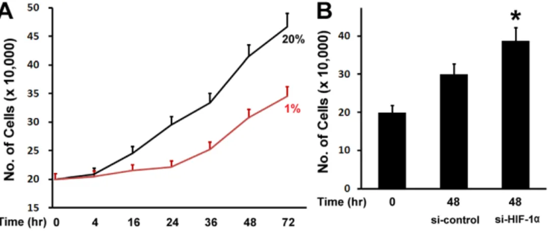
To examine the effect of hypoxia on osteoblast proliferation, MC3T3 osteoblastic cells were cultured in Alpha Minimum Essential Medium, and maintained for different time points in normoxic (20%O2) or hypoxia (1%O2) condition under a humidified hypoxia incubator. We observed that MC3T3 osteoblastic cells under hypoxia condition grew slower than those in normoxia condition (Fig. 1A). Hypoxia started to inhibit cell growth at 16 hr, and the inhibition remained at 72 hr. HIF-1ais a master regulator of cellular response to hypoxia. We then asked if the inhibitory effect of hypoxia on osteoblast proliferation is related to HIF-1a. To address this question, we used siRNA technology to knockdown the expression of HIF-1a under hypoxia. Osteoblast growth was then examined under hypoxia condition. As shown in Fig. 1B, compared with osteoblast growth in si-RNA control group, inhibition of HIF-1a expression by siRNA resulted in an increase of osteoblast growth under hypoxia. These experiments therefore indicate that HIF-1aparticipates in hypoxia-mediated inhibition of osteoblast growth.
Hypoxia led to downregulation of Wnt targets
To explore the possible mechanisms of hypoxia effect on osteoblast proliferation, we used quantitative real-time RT-PCR to examine the changes of gene expressions under hypoxia. It is well-known that Wnt pathway stimulates osteoblast proliferation. We asked whether hypoxia may inhibit osteoblast proliferation through inhibiting Wnt pathway. To address this question, we examined the effect of hypoxia on the expressions of Wnt target genes: Cyclin D1 and c-Myc. MC3T3 osteoblastic cells were cultured and maintained in normoxic (20%O2) or hypoxia (1%O2) condition under a humidified hypoxia incubator. Total RNA was purified 48 hr following culture in the presence or absence of hypoxia. The expressions of Cyclin D1 and c-Myc as well as HIF-1awere quantitated by real-time RT-PCR. As shown in Fig. 2A, HIF-1aRNA expression was enhanced by 1.8 fold under hypoxia compared with nomoxia, and western blotting experiments indicated that HIF-1a protein expression level also increased under hypoxia (Fig. 2B). These confirm the hypoxia-mediated upregulation of HIF-1a expression. We observed that the expressions of Cyclin D1 and c-Myc were inhibited by 54% and 32% respectively under hypoxia, compared with nomoxia (Fig. 2C). We then asked if the inhibitory effect of hypoxia on Cyclin D1 and c-Myc expressions is related to HIF-1a. To address this question, we used desferrioxamine (DFO) in this assay, a potent HIF-1a activator to increase the expression of HIF-1a under hypoxia. Cyclin D1 and c-Myc expressions were then examined under hypoxia. As shown in Fig. 2D, additions of DFO further inhibited Cyclin D1 and c-Myc expressions under hypoxia in a dose-dependent manner. These data suggest that HIF-1ais involved in hypoxia-mediated inhibition of Cyclin D1 and c-Myc expressions.
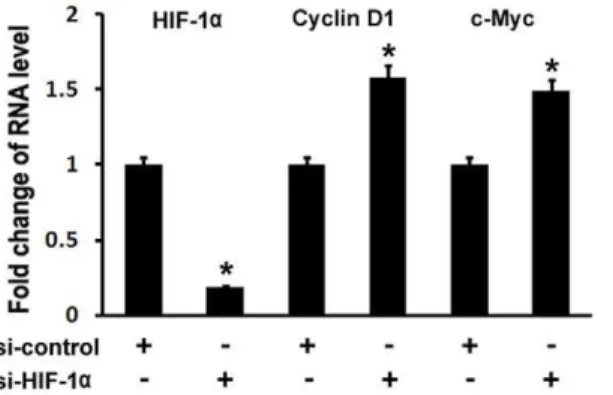
Inhibition of HIF-1aby siRNA resulted in upregulations of Cyclin D1 and c-Myc expressions in osteoblasts
To confirm the inhibitory of hypoxia/HIF-1a on Wnt target gene expressions, we used siRNA to knockdown HIF-1a expression in MC3T3 osteoblast cells. Real-time RT-PCR was performed to analyze Cyclin D1 and c-Myc expressions. As shown in Fig. 3, HIF-1a RNA expression was decreased by 81% using siRNA targeted against HIF-1a. Cyclin D1 RNA levels were increased by approximately 58%, and c-Myc RNA levels were
Figure 1. Hypoxia inhibits osteoblast proliferation.(A) Osteoblast number counts in the growth medium. MC3T3 osteoblastic cells were cultured in Alpha Minimum Essential Medium, and maintained for different time points as indicated from 4 hr to 72 hr in normoxic (20%O2) or
hypoxia (1%O2) condition. (B) Inhibition of HIF-1aexpression by siRNA resulted in an increase of osteoblast growth. MC3T3 osteoblastic cells were
transfected by siRNA, and cultured under hypoxia for 48 hr. si-control: si-RNA control; si-HIF-1a: si-RNA against HIF-1a. A pairedt-test was performed comparing si-control group and si-HIF-1agroup. *: A star indicates statistical significance compared to control group.
increased by approximately 49%. Therefore, these loss-of-function experiments support a role for HIF-1a in inhibiting Wnt target gene expression in osteoblasts.
HIF-1ainhibited Topflash reporter activity in a dose-dependent manner
We have shown that hypoxia led to downregulation of Wnt targets, and that enhanced expression of HIF-1aby DFO further inhibited Wnt target gene expressions while repression of HIF-1a by siRNA resulted in upregulation of Wnt target gene expressions. These data suggest that HIF-1a inhibits Wnt pathway in osteoblasts. Topflash reporter system is an established model in
vitro as a widely used indicator of Wnt/b-catenin signal transduction [15]. To examine Wnt pathway regulation by HIF-1a, we took advantage of this established model of b -catenin-induced Topflash activation. Here, we tested the effect of HIF-1a on b-catenin-induced Topflash activation. HEK293 cells were transiently transfected with Topflash reporter and b-catenin expression vector as indicated. Expression plasmid p1p2n HIF-1ais a HIF-1amutant which is constitutively active as previously used [27]. As shown in Fig. 4A, theb-catenin activated Topflash reporter expression as expected, and increasing amounts of HIF-1a transfection caused significantly lower expression of the Topflash reporter induced by b-catenin, indicating that HIF-1a Figure 2. Hypoxia leads to downregulation of Wnt targets.(A) Increase of HIF-1aexpression in RNA level in osteoblasts under hypoxia. RNA levels were normalized to heat shock protein 90 (HSP90). A pairedt-test was performed comparing control group (20% O2) and hypoxia group (1% O2). *: A star indicates statistical significance compared to control group. (B) Western blotting analysis of HIF-1aexpression in protein level in
osteoblasts under hypoxia. Heat shock protein 90 (HSP90) was used as a loading control. (C) RNA expression levels of cyclin D1 and c-Myc as determined by quantitative real-time RT-PCR. MC3T3 osteoblasts were cultured for 48 hr under hypoxia (1%O2). RNA was isolated and quantitated by
real-time RT-PCR. The RNA level from normoxic condition (20%O2) group was normalized to a value of 1. Values were presented as the mean6S.D. A
pairedt-test was performed comparing control group (20% O
2) and hypoxia group (1% O2). *: A star indicates statistical significance compared to
control group. (D) RNA expression levels of cyclin D1 and c-Myc treated with DFO as determined by quantitative real-time RT-PCR. MC3T3 osteoblasts were cultured for 48 hr under hypoxia (1%O2), and treated with desferrioxamine (DFO).+:100 uM;++:200 uM. The RNA level from normoxic condition
(20%O2) group was normalized to a value of 1. Values were presented as the mean6S.D. A pairedt-test was performed comparing control group
(20% O2) and hypoxia group (1% O2). *: A star indicates statistical significance compared to control group. A pairedt-test was also performed
comparing 1% O2group and DFO group (+and++). **: Two stars indicate statistical significance compared to 1% O2group.
inhibitedb-catenin-induced Topflash activation in a dose-depen-dent manner.
HIF-1acooperated with Osx to inhibit Wnt pathway activity
Both Osx and HIF-1a are important for endochondral ossification during bone formation. Our recent studies have demonstrated that Osx and HIF-1acooperatively regulate VEGF expression in osteoblasts [22]. Osx inhibits osteoblast proliferation through inhibiting Wnt pathway [15]. Here, we ask if there is any cooperation between HIF-1a and Osx to inhibit Wnt pathway. HEK293 cells were transiently cotransfected with Topflash reporter and b-catenin expression vector along with Osx expression plasmid. As shown in Fig. 4B, transfection of 100 ng HIF-1aor 25 ng Osx alone inhibitedb-catenin-induced Topflash reporter expression by 49% and 59%, respectively. Interestingly, cotransfection of such amounts of HIF-1a and Osx resulted in a further inhibition ofb-catenin-induced Topflash by 80%. These data indicate that there is a synergistic interplay between HIF-1a and Osx in Wnt pathway inhibition.
Discussion
Osx is an osteoblast-specific transcription factor that regulates the expression of essential genes needed for appropriate osteoblast differentiation and bone formation. The discovery that Osx inhibits the Wnt pathway highlights the potential for novel feedback control mechanisms involved in bone formation [15]. Our recent report has demonstrated that Osx and HIF-1a cooperatively regulate VEGF expression in osteoblasts [22]. In this study, we examined the role of hypoxia/HIF-1ain osteoblast proliferation and Wnt pathway. The findings presented here indicate that hypoxia/HIF-1ainhibits osteoblast proliferation and that HIF-1acooperates with Osx to inhibit Wnt pathway.
First, we showed that hypoxia inhibited osteoblast proliferation. This was supported by cell proliferation assay. MC3T3 osteoblas-tic cells grew slower under hypoxia than those in normoxia condition (Fig. 1A). HIF-1ais the crucial mediator of the adaptive response of cells to hypoxia. The oxygen dependent degradation of HIF-1a is controlled by a family of HIF prolyl hydroxylases.
Under normoxic conditions, HIF-1a is hydroxylated by prolyl hydroxylases that act as oxygen sensors. Hydroxylation of specific proline residues on HIF-1a is followed by proteasomal degrada-tion. Under hypoxic conditions, HIF-1ais stabilized, translocated to the nucleus, and forms a heterodimer with HIF-1bto regulate target genes. These target genes are involved in a variety of cellular processes including angiogenesis, energy metabolism, cell prolif-eration and survival, vasomotor control, and matrix metabolism [28]. To address whether HIF-1ais involved in hypoxia-mediated inhibition of osteoblast proliferation, siRNA technology was used to knockdown the expression of HIF-1a in this study. Fig. 1B demonstrated that inhibition of HIF-1aexpression by siRNA led to an increase of osteoblast growth compared with osteoblast growth in si-RNA control group. These experiments indicate that HIF-1aparticipates in hypoxia-mediated inhibition of osteoblast proliferation.
Figure 3. Inhibition of HIF-1aby siRNA results in upregulations of Cyclin D1 and c-Myc expressions in osteoblasts. MC3T3 osteoblasts were transfected with siRNA control or siRNA against HIF-1a. RNA was isolated 24 hr post-transfection and quantitated by quantitative real-time RT-PCR. The RNA level from the control siRNA group was normalized to a value of 1. Values were presented as the mean6S.D. si-control: si-RNA control; si-HIF-1a: si-RNA against HIF-1a. A pairedt-test was performed comparing si-control group and si-HIF-1a group. *: A star indicates statistical significance compared to control group.
doi:10.1371/journal.pone.0052948.g003
The current study also addresses possible mechanisms for hypoxia/HIF-1a to inhibit osteoblast proliferation. As a crucial mediator of hypoxia, HIF-1ais ubiquitous [29]. It is controversial how HIF-1a affects cell proliferation. Studies performed in cell lines or in ES cell-derived tumors indicate that HIF-1a can modulate tumor cell growth by controlling both metabolic functions and expression of angiogenic growth factors such as VEGF [30,31]. Despite an initial report showing that HIF-1a would act as a negative factor for the growth of ES cell-derived tumors, some studies support the model that the lack of HIF-1a inhibits tumor growth [31,32]. In this study, we demonstrated that hypoxia/HIF-1a inhibited osteoblast proliferation. We also investigated mechanisms that could mediate inhibitory action of hypoxia/HIF-1a in osteoblast proliferation. HIF-1 is a heterodi-mer that consists of HIF-1a, the oxygen sensitive subunit, and the constitutively expressed HIF-1b. HIF-1 activates target gene transcription by binding to the hypoxia-responsive elements in the proximal promoter region of the oxygen responsive genes. This study indicates that Wnt pathway is one of possible mechanisms for hypoxia/HIF-1a to inhibit osteoblast proliferation. This is supported by several evidences: 1) RT-PCR results revealed that Wnt target genes such as cyclin D1 and c-Myc were downregu-lated under hypoxia; 2) the treatment of HIF-1a activator DFO further downregulated expressions of cyclin D1 and c-Myc; 3) the inhibition of HIF-1aby siRNA in osteoblasts led to the expression increase of cyclin D1 and c-Myc; 4) our transfection assay showed that HIF-1a inhibited b-catenin-induced Topflash reporter activity. However, our study cannot rule out other possible mechanisms of the effect of hypoxia/HIF-1a on osteoblast
proliferation, such as hypoxia-induced PH value change, some other hypoxia-related factors, like vascular endothelial growth factor, insulin-like growth factor II, and transforming growth factor b1, etc. It has been reported that Osx inhibits osteoblast proliferation while it induces osteoblast differentiation [15]. We showed in that study that Osx negatively regulated b -catenin-induced Topflash activation in a dose-dependent manner. Since our recent studies indicate that Osx and HIF-1acollaboratively control VEGF expression in osteoblasts [22], it is interesting to explore the possibility whether Osx and HIF-1a may work together to control Wnt pathway. Indeed, our current results indicated that Osx and HIF-1a inhibited Wnt pathway in a synergistic manner (Fig. 4B). Because of the role of Wnt pathway in stimulating osteoblast proliferation, we speculate that hypoxia-induced inhibition of osteoblast proliferation may be at least partially through inhibition of Wnt pathway by HIF-1a.
In summary, we present here that hypoxia/HIF-1a inhibit osteoblast proliferation, and that HIF-1a has a synergistic effect with Osx on the inhibition of Wnt pathway. While additional studies need to address the role of synergistic inhibition of Wnt pathway by HIF-1a and Osx, these early stage studies revealed additional new information of the cooperation between HIF-1a and Osx in osteoblasts.
Author Contributions
Conceived and designed the experiments: CZ WT. Performed the experiments: DC YL ZZ YX YZ. Analyzed the data: CZ XZ WT. Wrote the paper: DC CZ.
References
1. Zhang C (2010) Transcriptional regulation of bone formation by the osteoblast-specific transcription factor Osx. J Orthop Surg Res 5: 37.
2. St-Jacques B, Hammerschmidt M, McMahon AP (1999) Indian hedgehog signaling regulates proliferation and differentiation of chondrocytes and is essential for bone formation. Genes Dev 13: 2072–2086.
3. Komori T, Yagi H, Nomura S, Yamaguchi A, Sasaki K, et al. (1997) Targeted disruption of Cbfa1 results in a complete lack of bone formation owing to maturational arrest of osteoblasts. Cell 89: 755–764.
4. Nakashima K, Zhou X, Kunkel G, Zhang Z, Deng JM, et al. (2002) The novel zinc finger-containing transcription factor osterix is required for osteoblast differentiation and bone formation. Cell 108: 17–29.
5. He X, Semenov M, Tamai K, Zeng X (2004) LDL receptor-related proteins 5 and 6 in Wnt/beta-catenin signaling: arrows point the way. Development 131: 1663–1677.
6. Krishnan V, Bryant HU, Macdougald OA (2006) Regulation of bone mass by Wnt signaling. J Clin Invest 116: 1202–1209.
7. Day TF, Guo X, Garrett-Beal L, Yang Y (2005) Wnt/beta-catenin signaling in mesenchymal progenitors controls osteoblast and chondrocyte differentiation during vertebrate skeletogenesis. Dev Cell 8: 739–750.
8. Hill TP, Spater D, Taketo MM, Birchmeier W, Hartmann C (2005) Canonical Wnt/beta-catenin signaling prevents osteoblasts from differentiating into chondrocytes. Dev Cell 8: 727–738.
9. Hu H, Hilton MJ, Tu X, Yu K, Ornitz DM, et al. (2005) Sequential roles of Hedgehog and Wnt signaling in osteoblast development. Development 132: 49– 60.
10. Rodda SJ, McMahon AP (2006) Distinct roles for Hedgehog and canonical Wnt signaling in specification, differentiation and maintenance of osteoblast progenitors. Development 133: 3231–3244.
11. Gong Y, Slee RB, Fukai N, Rawadi G, Roman-Roman S, et al. (2001) LDL receptor-related protein 5 (LRP5) affects bone accrual and eye development. Cell 107: 513–523.
12. Kato M, Patel MS, Levasseur R, Lobov I, Chang BH, et al. (2002) Cbfa1-independent decrease in osteoblast proliferation, osteopenia, and persistent embryonic eye vascularization in mice deficient in Lrp5, a Wnt coreceptor. J Cell Biol 157: 303–314.
13. Morvan F, Boulukos K, Clement-Lacroix P, Roman Roman S, Suc-Royer I, et al. (2006) Deletion of a single allele of the Dkk1 gene leads to an increase in bone formation and bone mass. J Bone Miner Res 21: 934–945.
14. Li J, Sarosi I, Cattley RC, Pretorius J, Asuncion F, et al. (2006) Dkk1-mediated inhibition of Wnt signaling in bone results in osteopenia. Bone 39: 754–766.
15. Zhang C, Cho K, Huang Y, Lyons JP, Zhou X, et al. (2008) Inhibition of Wnt signaling by the osteoblast-specific transcription factor Osterix. Proc Natl Acad Sci U S A 105: 6936–6941.
16. Erlebacher A, Filvaroff EH, Gitelman SE, Derynck R (1995) Toward a molecular understanding of skeletal development. Cell 80: 371–378. 17. Harper J, Klagsbrun M (1999) Cartilage to bone–angiogenesis leads the way.
Nat Med 5: 617–618.
18. Wan C, Shao J, Gilbert SR, Riddle RC, Long F, et al. (2010) Role of HIF-1alpha in skeletal development. Ann N Y Acad Sci 1192: 322–326. 19. Carano RA, Filvaroff EH (2003) Angiogenesis and bone repair. Drug Discov
Today 8: 980–989.
20. Wang Y, Wan C, Deng L, Liu X, Cao X, et al. (2007) The hypoxia-inducible factor alpha pathway couples angiogenesis to osteogenesis during skeletal development. J Clin Invest 117: 1616–1626.
21. Tang W, Yang F, Li Y, de Crombrugghe B, Jiao H, et al. (2012) Transcriptional Regulation of Vascular Endothelial Growth Factor (VEGF) by Osteoblast-specific Transcription Factor Osterix (Osx) in Osteoblasts. J Biol Chem 287: 1671–1678.
22. Chen D, Tian W, Li Y, Tang W, Zhang C (2012) Osteoblast-specific transcription factor Osterix (Osx) and HIF-1alpha cooperatively regulate gene expression of vascular endothelial growth factor (VEGF). Biochem Biophys Res Commun 424: 176–181.
23. Rajpurohit R, Koch CJ, Tao Z, Teixeira CM, Shapiro IM (1996) Adaptation of chondrocytes to low oxygen tension: relationship between hypoxia and cellular metabolism. J Cell Physiol 168: 424–432.
24. Tang W, Li Y, Osimiri L, Zhang C (2011) Osteoblast-specific Transcription Factor Osterix (Osx) Is an Upstream Regulator of Satb2 during Bone Formation. J Biol Chem 286: 32995–33002.
25. Zhang C, Dowd DR, Staal A, Gu C, Lian JB, et al. (2003) Nuclear coactivator-62 kDa/Ski-interacting protein is a nuclear matrix-associated coactivator that may couple vitamin D receptor-mediated transcription and RNA splicing. J Biol Chem 278: 35325–35336.
26. Zhang C, Tang W, Li Y, Yang F, Dowd DR, et al. (2011) Osteoblast-specific transcription factor Osterix increases vitamin D receptor gene expression in osteoblasts. PLoS One 6: e26504.
27. Zhang C, Yang F, Cornelia R, Tang W, Swisher S, et al. (2011) Hypoxia-inducible factor-1 is a positive regulator of Sox9 activity in femoral head osteonecrosis. Bone 48: 507–513.
29. Wiener CM, Booth G, Semenza GL (1996) In vivo expression of mRNAs encoding hypoxia-inducible factor 1. Biochem Biophys Res Commun 225: 485– 488.
30. Semenza GL (1999) Regulation of mammalian O2 homeostasis by hypoxia-inducible factor 1. Annu Rev Cell Dev Biol 15: 551–578.
31. Ryan HE, Poloni M, McNulty W, Elson D, Gassmann M, et al. (2000) Hypoxia-inducible factor-1alpha is a positive factor in solid tumor growth. Cancer Res 60: 4010–4015.