Active Immunization with an Octa-Valent
Staphylococcus aureus
Antigen Mixture in
Models of
S
.
aureus
Bacteremia and Skin
Infection in Mice
Sanne van den Berg1*‡, Dennis G. A. M. Koedijk2‡, Jaap Willem Back3, Jolanda Neef2, Annette Dreisbach2, Jan Maarten van Dijl2, Irma A. J. M. Bakker-Woudenberg1, Girbe Buist2
1Department of Medical Microbiology and Infectious Diseases, Erasmus University Medical Center, Rotterdam, the Netherlands,2Department of Medical Microbiology, University of Groningen, University Medical Center Groningen, Groningen, the Netherlands,3Pepscan Therapeutics BV, Lelystad, the Netherlands
‡These authors contributed equally to this work. *[email protected]
Abstract
Proteomic studies with differentStaphylococcus aureusisolates have shown that the cell surface-exposed and secreted proteins IsaA, LytM, Nuc, the propeptide of Atl (pro-Atl) and four phenol-soluble modulinsα(PSMα) are invariantly produced by this pathogen. There-fore the present study was aimed at investigating whether these proteins can be used for active immunization againstS.aureusinfection in mouse models of bacteremia and skin in-fection. To this end, recombinant His-tagged fusions of IsaA, LytM, Nuc and pro-Atl were isolated fromLactococcus lactisorEscherichia coli, while the PSMα1-4 peptides were chemically synthesized. Importantly, patients colonized byS.aureusshowed significant im-munoglobulin G (IgG) responses against all eight antigens. BALB/cBYJ mice were immu-nized subcutaneously with a mixture of the antigens at day one (5μg each), and boosted twice (25μg of each antigen) with 28 days interval. This resulted in high IgG responses against all antigens although the response against pro-Atl was around one log lower com-pared to the other antigens. Comcom-pared to placebo-immunized mice, immunization with the octa-valent antigen mixture did not reduce theS.aureusisolate P load in blood, lungs, spleen, liver, and kidneys in a bacteremia model in which the animals were challenged for 14 days with a primary load of 3 × 10⁵CFU. Discomfort scores and animal survival rates over 14 days did not differ between immunized mice and placebo-immunized mice upon bacteremia withS.aureusUSA300 (6 × 10⁵CFU). In addition, this immunization did not re-duce theS.aureusisolate P load in mice with skin infection. These results show that the tar-get antigens are immunogenic in both humans and mice, but in the used animal models do not result in protection againstS.aureusinfection.
OPEN ACCESS
Citation:van den Berg S, Koedijk DGAM, Back JW, Neef J, Dreisbach A, van Dijl JM, et al. (2015) Active Immunization with an Octa-ValentStaphylococcus aureusAntigen Mixture in Models ofS.aureus
Bacteremia and Skin Infection in Mice. PLoS ONE 10(2): e0116847. doi:10.1371/journal.pone.0116847
Academic Editor:Eliane Namie Miyaji, Instituto Butantan, BRAZIL
Received:September 5, 2014
Accepted:December 15, 2014
Published:February 24, 2015
Copyright:© 2015 van den Berg et al. This is an open access article distributed under the terms of the
Creative Commons Attribution License, which permits unrestricted use, distribution, and reproduction in any medium, provided the original author and source are credited.
Data Availability Statement:All relevant data are within the paper.
Funding:This study was financially supported by Top Institute Pharma project T4-502. The funders had no role in study design, data collection and analysis, decision to publish, or preparation of the manuscript.
Introduction
Staphylococcus aureusis a widespread Gram-positive bacterium that colonizes the skin and an-terior nares of about 20–30% of the healthy human population [1]. Although mainly a harm-less colonizer,S.aureuscan cause invasive diseases like skin and soft tissue infections, and can be responsible for severe infections in humans like pneumonia, endocarditis and osteomyelitis [1], which are frequently associated withS.aureusbacteremia [2].S.aureusin its methicillin-resistant form (MRSA) is the most important cause of antibiotic-methicillin-resistant health care-associat-ed infections worldwide [3,4]. In the case of MRSA, a single genetic element makesS.aureus
resistant to the most frequently prescribed class of antimicrobials—theβ-lactam antibiotics, in-cluding penicillins, cephalosporins, and carbapenems [5]. A high incidence of MRSA is en-countered in hospitals, resulting in prolonged hospital stays and in higher mortality rates [3,4], and limited effectiveness of alternative treatment regimens. Glycopeptides, especially vancomy-cin, are currently used as first-line treatment of MRSA infections. Unfortunately, this has led to the emergence of vancomycin-intermediate and vancomycin-resistant MRSA [6]. In addition, there is raising concern that the current first-line treatment for MRSA infection will become in-creasingly ineffective. Since in the past 25 years no novel small-molecule antibacterial drugs have been discovered [7], and the development pipeline of new antimicrobials remains lean [8], new ways of treatment ofS.aureusinfections such as immunization need to be explored. Several strategies of passive and active immunization inS.aureusinfections have been studied in experimental infection models, but until now none of these have been proved to be effective in clinical studies [9–11]. Insufficient power because of a low sample size in some clinical stud-ies [12,13], as well as the heterogeneity ofS.aureusstrains causing infections in humans [14,15] may contribute to the failure of treatment through immunization in patients. Despite the lack of success so far, immunization approaches are still worth pursuing especially in pa-tients admitted to the hospital for elective surgery. For this group of papa-tients, there may be enough time for immunization prior to surgery.
Novel target identification strategies have been applied to screen for new antigenic targets for immunization. Invariant immunogenic determinants of relevantS.aureusisolates have been successfully identified in previous studies using a combination of proteomics, genomics, bioinformatics and immunological approaches [15–18]. A complete inventory of predicted se-creted proteins of sequencedS.aureusstrains has been made [16]. Proteomic analysis of the exoproteomes of 25 clinicalS.aureusisolates showed that only seven of these secreted proteins (IsaA, Lip, LytM, Nuc, SA0620, SA2097, and SA2437) were produced by all clinical isolates studied [15]. In a proteolytic shaving approach ofS.aureuscells, multiple surface-exposed pro-teins were identified among which IsaA, Nuc, Atl and the phenol-soluble modulin (PSM)α1 peptide [17].
In the present study, we used IsaA, LytM, Nuc, pro-Atl, and the PSMα1-4 peptides as targets for active immunization inS.aureus-infected mice. TheseS.aureusantigens were all selected by the previous target identification strategies, and are all potential virulence factors ofS. aure-us. The PSMs are highly potent surfactants that facilitate the movement ofS.aureusover moist surfaces by colony spreading, and they are involved in the metastatic escape of staphylococcal cells from biofilms thereby representing an enormous risk factor for serious invasive disease [19,20]. High levels of PSM production are supposed to contribute to the high virulence and epidemic behavior of community-acquired MRSA lineages [21].
Staphylococcal major autolysin (Atl) is a bifunctional autolysin composed of a signal pep-tide for protein secretion, a propeppep-tide, and domains with amidase and glucosaminidase activi-ty. After proteolytic cleavage, the active domains are involved in cell separation and they have been shown to be cell surface-exposed [22]. It is believed that the propeptide of Atl (pro-Atl) adherence to PLOS ONE policies on sharing data
has a role in the folding and/or activation of Atl and that it would be degraded rapidly upon proteolytic processing of the Atl protein.
Immunodominant staphylococcal antigen A (IsaA) and autolysin M (LytM) have been shown to be peptidoglycan hydrolases with transglycosylase and glycyl-glycine endopeptidase specific activity, respectively [18]. The secreted nuclease (Nuc) has been shown to limit the for-mation of biofilms due to the degradation of extracellular DNA [23]. This protein also pro-motes resistance against neutrophil extracellular traps-mediated antimicrobial activity of neutrophils and thus contributes to disease pathogenesisin vivo[24].
In previous studies, IsaA, LytM, Nuc, pro-Atl and PSMα1-4 have all been shown to be im-munogenic. Using a multiplex assay to quantify antibody responses against 26 staphylococcal proteins, we found that mice with aS.aureusUSA300 pneumonia or skin infection showed good IgG responses against IsaA and Nuc, while anti-LytM levels were low [25]. In contrast, in mice immunized with monovalent staphylococcal vaccines containing IsaA, Nuc or LytM, the highest IgG responses were obtained against the latter antigen. Using the same multiplex assay, it has been shown that 75% of the patients with the genetic blistering disease epidermolysis bul-losa (EB), who were heavily colonized with multipleS.aureustypes, have increased IgG re-sponses against some secreted, cell wall and membrane-bound staphylococcal proteins [26,27]. Importantly, EB patients do not frequently suffer fromS.aureusbacteremia, which suggests that their high anti-staphylococcal antibody titers might be protective against invasiveS.aureus
infections. Within the EB patient group, the highest response was detected against IsaA, while for Nuc and LytM moderate responses were detected. These data show that both in mice and in human individuals a good IgG response is generated against IsaA, LytM and Nuc. Moreover, passive immunization of mice with murine anti-IsaA resulted in protection against staphylo-coccal infection both in a central venous catheter-related infection model and a sepsis survival model, most likely due to phagocytosis that resulted in killing ofS.aureus[28]. Holtfreter et al. showed in their review that Atl has been identified in different studies as a well-recognized tar-get of the human immune system [29]. Recently, we showed that pro-Atl is exposed on the out-side ofS.aureuscells and is recognized by antibodies from four out of six patients that were colonized withS.aureus(our unpublished observations). Within a patient group of hospital-ized adults with an invasiveS.aureusinfection, in those patients who did not develop sepsis, higher levels of immunoglobulin G (IgG) responses were detected against exotoxins among which PSMα3 [30].
In the present study in mice, we examined the protective efficacy of active immunization with an octa-valent antigen mixture containing IsaA-His6, LytM-His6, Nuc-His6, His6-pro-Atl
and the PMSα1–4 peptides. In mice immunized with a mixture of these antigens, the generated IgG response was determined by enzyme-linked immunosorbent assays (ELISA). Subsequent-ly, the protective capacity of this immunization strategy was studied in mouse models of methi-cillin-sensitiveS.aureus(MSSA) or MRSA bacteremia and in a mouse model of MSSA skin infection, which are clinically highly relevantS.aureusinfections [31].
Materials and Methods
Bacterial strains and growth conditions
Strains and plasmids used in this study are listed inTable 1.Lactococcus lactisstrains were grown at 30°C in M17 broth (Oxoid Ltd, Hampshire, UK) supplemented with 0.5% w/v glucose (GM17). When necessary the medium was supplemented with chloramphenicol (5μg/mL) for
previously analyzed by proteomics [15].S.aureusUSA300 is one of the most frequent causes of community-acquired infections in the United States of America [32].
Construction of His
6-pro-Atl expression plasmids
For cloning of the DNA fragment coding for the propeptide of Atl (pro-Atl) in the plasmid pNG4110, the primers atlpro.F1 (ATATGGATCCGCTGAGACGACACAAGATCAAACTAC-TAATAAAAACG) and atlpro.R2 (ATATGCGGCCGCTTAAGCGCTAAAAGTAGTTACTT-TAGGTGTCGCTTCAGTTTTAGC) were used with chromosomal DNA ofS.aureusstrain N315 as a template. Using this vector, a N-terminally His-tagged fusion protein can be secreted fromL.lactis[33]. PCR product and vector were digested usingBamHI andNotI (cleavage sites are underlined in the primers sequences). After ligation the resulting plasmids were transferred intoL.lactisPA1001 by electro transformation with selection on chloramphenicol. A nucleotide sequence analysis of the cloned inserts was performed by Eurofins DNA (Ebersberg, Germany).
Protein isolation, purification and quantification
For the production and isolation of IsaA-His6, an overnight culture ofE.coliBL21DE3
(pET24d::isaA::his6) was diluted 1:100 in fresh lysogeny broth with 50μg/mL kanamycin [34].
Induction was performed at OD600~ 0.5 for 4 h by adding 1 mM isopropylβ
-D-1-thiogalacto-pyranoside (IPTG) (Duchefa, Haarlem, the Netherlands). The culture was centrifuged and the pellet was resolved in binding buffer (20 mM Na-Phosphate, pH 7.4, 0.5 M NaCl2, 60 mM
im-idazole) containing 6 M urea. After sonification (Sonicator S-4000, Misonix, Farmingdale, USA) the supernatant was mixed with binding buffer (1:1) and HisLink beads (Promega, Madison, USA) for 1 h at 4°C under shaking conditions. Protein elution was performed with binding buffer containing 500 mM imidazole.
For the production and isolation of LytM-His6[25], Nuc-His6[25] and His6-pro-Atl,
over-night cultures ofL.lactisPA1001 were diluted 1:2 in GM17 medium. Nisin induction for pro-tein expression was performed at OD600~ 0.5 for 16 h by adding the culture supernatant
(1:1000) from an overnight culture ofL.lactisNZ97000. For analysis of the extracellular pro-duction of His6-pro-Atl byL.lactisPA1001 (pNG4110::proAtl), proteins from the culture
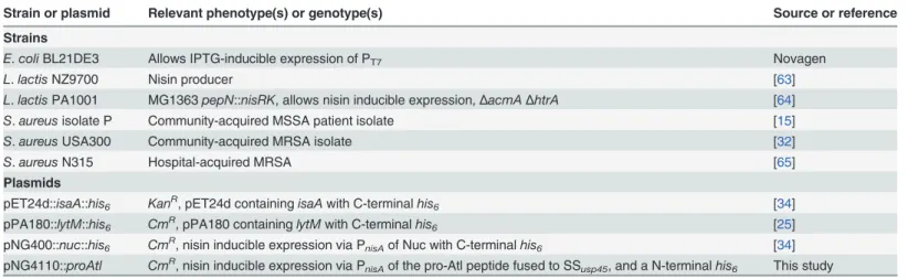
Table 1. Bacterial strains and plasmids used in this study.
Strain or plasmid Relevant phenotype(s) or genotype(s) Source or reference
Strains
E.coliBL21DE3 Allows IPTG-inducible expression of PT7 Novagen
L.lactisNZ9700 Nisin producer [63]
L.lactisPA1001 MG1363pepN::nisRK, allows nisin inducible expression,ΔacmAΔhtrA [64]
S.aureusisolate P Community-acquired MSSA patient isolate [15]
S.aureusUSA300 Community-acquired MRSA isolate [32]
S.aureusN315 Hospital-acquired MRSA [65]
Plasmids
pET24d::isaA::his6 KanR, pET24d containingisaAwith C-terminalhis6 [34] pPA180::lytM::his6 CmR, pPA180 containinglytMwith C-terminalhis6 [25] pNG400::nuc::his6 CmR, nisin inducible expression via PnisAof Nuc with C-terminalhis6 [34] pNG4110::proAtl CmR, nisin inducible expression via P
nisAof the pro-Atl peptide fused to SSusp45, and a N-terminalhis6 This study
KanR, kanamycin resistance gene;CmR, chloramphenicol resistance gene; PT7, IPTG inducible T7-promoter; PnisA, nisin inducible promoter;his6, 6 histidine-tag; SSusp45, signal sequence ofusp45.
supernatant were precipitated using trichloroacetic acid (TCA; 10% w/v). The culture was cen-trifuged and the pellet was washed with acetone and dried. For the isolation of Nuc-His6,from
the supernatant fraction of a culture ofL.lactisPA1001 (pNG400::nuc::his6), HisLink beads were added for 1 h at 4°C under shaking conditions after which HisLink beads were collected. For isolation of LytM-His6cells ofL.lactisPA1001 (pPA180::lytM::his6) were harvested and disrupted in binding buffer using a Sonicator S-4000. Purification was performed with Mag beads (GE Healthcare, Uppsala, Sweden) for 1.5 h under shaking conditions at 4°C. Elution of Nuc-His6and LytM-His6was done with binding buffer containing 500 mM imidazole. The
flow-through, wash and elution fractions were analyzed by LDS-PAGE. Protein samples were mixed with LDS buffer and incubated at 95°C for 10 min, separated by LDS-PAGE using pre-cast 10% NuPage gels (Life Technologies, Bleiswijk, the Netherlands) and stained with Simply Blue™Safe Stain (Life Technologies).
The fractions containing the purified His-tagged proteins of IsaA-His6, LytM-His6,
Nuc-His6or His6-pro-Atl were pooled and dialyzed (G2-Float-A-Lyzer, Spectrum Europe BV,
Breda, the Netherlands) against PBS and concentrated with the Speedvac Concentrator Plus (Eppendorf Nederland BV, Nijmegen, the Netherlands).
Protein concentrations were determined with the DC Protein Assay (Bio-Rad, Veenendaal, the Netherlands) according to the instructions of the supplier, using Bovine Serum Albumin (Sigma-Aldrich, Zwijndrecht, the Netherlands) as a standard, or with the Nanodrop ND-1000 (Thermo Fisher Scientific, Wilmington, Delaware USA) using the extinction coefficient calcu-lated for each of the proteins.
PSMα1-4 were synthesized as described previously [19], with the addition of a GGG-Lys(ε -biotin). The peptides were mixed in a 1:1:1:1 molar ratio, and incubated in PBS in a stoichio-metric ratio to avidin (Thermo Fisher Scientific Inc., Rockford, USA).
Human plasma
Whole blood donations from EB patients were collected under the approval of the medical ethics committee of the University Medical Center Groningen (approval no. NL27471,042,09) upon writ-ten informed patient consent, and with adherence to the Helsinki Guidelines [26]. The Indepen-dent Ethics Committee of the Foundation‘Evaluation of Ethics in Biomedical Research’(Assen, the Netherlands), approved the protocol for blood donations from healthy volunteers. This proto-col is registered by QPS Groningen (code 04132-CS011). The required written informed consent was obtained from all EB patients and healthy volunteers included in the present studies.
Protein detection and activity assays
For Western blot analyses, proteins separated by LDS-PAGE were blotted onto a nitrocellulose membrane (Protran, Schleicher & Schuell BioScience, Dassel, Germany). Immunodetection was performed using anti-His-tag antibodies (Invitrogen, Life Technologies) and rabbit poly-clonal antibodies raised against the amidase or glucosaminidase domains of Atl (gift from Motoyuki Sugai, Hiroshima University, Japan [35]). Dilutions (1:1000) of the collected human plasma [26] were used to detect the IgG responses against IsaA-His6, LytM-His6and Nuc-His6.
Equal amounts of the three proteins were loaded on the gel and after blotting strips of the blot were incubated with the different plasma samples. Bound primary antibodies were visualized using specific fluorescently labeled secondary antibodies (IRDye 800 CW, Li-Cor Biosciences, Lincoln, USA). Membranes were scanned for fluorescence at 800 nm using the Odyssey Infra-red Imaging System (Li-Cor Biosciences, Nebraska, USA).
overnight culture, an aliquot of 2μL was spotted in the middle of a TSA plate, which was then
in-cubated overnight at 37°C. The spreading assay was performed as described [19,36].
Peptidoglycan hydrolase activity of IsaA-His6and LytM-His6was detected by a zymogram
staining technique using SDS-polyacrylamide (12.5%) gels containing 0.1% (w/v) autoclaved cell wall fragments ofS.aureusRN4220 isolated as described previously [37]. After electropho-resis, the gels were gently shaken at room temperature for 24 h in three to five changes of 100 mL of 25 mM Tris-HCl (pH 6.0) containing 1% (v/v) Triton X-100 for protein renaturation.
Pepscan analysis
To determine whether regions of theS.aureusPSMα1-4 peptides were recognized by human IgGs, libraries of linear 15-mer peptides were synthesized with an overlap on solid support (Pepscan), as previously described [38]. The peptide libraries were probed with heat-inacti-vated human sera, in a dilution of 1:1000, with goat-anti-human-HRP conjugate (SouthernBio-tech, Birmingham, USA) as a secondary antibody, and developed with 2,2'-azino-bis(3-ethylbenzothiazoline-6-sulphonic acid (Sigma-Aldrich). A charge-coupled device camera was used to register absorbance at 405 nm. For every single Pepscan dataset, the data were normal-ized to the average signal intensity of the analysis. Furthermore, the signals for every single pro-tein were normalized to the median of the corresponding propro-tein. In addition the standard deviations of the normalized data sets were calculated for each protein. Peptides with a signal exceeding the median plus twice the standard deviation and normalized signal intensity higher than three were regarded as being immunogenic domains.
Animals
Specified pathogen-free female BALB/cBYJ mice were obtained from Charles River (Saint-Ger-main-sur-l’Arbresle, France). Mice were housed in individually ventilated cages, 3–4 mice per cage. Animals were 11–13 weeks old at the day of infection, and were given food and water ad libitum. All animal experiments were performed in accordance with the rules laid down in the Dutch Ani-mal Experimentation Act and the EU AniAni-mal Directive 2010/63/EU (permit number: EMC2694).
Immunization procedure
Purified Nuc-His6, LytM-His6, IsaA-His6, His6-pro-Atl, and PSMα1-4 were emulsified 1:1 with
TiterMax Gold adjuvant (Sigma-Aldrich). Mice were immunized subcutaneously in the flank with 100μL formulated vaccine on days -70 (5μg of each antigen), days -42, and -14 (25μg of
each antigen). PSMα1-4 was considered as a single antigen, and therefore a total of 5 or 25μg
of this 1:1:1:1 mixture was used per immunization. Control mice received 100μL PBS
emulsi-fied with adjuvant. Mice were randomly allocated to either the vaccine or the placebo group. At days -71 and -1, blood was withdrawn from the tail artery. Sera were examined by ELISA for IgG titers with specific antigen-binding activity.
ELISA
(Sigma-Aldrich) for 30 min at room temperature. The reaction was stopped by adding 2 M H2SO4. The plates were measured at 492 nm in a plate reader (Biotek Powerwave XS2, Beun de
Ronde, Abcoude, the Netherlands).
Infection model of
S
.
aureus
bacteremia
Immunized mice (n = 8 per group) were challenged on day 0 by intravenous inoculation of 100μL ofS.aureusisolate P (3 × 10⁵CFU) orS.aureusUSA300 (6 × 10⁵CFU) as described
previously [39]. Discomfort and animal survival rate over 14 days after infection were moni-tored. For discomfort score, clinical signs of illness in each mouse were evaluated at least twice daily as described before [39]. Mice were scored -1 directly after bacterial inoculation. Mice with bad fur were scored -2. Mice with bad fur and hunched back were scored -3. Mice with bad fur and hunched back and that were instable were scored -4. These mice showed severe signs of ill-ness and were euthanized by CO2exposure. At day 14 after infection in surviving mice,S.aureus
load in blood, lungs, spleen, liver and kidneys was determined. Mice were sacrificed by CO2
expo-sure and exsanguinated by cardiac puncture. Blood was collected in a vial containing Lithium Heparin (Sarstedt, Etten-Leur, the Netherlands). Organs were removed aseptically and homoge-nized using a gentleMACS™Dissociator (Miltenyi Biotec, Leiden, the Netherlands) in 2 mL of sa-line. CFUs of (un-)diluted blood and organ homogenates were determined after overnight growth on trypticase soy agar with 5% sheep blood (Becton Dickinson).
Infection model of
S
.
aureus
skin infection
Immunized mice (n = 4 per group) were challenged on day 0 by intradermal inoculation ofS.
aureusisolate P. The method of induction ofS.aureusskin infection in mice was adapted from Brown et al. [40]. In short, the lower back of the mice was shaved and cleaned with 70% ethanol under general anesthesia after using a mixture of medetomidine (Sedator1, 0.5 mg/kg; Eurovet Animal Health, Bladel, the Netherlands), midazolam (Midazolam, 5 mg/kg; Actavis, Baarn, the Netherlands) and fentanyl (Fentanyl, 0.05 mg/kg; Hameln Pharmaceuticals, Hameln, Ger-many).S.aureusisolate P (3 × 10⁷CFU) was injected intradermally (50μL). The mice were
an-tagonized using a mixture of atipamezole (Antisedan1, 2.5 mg/kg; Orion Corporation, Espoo, Finland), flumazenil (Flumazenil, 0.5 mg/kg; Pharmachemie, Haarlem, the Netherlands) and naloxon (Naloxon, 1.2 mg/kg; Orpha-Devel Handels und Vertriebs, Purkersdorf, Germany). Anesthetic and antagonistic agents were administered intraperitoneally, in a total volume of 175 and 250μL, respectively. Animal body weight and lesion size over 7 days after infection
were monitored. Lesions were measured with a caliper. Lesion size was calculated by using the formulaA=π(L×W)/2, whereLis length andWis width of the lesion [41]. At day 7 after
in-fection, mice were sacrificed by CO2exposure. A circular area (diameter 14 mm) of the skin
le-sion was removed aseptically and homogenized using a gentleMACT™Dissociator in 2 mL saline. Serial dilutions of skin homogenates were cultured on phenol-red mannitol salt agar (Becton Dickinson). Culture plates were incubated at 35°C for 48 h and at room temperature for 5 days. Identification ofS.aureuswas based upon colony morphology on the PHMA. Sus-pected colonies were cultured overnight on trypticase soy agar with 5% sheep blood (Becton Dickinson). A latex agglutination test (Staph Plus Latex; DiaMondial, Vienna, Austria) was then performed. The primary outcome measure was a reduction inS.aureusload in the skin le-sion, secondary outcome measures were animal body weight and skin lesion size over time.
Statistical analysis
rate between groups. Correlations between IgG titers and time-to-death were assessed using Pear-son’s correlation coefficient. GraphPad Prism 5 for Windows (GraphPad Software Inc., La Jolla, CA) was used for these statistical analyses. Quade’s rank analysis of covariance was used to com-pare discomfort score, skin lesion size and body weight in different groups over time. These statis-tical analyses were performed using the Statisstatis-tical Package of Social Sciences version 17.0 for Windows (SPSS Inc., Chicago, IL).P-values<0.05 were considered to be statistically significant.
Results
Isolation and purification of
S
.
aureus
antigens
In a recent study on epitope mapping of surface proteins ofS.aureus, the N-terminal propep-tide of Atl (pro-Atl) was shown to be cell surface exposed (our unpublished observations). Therefore, this protein domain was chosen as a candidate target for the screening of human an-tibodies againstS.aureus. After cloning, the His6-pro-Atl fusion protein was produced byL.
lactisand expression and secretion of the peptide was proven by comparing the cell and growth medium fractions of nisin-induced and non-induced cultures using LDS-PAGE analysis as pre-viously described [34] (data not shown). Western detection confirmed that this recombinant protein contained the His-tag needed for its isolation (data not shown).
TheS.aureusantigen IsaA-His6was isolated fromE.coli, whereas LytM-His6, Nuc-His6,
and His6-pro-Atl were isolated fromL.lactisto avoid possible cloning, degradation and
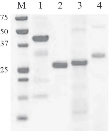
expres-sion problems [34]. The isolated and purified His-tagged proteins were verified using LDS-PAGE and subsequent protein staining (Fig. 1). Using zymographic analysis it was shown that purified LytM-His6and IsaA-His6had retained their enzymatic peptidoglycan-degrading
ac-tivity while Nuc-His6was still able to hydrolyze DNA [34]. The individual and pooled synthetic
PSMα1-4 peptides were shown to facilitate the spreading motility of a non-motileS.aureus
strain that cannot produce these peptides [36], indicating that they are biologically active [19]. These data show that the antigens were successfully isolated or synthesized and that the IsaA-His6, LytM-His6, and Nuc-His6fusions had retained their enzymatic activities.
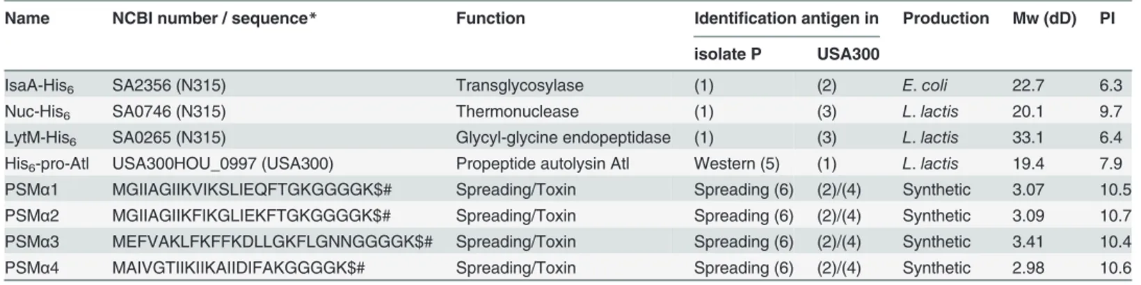
Production of antigens by S. aureus isolate P and USA300
To show the production of the selected antigens by theS.aureusisolates P (MSSA) and USA300 (MRSA) that were used in thein vivostudies, different protein detection methods were used. ForS.aureusUSA300 all antigens used in this study had been identified in earlier (proteomics) studies (Table 2). Also for theS.aureusisolate P, most of the antigens were iden-tified by proteomics in earlier studies (Table 2). Expression of the Atl protein byS.aureus iso-late P was detected using specific polyclonal antibodies as mentioned in the Materials and Methods (data not shown). As pro-Atl is the propeptide of the autolysin Atl, which was de-tected by proteomic studies, pro-Atl was concluded to be expressed by bothS.aureusstrains (our unpublished observations). The expression of the PSMα1-4 peptides has been detected in-directly by using a plate assay in which cells ofS.aureusisolates P or USA300 showed a spread-ing phenotype on a soft agar plate, indicatspread-ing that both strains produce the PSM peptides (Table 2, data not shown). These data show that all antigens used in this study are produced and secreted byS.aureusisolates P and USA300.
Detection of IgG responses against the selected antigens in EB patients
To assess the IgG responses against IsaA-His6, LytM-His6and Nuc-His6,immunodetection
three proteins in comparison to the healthy donors. The Atl propeptide was shown to be recog-nized by antibodies present in sera of EB patients (our unpublished observations).
As the PSMα1 has previously been shown to be exposed on the cell surface ofS.aureus, we included the PSMα1,α2,α3, andα4 in an epitope mapping approach using the PepScan tech-nology [38]. This analysis showed that 5 out of 7 EB patients have an above average titer of IgGs against the peptides tested (Fig. 3). Three out of 7 plasma samples from EB patients showed a very high reactivity against the N-terminal regions of the PSMα1,α2, andα3 pep-tides. These results show that besides the IsaA-His6, LytM-His6and Nuc-His6proteins, the
PSM peptides are also well recognized by the humoral immune system.
Immunization with an octa-valent mixture of
S
.
aureus
antigens does not
reduce bacterial load in mice with
S
.
aureus
isolate P bacteremia
Groups of mice were immunized by subcutaneous injection with 5μg of purified IsaA-His6,
LytM-His6, Nuc-His6, His6-pro-Atl, and PSMα1-4 each, emulsified in TiterMax Gold adjuvant
Fig 1. LDS-PAGE detection of the purified and dialyzedS.aureusantigens.Fifteenμg of LytM-His6 (1), Nuc-His6 (2), IsaA-His6 (3) or His6-pro-Atl (4) was loaded. The molecular weight of marker proteins is indicated (in kDa).
on day -70. On days -42 and -14, they were boosted with this mixture containing 25μg of each
purified antigen. Blood samples were collected before and after immunization, and specific serum IgG levels were determined by ELISA, demonstrating that these antigens generated hu-moral immune responses to immunization (Fig. 4). The response against His6-pro-Atl was on
average 10-fold lower compared to the other 4 antigens.
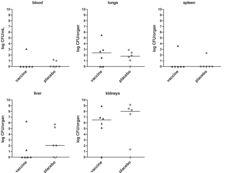
Mice were challenged at day 0 with mildS.aureusisolate P bacteremia (n = 8 immunized mice, n = 8 placebo-immunized mice), and animal discomfort and survival rate over 14 days after infection were monitored. At day 14, surviving mice were sacrificed, and blood and organs were removed for assessment of the bacterial load. In placebo-immunized mice, discomfort in-creased, while animal survival declined over time. At day 14, 6 out of 8 placebo-immunized mice were still alive.S.aureusload in kidneys of these mice was high, while bacterial load in lungs and liver was low, and blood and spleen were culture negative (Fig. 5).
Compared to placebo-immunized animals, immunization with the octa-valent antigen mix-ture did not reduce theS.aureusload in blood, lungs, spleen, liver, and kidneys (Fig. 5). Fur-thermore, discomfort score and animal survival rate over 14 days did not differ between immunized and placebo-immunized mice (data not shown).
Table 2. Proteins and peptides used in this study.
Name NCBI number / sequence* Function Identification antigen in Production Mw (dD) PI
isolate P USA300
IsaA-His6 SA2356 (N315) Transglycosylase (1) (2) E.coli 22.7 6.3
Nuc-His6 SA0746 (N315) Thermonuclease (1) (3) L.lactis 20.1 9.7
LytM-His6 SA0265 (N315) Glycyl-glycine endopeptidase (1) (3) L.lactis 33.1 6.4
His6-pro-Atl USA300HOU_0997 (USA300) Propeptide autolysin Atl Western (5) (1) L.lactis 19.4 7.9 PSMα1 MGIIAGIIKVIKSLIEQFTGKGGGGK$# Spreading/Toxin Spreading (6) (2)/(4) Synthetic 3.07 10.5 PSMα2 MGIIAGIIKFIKGLIEKFTGKGGGGK$# Spreading/Toxin Spreading (6) (2)/(4) Synthetic 3.09 10.7 PSMα3 MEFVAKLFKFFKDLLGKFLGNNGGGGK$# Spreading/Toxin Spreading (6) (2)/(4) Synthetic 3.41 10.4 PSMα4 MAIVGTIIKIIKAIIDIFAKGGGGK$# Spreading/Toxin Spreading (6) (2)/(4) Synthetic 2.98 10.6
*the sequences GGK$# stand for the addition of the GGG-Lys(ε-biotin) to each of the peptides (1) Identified by proteomics [15]
(2) Identified by proteomics [17]
(3) Identified by proteomics (our unpublished observations) (4) Identified using spreading assay [19]
(5) detection of Atl using specific antibodies (seeMaterials and Methods) (6) Spreading as determined by plate assay (results not shown)
doi:10.1371/journal.pone.0116847.t002
Fig 2. Western detection of immune responses against the antigens IsaA-His6, LytM-His6and Nuc-His6.Sera of healthy volunteers (T7-1 till T7-6) and EB patients (EB51, 15, 01, 11, 09, 55) were used.
Immunization with an octa-valent
S
.
aureus
antigen mixture is not
protective against mortality due to
S
.
aureus
USA300 bacteremia in mice
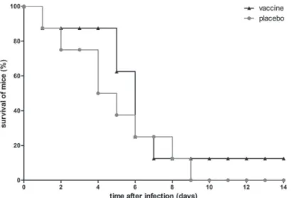
To assess whether the lack of protective effect of immunization with the octa-valent mixture wasS.aureusstrain-dependent, we included a lethal mouse model of MRSA bacteremia as well. As immunized mice showed excellent IgG titers, the immunization schedule was not adapted. In this model, mice were challenged with severeS.aureusUSA300 bacteremia (n = 8 immunized mice, n = 8 placebo-immunized mice), and animal discomfort and survival rate over 14 days after infection were monitored. In placebo-immunized mice, discomfort in-creased, while animal survival declined gradually over time, and at day 9, all placebo-immu-nized mice had died (Fig. 6).
Fig 3. Heatmap of anti-PSMα1-4 reactivity.Plot of IgG responses from sera of volunteers (T7-1 till T7-4) and EB patients (EB01, 09, 51, 11, 15, 53, 02)
against the N- (1 to 15) and C- (7 to 21) terminal parts of the PSMα1-4 peptides. Colors represent a gradient of reactivity against the various peptides (green is low and red is high). Peptides with a signal exceeding the median plus twice the standard deviation are boxed.
doi:10.1371/journal.pone.0116847.g003
Fig 4. IgG titers in serum of mice after immunization with the octa-valent mixture.Mice (n = 20) were immunized subcutaneously with the mixture containing IsaA-His6, LytM-His6, Nuc-His6, His6-pro-Atl, and PSMα1-4 at days -70 (5μg of each antigen), days -42, and -14 (25μg of each antigen). IgG titerson day -1 were assessed. Each symbol represents a single mouse. Median values are indicated by horizontal lines.
In this model of severe MRSA bacteremia, immunization with the octa-valent mixture con-taining IsaA-His6, LytM-His6, Nuc-His6, His6-pro-Atl, and PSMα1-4 did not protect against
mortality dueS.aureusUSA300 bacteremia (Fig. 6). Furthermore, the discomfort score over 14 days was not reduced, and higher IgG levels did not correlate with increased time-to-death of mice (data not shown).
Immunization with an octa-valent
S
.
aureus
antigen mixture does not
protect against S. aureus isolate P skin infection
Absence of protection of the octa-valent mixture inS.aureusbacteremia does not exclude a possible (lack of) protection in other types ofS.aureusinfections. To assess whether this anti-gen mixture elicits a protective immune response againstS.aureusskin infection, immunized mice were challenged withS.aureusisolate P via the intradermal route (n = 4 immunized mice, n = 4 placebo-immunized mice). Animal body weight and lesion size over 7 days after
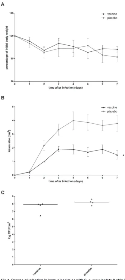
Fig 5. Bacterial load in immunized mice withS.aureusisolate P bacteremia.Mice (n = 8) were immunized with the octa-valent vaccine containing IsaA-His6, LytM-His6, Nuc-His6, His6-pro-Atl, and PSMα1-4, or with placebo. Animals were infected by intravenous inoculation ofS.aureusisolate P (3 × 10⁵ CFU). At day 14, surviving mice were sacrificed and quantitative cultures of blood, lungs, spleen, liver, and kidneys were performed. Each symbol represents a single mouse. Median values are indicated by horizontal lines. Statistically significant differences inS.aureusload were not observed (P>0.05; Mann-Whitney U test).
infection were monitored. At day 7, mice were sacrificed and theS.aureusload in skin lesions was assessed. In placebo-immunized mice, body weight loss over 7 days was minor, with a maximum of 6.5%. Size of the skin lesion increased over time until 2.8–5.2 cm2at day 7. At this time point, medianS.aureusload in the skin lesion was 2 × 10⁸CFU/cm2(Fig. 7).
Compared to placebo-immunized mice, immunization with the octa-valent antigen mixture did not limit the body weight loss over time (Fig. 7A), while size of the skin lesion was reduced in immunized mice (Fig. 7B).S.aureusload in skin lesions at day 7 was not reduced in immu-nized mice (Fig. 7C). As reduction of the bacterial load in the skin lesion was our primary out-come measure, we concluded that immunization with the octa-valentS.aureusantigen mixture did not protect againstS.aureusisolate P skin infection.
Discussion
In view of the high mortality rates ofS.aureusinfections [42–45], the emergence of antibiotic-resistantS.aureusstrains [6] and the lack of new antimicrobials in the development pipeline [8], alternative treatment strategies forS.aureusinfections are urgently needed. One approach is treatment through immunization targetingS.aureusantigens. This may be an interesting substitute for or additive to the currently used antibiotics. A role of certain anti-staphylococcal antibodies in protection againstS.aureusinfection-related death is suggested by a number of studies in humans [14,26,46,47]. Next to these suggestions from clinical studies, a number of studies inS.aureus-infected experimental animals showed protective effects of active or passive immunization. Notwithstanding these promising results obtained in experimental animals, no efficacy of immunization is observed in clinical studies [9–11].
The lack of protective capacity of anti-staphylococcal immunization may be explained by, for example, a lack of power of several clinical studies [12,13], the limited number ofS.aureus
antigens targeted in these studies (mostly one or two), or theS.aureusinfection studied (most-ly bacteremia/sepsis). In addition, the apparent lack of success of immunization targeting for example CP5 and CP8 [48,49], IsdB [50], or ClfA [51] inS.aureus-infected patients may be re-lated to the choice of targets for immunization. Therefore, in the present study, we selectedS.
Fig 6. Survival of immunized mice withS.aureusUSA300 bacteremia.Mice (n = 8) were immunized with the octa-valent vaccine containing IsaA-His6, LytM-His6, Nuc-His6, His6-pro-Atl, and PSMα1-4, or with placebo. Animals were infected by intravenous inoculation ofS.aureusUSA300 (6 × 10⁵CFU), and were monitored for 14 days. A statistically significant difference in animal survival rates was not observed (P>0.05; log rank test).
aureusantigens that are accessible for antibodies based on proteomic analysis of exoproteomes ofS.aureus[15] and a proteolytic shaving approach ofS.aureus[17]. This strategy resulted in the selection of IsaA, LytM, Nuc, pro-Atl, and PSMα1-4 as targets for active immunization. Previous studies showed that these eightS.aureusantigens are immunogenic in humans [14,26,29,30], and may therefore be applicable in the clinical setting. Moreover, monoclonal antibodies against IsaA enhance the killing ofS.aureusin whole blood samples from healthy subjects and patients prone to staphylococcal infections [52], and passive immunization with these monoclonal antibodies can lead to protection againstS.aureusinfections in mice [28,53].
Using the hostsL.lactisandE.coli, all selectedS.aureusantigens (IsaA, LytM, Nuc, and pro-Atl) were successfully isolated. No major degradation products were observed after isola-tion, and IsaA-His6, LytM-His6, and Nuc-His6fusion proteins were all biologically active after
purification, indicating they had retained their natural conformation. In addition, these three fusion proteins as well as the PSMα1-4 peptides were well recognized by IgG present in plasma of healthy volunteers and EB patients. These results indicate that His-tagged fusions did not af-fect binding of IgG to these antigens. In addition, in a previous study, it has been shown that the Atl propeptide is recognized by antibodies present in sera of EB patients (our unpublished data).
The potentially protective capacity of immunization was evaluated in mice immunized with a mixture of IsaA, LytM, Nuc, pro-Atl and PSMα1-4. In the use of an octa-valent antigen cock-tail we followed the approach of Stranger-Jones et al. [54], who immunized mice with IsdA, IsdB, SdrD, and SdrE, either in a combination or individually. They observed that single anti-gen immunization elicited no or only very modest protection againstS.aureusabscess forma-tion orS.aureuslethal challenge, whereas immunization with the cocktail completely protected in bothS.aureusinfections. In addition, Bagnoli et al. proposed a model in which vaccine efficacy is gained via antibodies that directly inhibit bacterial viability and/or toxicity, via antibodies to mediate opsonophagocytosis, and via cell-mediated immunity to stimulate re-cruitment of phagocytes at the site of infection [55]. For this purpose, multipleS.aureus anti-gens with different functions should be targeted. In the present study immunization of mice with the octa-valent antigen mixture of IsaA, LytM, Nuc, pro-Atl and PSMα1-4 resulted in de-tectable IgG responses against all eight antigens. Although anti-pro-Atl levels were slightly lower, all observed IgG levels were in the expected range [54,56]. This observation indicated that all antigens were recognized well by the immune system of the mice.
After the final booster immunization, mice were infected withS.aureus. Clinically relevant models were used: mild bacteremia byS.aureusisolate P (MSSA), lethal bacteremia byS. aure-usUSA300 (MRSA), and transientS.aureusisolate P skin infection. It was shown that active immunization with the octa-valent mixture of IsaA-His6, LytM-His6, Nuc-His6, His6-pro-Atl,
and PSMα1-4, although resulting in high anti-staphylococcal IgG levels, did not protect mice against mildS.aureusisolate P bacteremia, severeS.aureusUSA300 bacteremia, orS.aureus
isolate P skin infection.
The lack of protection by active immunization with the octa-valentS.aureusantigen mix-ture in our mouse infection experiments may be related to the study design or the presumed role of anti-staphylococcal antibodies in protection against theseS.aureusinfections. Regard-ing the study design, theS.aureusisolates used and the type ofS.aureusinfections studied are
PSMα1-4, or with placebo. Animals were infected by intradermal inoculation ofS.aureusisolate P (3 × 10⁷ CFU), and were monitored for 7 days. (A) Animal body weight over time afterS.aureusskin infection was not affected by vaccination (P>0.05; Quade’s rank analysis of covariance). (B) Lesion size was significantly reduced by vaccination, as indicated by the asterisk (P= 0.005; Quade’s rank analysis of covariance). (C)S.
aureusload in the skin lesion at day 7 was not reduced by vaccination (P>0.05; Mann-Whitney U test).
clinically relevant [15,31,32]. Other investigators used these infection models as well, and dem-onstrated that active or passive immunization targetingS.aureusantigens can lead to protec-tion in mice [9–11]. In addition, bothS.aureusstrains produced all eightS.aureusantigens. It should be noted that preclinical animal models ofS.aureusinfection do not fully mimic the naturalS.aureusinfection process in humans due to various differences among which differ-ences in host cell proteins such as hemoglobin [57] and the requirement for high bacterial inoc-ula [58] in the non-human host. Despite these limitations of experimental animal models, we conclude that failure to show protective activity upon active immunization can presumably not be explained by the choice ofS.aureusstrains or the choice of infection models. As the ob-served differences between immunized mice and placebo-immunized mice were not signifi-cant, not even borderline, increasing the group sizes will probably not result in significant differences. Therefore, this cannot clarify the failure of immunization in ourin vivomodels.
A more plausible explanation for the lack of protection of the octa-valent antigen mixture in the present study may be related to the presumed role of anti-staphylococcal antibodies in pro-tection againstS.aureusinfection. Although previous studies suggested a role of anti-staphylo-coccal antibodies in protection against death inS.aureuscarriers [14,26,46,47,56,59], their role may be overestimated. Possibly, IgG against IsaA-His6, LytM-His6, Nuc-His6, His6-pro-Atl,
and PSMα1-4, or anti-staphylococcal IgG in general, has no or only a limited role in protection againstS.aureusinfections. It may be conceivable that antibodies againstS.aureusantigens in humans are just a result of prior exposure in carriers and non-carriers, viaS.aureus coloniza-tion or previous (sub)clinicalS.aureusinfection, while their protective capacity is limited. Pre-vious exposure toS.aureusin carriers may also result in improved cellular immunity, which could also protect againstS.aureusinfection-related death, as was already suggested by Joshi et al. [60]. Beside this, the production of virulence factors such as toxins and immune evasion proteins byS.aureus[61,62] might overwhelm the generated humoral immune response. An-other reason for the failure of the octa-valent antigen mixture to protect mice againstS.aureus
infection might be related to a potential immunosuppression induced by one of the antigens in the cocktail. As other combinations ofS.aureusantigens were not tested, conclusions in this re-spect cannot be drawn.
Although passive immunization with monoclonal antibodies against IsaA protected mice againstS.aureusinfection [28,53], in the present study no protection was obtained after active immunization with a mixture including IsaA. A possible explanation for this discrepancy may be related to insufficient binding of polyclonal antibodies induced by active immunization to relevant epitopes of IsaA in order to provide protection againstS.aureusinfection, in contrast to the monoclonal antibodies administered by passive immunization, clearly binding to rele-vant epitopes of IsaA.
In conclusion, active immunization with an octa-valent mixture containing IsaA-His6,
LytM-His6, Nuc-His6, His6-pro-Atl and the PMSα1–4 peptides does not protect mice against
S.aureusbacteremia andS.aureusskin infection. The observations suggest that these polyclon-al anti-staphylococcpolyclon-al antibodies do not provide protection againstS.aureusinfection. Conse-quence of the present study should not be abandoning of research focusing on immunization inS.aureusinfections, as other investigators obtained promising results in this respect. Instead, future research should focus on novel treatment strategies combining immunization with anti-biotic treatment and/or cytokine administration.
Acknowledgments
Author Contributions
Conceived and designed the experiments: SvdB DK JWB JMvD IBW GB. Performed the exper-iments: SvdB DK JWB JN GB. Analyzed the data: SvdB DK JWB JN AD JMvD IBW GB. Con-tributed reagents/materials/analysis tools: SvdB DK JWB AD JMvD IBW GB. Wrote the paper: SvdB DK JWB JMvD IBW GB.
References
1. Wertheim HF, Melles DC, Vos MC, van Leeuwen W, van Belkum A, et al. (2005) The role of nasal car-riage inStaphylococcus aureusinfections. Lancet Infect Dis 5: 751–762. PMID:16310147
2. Klevens RM, Morrison MA, Nadle J, Petit S, Gershman K, et al. (2007) Invasive methicillin-resistant
Staphylococcus aureusinfections in the United States. JAMA 298: 1763–1771. PMID:17940231 3. Cosgrove SE (2006) The relationship between antimicrobial resistance and patient outcomes:
mortali-ty, length of hospital stay, and health care costs. Clin Infect Dis 42 Suppl 2: S82–89. PMID:16355321 4. Cosgrove SE, Qi Y, Kaye KS, Harbarth S, Karchmer AW, et al. (2005) The impact of methicillin
resis-tance inStaphylococcus aureusbacteremia on patient outcomes: mortality, length of stay, and hospital charges. Infect Control Hosp Epidemiol 26: 166–174. PMID:15756888
5. Ito T, Hiramatsu K, Tomasz A, de Lencastre H, Perreten V, et al. (2012) Guidelines for reporting novel
mecAgene homologues. Antimicrob Agents Chemother 56: 4997–4999. PMID:22869575
6. Chambers HF, DeLeo FR (2009) Waves of resistance:Staphylococcus aureusin the antibiotic era. Nat Rev Microbiol 7: 629–641. doi:10.1038/nrmicro2200PMID:19680247
7. Silver LL (2011) Challenges of antibacterial discovery. Clin Microbiol Rev 24: 71–109. doi:10.1128/ CMR.00030-10PMID:21233508
8. Boucher HW, Talbot GH, Bradley JS, Edwards JE, Gilbert D, et al. (2009) Bad bugs, no drugs: no ESKAPE! An update from the Infectious Diseases Society of America. Clin Infect Dis 48: 1–12. doi:10. 1086/595011PMID:19035777
9. Ohlsen K, Lorenz U (2010) Immunotherapeutic strategies to combat staphylococcal infections. Int J Med Microbiol 300: 402–410. doi:10.1016/j.ijmm.2010.04.015PMID:20547101
10. Verkaik NJ, van Wamel WJ, van Belkum A (2011) Immunotherapeutic approaches against Staphylo-coccus aureus. Immunotherapy 3: 1063–1073. doi:10.2217/imt.11.84PMID:21913829
11. Daum RS, Spellberg B (2012) Progress toward aStaphylococcus aureusvaccine. Clin Infect Dis 54: 560–567. doi:10.1093/cid/cir828PMID:22186773
12. Rupp ME, Holley HP Jr, Lutz J, Dicpinigaitis PV, Woods CW, et al. (2007) Phase II, randomized, multi-center, double-blind, placebo-controlled trial of a polyclonal anti-Staphylococcus aureuscapsular poly-saccharide immune globulin in treatment ofStaphylococcus aureusbacteremia. Antimicrob Agents Chemother 51: 4249–4254. PMID:17893153
13. Benjamin DK, Schelonka R, White R, Holley HP, Bifano E, et al. (2006) A blinded, randomized, multi-center study of an intravenousStaphylococcus aureusimmune globulin. J Perinatol 26: 290–295. PMID:16598296
14. den Reijer PM, Lemmens-den Toom N, Kant S, Snijders SV, Boelens H, et al. (2013) Characterization of the humoral immune response duringStaphylococcus aureusbacteremia and global gene expres-sion byStaphylococcus aureusin human blood. PLoS One 8: e53391. doi:10.1371/journal.pone. 0053391PMID:23308212
15. Ziebandt AK, Kusch H, Degner M, Jaglitz S, Sibbald MJ, et al. (2010) Proteomics uncovers extreme heterogeneity in theStaphylococcus aureusexoproteome due to genomic plasticity and variant gene regulation. Proteomics 10: 1634–1644. doi:10.1002/pmic.200900313PMID:20186749
16. Sibbald MJ, Ziebandt AK, Engelmann S, Hecker M, de Jong A, et al. (2006) Mapping the pathways to staphylococcal pathogenesis by comparative secretomics. Microbiol Mol Biol Rev 70: 755–788. PMID: 16959968
17. Dreisbach A, Hempel K, Buist G, Hecker M, Becher D, et al. (2010) Profiling the surfacome of Staphylo-coccus aureus. Proteomics 10: 3082–3096. doi:10.1002/pmic.201000062PMID:20662103
18. Dreisbach A, van Dijl JM, Buist G (2011) The cell surface proteome ofStaphylococcus aureus. Proteo-mics 11: 3154–3168. doi:10.1002/pmic.201000823PMID:21630461
19. Tsompanidou E, Sibbald MJ, Chlebowicz MA, Dreisbach A, Back JW, et al. (2011) Requirement of the
20. Tsompanidou E, Denham EL, van Dijl JM (2013) Phenol-soluble modulins, hellhounds from the staphy-lococcal virulence-factor pandemonium. Trends Microbiol 21: 313–315. doi:10.1016/j.tim.2013.04.007 PMID:23684152
21. Li M, Cheung GY, Hu J, Wang D, Joo HS, et al. (2010) Comparative analysis of virulence and toxin ex-pression of global community-associated methicillin-resistantStaphylococcus aureusstrains. J Infect Dis 202: 1866–1876. doi:10.1086/657419PMID:21050125
22. Baba T, Schneewind O (1998) Targeting of muralytic enzymes to the cell division site of Gram-positive bacteria: repeat domains direct autolysin to the equatorial surface ring ofStaphylococcus aureus. EMBO J 17: 4639–4646. PMID:9707423
23. Beenken KE, Spencer H, Griffin LM, Smeltzer MS (2012) Impact of extracellular nuclease production on the biofilm phenotype ofStaphylococcus aureusunderin vitroandin vivoconditions. Infect Immun 80: 1634–1638. doi:10.1128/IAI.06134-11PMID:22354028
24. Berends ET, Horswill AR, Haste NM, Monestier M, Nizet V, et al. (2010) Nuclease expression by Staph-ylococcus aureusfacilitates escape from neutrophil extracellular traps. J Innate Immun 2: 576–586. doi:10.1159/000319909PMID:20829609
25. van den Berg S, Bowden MG, Bosma T, Buist G, van Dijl JM, et al. (2011) A multiplex assay for the quantification of antibody responses inStaphylococcus aureusinfections in mice. J Immunol Methods 365: 142–148. doi:10.1016/j.jim.2010.12.013PMID:21185300
26. van der Kooi-Pol MM, de Vogel CP, Westerhout-Pluister GN, Veenstra-Kyuchukova YK, Duipmans JC, et al. (2013) High anti-staphylococcal antibody titers in patients with epidermolysis bullosa relate to long-term colonization with alternating types ofStaphylococcus aureus. J Invest Dermatol 133: 847– 850. doi:10.1038/jid.2012.347PMID:23014336
27. Swierstra J, Debets S, de Vogel C, Lemmens-den Toom N, Verkaik N, et al. (2014) IgG4 subclass-spe-cific responses toStaphylococcus aureusantigens shed new light on host-pathogen interaction. Infect Immun.
28. Lorenz U, Lorenz B, Schmitter T, Streker K, Erck C, et al. (2011) Functional antibodies targeting IsaA of
Staphylococcus aureusaugment host immune response and open new perspectives for antibacterial therapy. Antimicrob Agents Chemother 55: 165–173. doi:10.1128/AAC.01144-10PMID:20956605 29. Holtfreter S, Kolata J, Broker BM (2010) Towards the immune proteome ofStaphylococcus aureus—
The anti-S.aureusantibody response. Int J Med Microbiol 300: 176–192. doi:10.1016/j.ijmm.2009.10. 002PMID:19889576
30. Adhikari RP, Ajao AO, Aman MJ, Karauzum H, Sarwar J, et al. (2012) Lower antibody levels to Staphy-lococcus aureusexotoxins are associated with sepsis in hospitalized adults with invasiveS.aureus in-fections. J Infect Dis 206: 915–923. doi:10.1093/infdis/jis462PMID:22807524
31. Lowy FD (1998)Staphylococcus aureusinfections. N Engl J Med 339: 520–532. PMID:9709046 32. McDougal LK, Steward CD, Killgore GE, Chaitram JM, McAllister SK, et al. (2003) Pulsed-field gel
elec-trophoresis typing of oxacillin-resistantStaphylococcus aureusisolates from the United States: estab-lishing a national database. J Clin Microbiol 41: 5113–5120. PMID:14605147
33. Neef J, Milder F, Koedijk DGAM, Klaassens M, Heezius E, et al. Straightforward one-step cloning and overexpression system for flexible, secretable and removable his-tagged fusions of heterologous pro-teins forEscherichia coliandLactococcus lactis. In press.
34. Neef J, Koedijk DG, Bosma T, van Dijl JM, Buist G (2014) Efficient production of secreted staphylococ-cal antigens in a non-lysing and proteolytistaphylococ-cally reducedLactococcus lactisstrain. Appl Microbiol Bio-technol Accepted.
35. Yamada S, Sugai M, Komatsuzawa H, Nakashima S, Oshida T, et al. (1996) An autolysin ring associat-ed with cell separation ofStaphylococcus aureus. J Bacteriol 178: 1565–1571. PMID:8626282 36. Tsompanidou E, Denham EL, Becher D, de Jong A, Buist G, et al. (2013) Distinct roles of
phenol-solu-ble modulins in spreading ofStaphylococcus aureuson wet surfaces. Appl Environ Microbiol 79: 886– 895. doi:10.1128/AEM.03157-12PMID:23183971
37. Buist G, Kok J, Leenhouts KJ, Dabrowska M, Venema G, et al. (1995) Molecular cloning and nucleotide sequence of the gene encoding the major peptidoglycan hydrolase ofLactococcus lactis, a murami-dase needed for cell separation. J Bacteriol 177: 1554–1563. PMID:7883712
38. Timmerman P, Van Dijk E, Puijk W, Schaaper W, Slootstra J, et al. (2004) Mapping of a discontinuous and highly conformational binding site on follicle stimulating hormone subunit-beta (FSH-beta) using domain Scan and Matrix Scan technology. Mol Divers 8: 61–77. PMID:15209158
40. Brown EL, Dumitrescu O, Thomas D, Badiou C, Koers EM, et al. (2009) The Panton-Valentine leukoci-din vaccine protects mice against lung and skin infections caused byStaphylococcus aureusUSA300. Clin Microbiol Infect 15: 156–164. doi:10.1111/j.1469-0691.2008.02206.xPMID:19281461
41. Bunce C, Wheeler L, Reed G, Musser J, Barg N (1992) Murine model of cutaneous infection with gram-positive cocci. Infect Immun 60: 2636–2640. PMID:1612733
42. Mylotte JM, McDermott C, Spooner JA (1987) Prospective study of 114 consecutive episodes of Staph-ylococcus aureusbacteremia. Rev Infect Dis 9: 891–907. PMID:3317734
43. Lesens O, Methlin C, Hansmann Y, Remy V, Martinot M, et al. (2003) Role of comorbidity in mortality re-lated toStaphylococcus aureusbacteremia: a prospective study using the Charlson weighted index of comorbidity. Infect Control Hosp Epidemiol 24: 890–896. PMID:14700403
44. Cosgrove SE, Sakoulas G, Perencevich EN, Schwaber MJ, Karchmer AW, et al. (2003) Comparison of mortality associated with methicillin-resistant and methicillin-susceptibleStaphylococcus aureus bac-teremia: a meta-analysis. Clin Infect Dis 36: 53–59. PMID:12491202
45. Lambert ML, Suetens C, Savey A, Palomar M, Hiesmayr M, et al. (2011) Clinical outcomes of health-care-associated infections and antimicrobial resistance in patients admitted to European intensive-care units: a cohort study. Lancet Infect Dis 11: 30–38. doi:10.1016/S1473-3099(10)70258-9PMID: 21126917
46. Verkaik NJ, Dauwalder O, Antri K, Boubekri I, de Vogel CP, et al. (2010) Immunogenicity of toxins dur-ingStaphylococcus aureusinfection. Clin Infect Dis 50: 61–68. doi:10.1086/648673PMID:19947854 47. Kolata J, Bode LG, Holtfreter S, Steil L, Kusch H, et al. (2011) Distinctive patterns in the human
anti-body response toStaphylococcus aureusbacteremia in carriers and non-carriers. Proteomics 11: 3914–3927. doi:10.1002/pmic.201000760PMID:21805632
48. Shinefield H, Black S, Fattom A, Horwith G, Rasgon S, et al. (2002) Use of aStaphylococcus aureus
conjugate vaccine in patients receiving hemodialysis. N Engl J Med 346: 491–496. PMID:11844850 49. Fattom AI, Horwith G, Fuller S, Propst M, Naso R (2004) Development of StaphVAX, a polysaccharide
conjugate vaccine againstS.aureusinfection: from the lab bench to phase III clinical trials. Vaccine 22: 880–887. PMID:15040941
50. Fowler VG, Allen KB, Moreira ED, Moustafa M, Isgro F, et al. (2013) Effect of an investigational vaccine for preventingStaphylococcus aureusinfections after cardiothoracic surgery: a randomized trial. JAMA 309: 1368–1378. doi:10.1001/jama.2013.3010PMID:23549582
51. Weems JJ Jr, Steinberg JP, Filler S, Baddley JW, Corey GR, et al. (2006) Phase II, randomized, dou-ble-blind, multicenter study comparing the safety and pharmacokinetics of tefibazumab to placebo for treatment ofStaphylococcus aureusbacteremia. Antimicrob Agents Chemother 50: 2751–2755. PMID:16870768
52. Oesterreich B, Lorenz B, Schmitter T, Kontermann R, Zenn M, et al. (2014) Characterization of the bio-logical anti-staphylococcal functionality of hUK-66 IgG1, a humanized monoclonal antibody as substan-tial component for an immunotherapeutic approach. Hum Vaccin Immunother 10: 926–937. PMID: 24495867
53. van den Berg S, Bonarius HPJ, van Kessel KPM, Elsinga GS, Kooi N, et al. (2015) A human monoclo-nal antibody targeting the conserved staphylococcal antigen IsaA protects mice against Staphylococ-cus aureusbacteremia. Int J Med Microbiol.In press.
54. Stranger-Jones YK, Bae T, Schneewind O (2006) Vaccine assembly from surface proteins of Staphylo-coccus aureus. Proc Natl Acad Sci U S A 103: 16942–16947. PMID:17075065
55. Bagnoli F, Bertholet S, Grandi G (2012) Inferring reasons for the failure ofStaphylococcus aureus vac-cines in clinical trials. Front Cellul Infect Microbiol 2: 16.
56. Clarke SR, Brummell KJ, Horsburgh MJ, McDowell PW, Mohamad SA, et al. (2006) Identification ofin vivo-expressed antigens ofStaphylococcus aureusand their use in vaccinations for protection against nasal carriage. J Infect Dis 193: 1098–1108. PMID:16544250
57. Pishchany G, McCoy AL, Torres VJ, Krause JC, Crowe JE Jr, et al. (2010) Specificity for human hemo-globin enhancesStaphylococcus aureusinfection. Cell Host Microbe 8: 544–550. doi:10.1016/j.chom. 2010.11.002PMID:21147468
58. Kiser KB, Cantey-Kiser JM, Lee JC (1999) Development and characterization of aStaphylococcus au-reusnasal colonization model in mice. Infect Immun 67: 5001–5006. PMID:10496870
59. Dryla A, Prustomersky S, Gelbmann D, Hanner M, Bettinger E, et al. (2005) Comparison of antibody repertoires againstStaphylococcus aureusin healthy individuals and in acutely infected patients. Clin Diagn Lab Immunol 12: 387–398. PMID:15753252
61. Spaan AN, Surewaard BG, Nijland R, van Strijp JA (2013) Neutrophils versusStaphylococcus aureus: a biological tug of war. Annu Rev Microbiol 67: 629–650. doi:10.1146/annurev-micro-092412-155746 PMID:23834243
62. Foster TJ, Geoghegan JA, Ganesh VK, Hook M (2014) Adhesion, invasion and evasion: the many func-tions of the surface proteins ofStaphylococcus aureus. Nature Reviews Microbiology 12: 49–62. doi: 10.1038/nrmicro3161PMID:24336184
63. Kuipers OP, Beerthuyzen MM, Siezen RJ, De Vos WM (1993) Characterization of the nisin gene cluster
nisABTCIPRofLactococcus lactis. Requirement of expression of thenisAandnisIgenes for develop-ment of immunity. Eur J Biochem 216: 281–291. PMID:7689965
64. Bosma T, Kanninga R, Neef J, Audouy SA, van Roosmalen ML, et al. (2006) Novel surface display sys-tem for proteins on non-genetically modified gram-positive bacteria. Appl Environ Microbiol 72: 880– 889. PMID:16391130