TATHYANE RAMALHO SANTOS GIONBELLI
NUTRIÇÃO MATERNA E SEXO FETAL SOBRE O DESENVOLVIMENTO PRÉ-NATAL DE BOVINOS MESTIÇOS HOLANDÊS × GIR
VIÇOSA
MINAS GERAIS – BRASIL 2015
Ficha catalográfica preparada pela Biblioteca Central da Universidade Federal de Viçosa - Câmpus Viçosa
T
Gionbelli, Tathyane Ramalho Santos, 1984-G496n
2015
Nutrição materna e sexo fetal sobre o desenvolvimento pré-natal de bovinos mestiços Holandês × Gir / Tathyane Ramalho Santos Gionbelli. – Viçosa, MG, 2015.
xii, 69f. : il. ; 29 cm.
Orientador: Cristina Mattos Veloso.
Tese (doutorado) - Universidade Federal de Viçosa. Inclui bibliografia.
1. Vaca - Nutrição. 2. Vaca - Gestação. 3. Feto - Sistema musculoesquelético. 4. Feto - Intestino - Morfologia.
I. Universidade Federal de Viçosa. Departamento de Zootecnia. Programa de Pós-graduação em Zootecnia. II. Título.
ii TATHYANE RAMALHO SANTOS GIONBELLI
NUTRIÇÃO MATERNA E SEXO FETAL SOBRE O DESENVOLVIMENTO PRÉ-NATAL DE BOVINOS MESTIÇOS HOLANDÊS × GIR
APROVADA: 12 de março de 2015.
iii Em memória ao meu pai, Antônio Jarbas Ramalho dos Santos.
A meu marido Mateus, meu filho Antônio, minha mãe Maria das Graças, meu irmão Talles Antônio e minha irmã Tane Cristina, alicerces da minha vida.
Em especial a Mateus Pies Gionbelli, meu anjo da guarda.
iv
AGRADECIMENTOS
Em primeiro lugar, agradeço a Deus por se fazer presente em todos os dias de minha vida, por ser meu amigo, meu amparo e minha força. Obrigada senhor, por fazer parte de minha vida!
À Universidade Federal de Viçosa, pela oportunidade e, em especial, ao Departamento de Zootecnia, pelo apoio na realização deste curso.
Ao Concelho Nacional de Desenvolvimento Científico e Tecnológico (CNPq), pela concessão da bolsa de doutorado.
À professora Cristina Mattos Veloso, pela oportunidade, pela excelente orientação, confiança, simplicidade, paciência e amizade.
Ao pesquisador Bruno Campos de Carvalho, pela confiança e auxílio na construção deste trabalho.
A pesquisadora Bruna Rios Coelho Alves, pela disposição para participar da banca de defesa de tese.
Ao professor Marcos Inácio Marcondes, pelo apoio, confiança e disposição em ajudar.
Ao professor José Domingos Guimarães, pelo apoio, atenção e disposição em ajudar.
Ao professor Sebastião de Campos Valadares Filho, pelo apoio irrestrito, pela confiança e disposição em ajudar.
Ao professor Marcio Duarte, pela atenção, pelos ensinamentos, pelas excelentes ideias, pela paciência e amizade.
À professora Simone, pela atenção e apoio.
v Aos funcionários da Fábrica de Ração, pelo auxílio na confecção das rações.
Aos demais professores e funcionários do Departamento de Zootecnia, pelo apoio, convívio e amizade.
A todos os estagiários, pela ajuda, apoio, amizade e companheirismo. Sem vocês, nada disso seria possível.
À Polyana, pelo companheirismo, confiança, ajuda e amizade.
À turma de Zootecnia 2005, pelos inesquecíveis momentos de alegria, pela amizade. Turma da qual sempre terei orgulho de ter feito parte.
Ao Mateus (Xuxu), por todo o aprendizado, companheirismo, amizade, carinho, dedicação, ajuda, força e amor. Juntos para sempre.
Ao meu filho Antônio, pela inocência, amor e por me fazer a pessoa mais feliz do mundo.
Aos meus pais, Maria das Graças e Antônio Jarbas (em memória), pelos ensinamentos de vida, pelo amor, pela dedicação, confiança, apoio e compreensão.
Ao meu irmão, Talles Antônio, pela alegria de viver, coragem, amizade, simplicidade e carinho.
A minha irmã Tane, Cristina, pelo carinho, companheirismo, ensinamentos, consultas e amizade.
A minha sogra, Ceci, meu sogro Valdir, meu cunhado Cristian (em memória) e minha cunhada Mariana, pela amizade, carinho, paciência, confiança, ajuda, apoio e exemplo de família.
A todos os meus familiares, que sempre me apoiaram e torceram por mim.
vi BIOGRAFIA
TATHYANE RAMALHO SANTOS GIONBELLI, filha de Maria das Graças Santos Ramalho e Antônio Jarbas Ramalho dos Santos, nasceu em Teófilo Otoni, Estado de Minas Gerais, em 04 de março de 1984.
Em 2005, ingressou na Universidade Federal de Viçosa-UFV, onde obteve título de bacharel em Zootecnia, colando grau em 25 de julho de 2009.
Em agosto de 2009, iniciou o curso de Mestrado em Zootecnia na mesma instituição, concentrando seus estudos na área de Fisiologia da Produção Animal. Em julho de 2011 tornou-se mestre em Zootecnia.
vii SUMÁRIO
RESUMO ... vii
ABSTRACT ... x
INTRODUÇÃO GERAL ... 1
LITERATURA CITADA ... 9
Capítulo 1 – MATERNAL NUTRITION INTENSIFICATION AND FETAL SEX ON DEVELOPMENT OF SKELETAL MUSCLE OF BOVINE FETUSES ALONG GESTATION Abstract ... 14
Resumo ... 16
/body ... 18
Results ... 20
Discussion ... 24
Materials and methods ... 27
References ... 31
Capítulo 2 – DESENVOVIMENTO INTESTINAL FETAL AO LONGO DA GESTAÇÃO É AFETADO PELO SEXO E NUTRIÇÃO MATERNA EM BOVINOS Resumo ... 43
Abstract ... 45
Introdução ... 47
Material e métodos ... 48
Resultados ... 53
Discussão ... 56
Conclusão ... 61
Literatura citada ... 61
viii RESUMO
GIONBELLI, Tathyane Ramalho Santos, D.Sc., Universidade Federal de Viçosa, março de 2015. Nutrição materna e sexo fetal sobre o desenvolvimento pré-natal de bovinos mestiços Holandês × Gir. Orientadora: Cristina Mattos Veloso. Coorientadores: Marcos Inácio Marcondes e Bruno Campos de Carvalho.
xi ABSTRACT
GIONBELLI, Tathyane Ramalho Santos D.Sc., Universidade Federal de Viçosa, March, 2015. Maternal nutrition and fetal sex on fetal development of Holstein × Gyr cattle. Adviser: Cristina Mattos Veloso. Co-advisers: Marcos Inácio Marcondes and Bruno Campos de Carvalho.
1 INTRODUÇÃO GERAL
Apesar de ser um país predominantemente tropical, o Brasil possui grande variabilidade climática, refletindo nos regimes pluviométricos e, consequentemente, na quantidade e qualidade de alimentos disponíveis. Esse se torna um dos principais problemas da pecuária no Brasil e na América do Sul, sendo um dos pontos críticos na redução da eficiência de produção. Essa variação sazonal afeta grande parte das regiões brasileiras, consequentemente, vacas gestantes criadas em pasto, frequentemente, são submetidas à variação na oferta e qualidade da forragem, principalmente, na época seca do ano.
Buscando contornar esse problema, alguns pecuaristas têm adotado estratégias de suplementação para vacas gestantes, especialmente no terço final da gestação, quando ocorre a maior parte do crescimento fetal (Ferrell et al., 1976). Entretanto, alguns estudos recentes mostraram que a deficiência nutricional materna durante o início e meio da gestação, em ruminantes, pode causar redução do número de fibras e massa muscular da progênie, afetando o desempenho produtivo durante toda a vida do animal, mesmo que não tenha sido notada redução do peso ao nascimento (Du et al., 2010; Wu et al., 2006). Cabe ressaltar que é nesta fase que ocorre crescimento placentário máximo e organogénese fetal, os quais são acontecimentos críticos para o desenvolvimento do concepto (Funston et al., 2010).
Nos anos recentes, o aumento do número de estudos desenvolvidos nessa área levou à aplicação do tema “desenvolvimento fetal programado” ou “programação fetal”
2 (Barker et al., 2002). Esse conceito pode ser entendido como o resultado de mudanças específicas nos mamíferos, as quais ocorrem durante o desenvolvimento intra-uterino e alteram quantitativa e/ou qualitativamente a trajetória do seu desenvolvimento, com resultados que persistem por toda a vida do indivíduo (Duarte et al., 2012).
Nesse contexto, a compreensão da fisiologia do desenvolvimento fetal é de extrema importância. Por isso, estudos têm sido realizados com o objetivo de compreender os processos envolvidos no crescimento e desenvolvimento dos tecidos, uma vez que a produção de carne visa maximização do sistema. A partir desse conhecimento, torna-se possível a adoção de estratégias alimentares, durante os diferentes estágios da gestação, que possam resultar em incremento do desempenho da progênie, bem como da melhoria da qualidade da carne destes animais.
O componente principal da carne bovina é o tecido muscular, e o principal componente de um músculo são as fibras que o constituem. A massa muscular é, portanto, em grande parte, determinada pelo número (hiperplasia) e tamanho das fibras musculares (hipetrofia). A pesquisa sugere que os animais com um maior número de fibras musculares, de tamanho moderado conseguem produzir uma maior quantidade e qualidade de carne (Rehfeldt et al., 2004). O número de fibras musculares de um animal é definido, exclusivamente, durante o período pré-natal (Cossu & Borello, 1999), uma vez que as fibras são formadas durante a fase embrionária e fetal, época em que fatores de crescimento e de transcrição interagem, resultando no tecido muscular.
3 (Beermann et al., 1978). A regulação deste processo envolve a ativação, proliferação e diferenciação de várias linhagens de células miogênicas e depende da expressão e atividade de fatores transcricionais, conhecidos como fatores de regulação miogênica (MRF). No embrião, o comprometimento das células do mesoderma somítico com a linhagem muscular, inicia-se com ação sinalizadora oriunda de tecidos circundantes, tais como a notocorda e o tubo neural (Charge & Rudiniki, 2004). Estes sinais são responsáveis pela ativação de genes capazes de transformar células não musculares em células com fenótipo muscular.
No embrião a ação sinalizadora para o direcionamento das células do mesoderma pars a linhagem muscular inicia com as proteínas Wnt/ -catenina. Este sistema Wnt/ -catenin é formado por várias proteínas, que transmitem sinais externos para dentro da célula, por meio de ligação ao receptor de membrana. Este complexo é responsável pela regulação dos fatores trascricionais Pax3 e Pax7. A ativação destes fatores promoverá a subsequente expressão dos genes responsáveis pela produção dos fatores reguladores da miogênese: Myf5 e Myod (Buckingham, 2001); que induzirão as células à linhagem miogênica. Parte dessas células (mioblastos) que expressam Myf5 e MyoD, saem do ciclo celular, tornando-se miócitos diferenciados e iniciam a expressão dos MRFs miogenina e MRF4, os quais regulam a diferenciação dessas células em fibras musculares (Keren et al., 2006; Kollias & McDermott, 2008). Assim, ao final do processo de miogênese, os miócitos mononucleados se fundem para formar os miotubos e, no animal adulto, o músculo esquelético torna-se um tecido estável, caracterizado por fibras musculares multinucleadas (Schmalbruch & Lewis, 2000).
4 (Charge & Rudiniki, 2004). Quando estimulada, a célula satélite é ativada, prolifera e funde-se com a fibra muscular pré-existente. Os núcleos derivados das células satélites começam a sintetizar proteínas musculares específicas que aumentam o volume das fibras musculares por meio da formação de novos sarcômeros, em posição externa às miofibrilas existentes, culminando no aumento da massa muscular por hipertrofia (Goldspink et al., 1972). Entretanto, além de células miogênicas, uma porcentagem das células satélites pode se diferenciar em adipócitos ou fibroblastos (Aguiari et al., 2008; Kuang et al., 2007).
Alguns trabalhos de pesquisa reportaram a nutrição materna como sendo um dos principais fatores que afetam a miogênese em ruminantes e, consequentemente, o crescimento e desenvolvimento muscular fetal, com efeitos que persistem por toda vida do animal (Godfrey & Barker, 2000; Quigley et al., 2005; Wu et al., 2006; Zhu et al., 2006), mesmo quando não é verificada alteração no peso ao nascimento (Ford et al., 2007; Martin et al., 2007; Larson et al., 2009). O músculo esquelético tem menor prioridade na partição de nutrientes durante o desenvolvimento fetal, quando comparado
a órgãos como cérebro, coração e fígado fazendo com que este tecido apresente grande
vulnerabilidade quanto à disponibilidade de nutrientes (Zhu et al., 2006)
Em pesquisa realizada por Zhu et al. (2006) quando se restringiu em 50% das
exigências de ovinos em gestação, de acordo com o NRC (1985) entre o 280 e o 780 dias
de gestação, foi observada redução do número total de fibras musculares no músculo
Longissimus dorsi de cordeiros abatidos aos oito meses de idade.
intra-5 muscular, em bovinos, também tem início durante a fase pré-natal (Du et al., 2010) e conforme abordado anteriormente, os adipócitos tem origem do mesmo pool de células mesenquimais que as fibras musculares, sendo que parte dessas células presentes no tecido muscular esquelético fetal diferencia-se em adipócitos, formando sítios para deposição de gordura no músculo do neonato (Tong et al., 2009). A adipogênese é regulada por fatores de transcrição, tais como C/EBP (Enhacer binding protein) e PPAR (Peroxissome proliferator-activated receptor ). Alguns trabalhos de pesquisa
avaliaram esses fatores transcricionais e evidenciaram que a adipogênese, em animais ruminantes, inicia-se concomitantemente com a miogênese secundária, no terço médio da gestação (Fève, 2005; Gnanalingham et al., 2005; Muhlhausler et al., 2007; Du et al., 2010). Sendo assim, a adoção de nutrição materna adequada, durante a gestação, pode resultar em maior número de adipócitos, em função do aumento do comprometimento de células mesenquimais com a adipogênese, acarretando maior quantidade de gordura de marmoreio na carne da progênie. Em um estudo recente realizado por Underwood et al.,(2010), foi avaliada a qualidade da carne de novilhos oriundos de matrizes mantidas
em pastagem, e que foram submetidas ou não a estresse nutricional por sessenta dias,
durante o terço médio da gestação. Os autores verificaram maior peso ao abate, maior
presença de gordura subcutânea na carcaça, maior teor de gordura intramuscular e
menores valores de força de cisalhamento na carne de novilhos nascidos de matrizes
que não sofreram estresse nutricional durante a gestação.
6 regulatórios da diferenciação destas células constituem-se num dos pontos-chave para a avaliação e o entendimento do desenvolvimento do tecido muscular e adiposo, durante a fase pré-natal.
A técnica de PCR (reação em cadeia da polimerase) quantitativo em tempo real (qPCR) tem sido amplamente utilizada, com grande eficiência, para avaliar a ação dos fatores transcricionais, sendo está um dos mais confiáveis métodos para análise de expressão gênica (Yuan el al., 2006)
Seus resultados baseiam-se na medida de produtos gerados durante cada um dos ciclos do processo de PCR, os quais são diretamente proporcionais à quantidade do molde presente no início do processo. Por meio dele é possível medir a quantidade do produto de PCR ainda na fase, exponencial, pois, somente nessa fase é possível estimar a quantidade do molde utilizado inicialmente (Ginzinger, 2002). Dois diferentes métodos de análise são utilizados para análises de PCR em tempo real: quantificação absoluta e quantificação relativa. A primeira determina o número de cópias do transcrito de interesse, baseado numa curva padrão, enquanto o segundo descreve a diferença de expressão entre dois grupos, no qual, geralmente, se usa um grupo controle ou referência (Livak & Shimittigen, 2001). No caso de quantificação relativa, a escolha correta dos genes de referência para normalizar a expressão do gene-alvo em PCR em tempo real é essencial para refletir verdadeiramente o processo biológico (Robinson et al., 2007). Em trabalho recente de validação de genes de referência especificamente em músculo bovino. Perez et al. (2008) testaram 10 genes (18S, GAPDH, ACTB, B2M, RPII, UBC, CASC3, HMBS, SF3A1, EEF1A2) e verificaram que os três últimos apresentaram-se mais consistentes para uso como genes para controle endógeno.
7 explicadas não somente por alterações do desenvolvimento do tecido muscular e adiposo, mas, também, por alterações do desenvolvimento do trato gastrointestinal (Duarte et al., 2013; Wang et al., 2008; Wu et al., 2006).
Em ruminantes, a absorção de macromoléculas intactas de imunoglobulina do leite materno representa a principal forma de aquisição de imunidade pós-natal. Essa absorção ocorre através do epitélio intestinal, sendo possível, somente, por cerca de 24 horas após o nascimento do neonato. Dessa forma, o adequado desenvolvimento do trato gastrintestinal, durante as fases intra-uterinas, é fundamental para se reduzir a morbidade e mortalidade neonatal. Reduzida aquisição de imunidade, bem como alterações no desenvolvimento do trato gastrintestinal, que prejudiquem a absorção de nutrientes, podem afetar, permanentemente, o desempenho da progênie e a eficiência do uso de nutrientes (Duarte et al., 2013).
Alguns estudos têm demonstrado que os fetos oriundos de matrizes submetidas à restrição alimentar, durante o início e meio da gestação, diminuíram o crescimento do trato gastrintestinal (Trahair et al., 1997; Wang et al., 2008) e, em alguns casos, mesmo com intervenção nutricional pós-natal, o crescimento reduzido do trato gastrintestinal provocou alterações permanentes nas funções gastrintestinais, tais como a permeabilidade epitelial, por toda a vida do animal (Trahair et al., 1997).
8 celular, que ocorrem ao longo do desenvolvimento fetal devido às alterações ambientais, tais como a nutrição materna, em diferentes estágios da gestação. Embora existam alguns trabalhos envolvendo programação fetal em bovinos na literatura mundial, nenhum utilizou animais mestiços europeu × zebu criados em condições tropicais, além de que os resultados obtidos ainda são inconclusivos para explicar com clareza, os mecanismos de controle do desenvolvimento celular na fase pré-natal (Duarte et al., 2012). Da mesma forma, os trabalhos nos quais o desenvolvimento do trato gastrintestinal foi avaliado como função da nutrição materna, em bovinos (Duarte et al., 2013; Meyer et al., 2010), apesar de evidenciarem algumas diferenças no desenvolvimento do trato gastrintestinal fetal como função da nutrição materna, não definem estratégias de manejo nutricional materno durante a gestação e salientam a necessidade da realização de mais estudos na área.
Assim sendo, o conhecimento e entendimento dos mecanismos que afetam o desenvolvimento dos tecidos muscular e adiposo e do trato gastrintestinal, durante a fase pré-natal, como função do nível alimentar a que são submetidas as matrizes durante a gestação, poderão oferecer suporte ao desenvolvimento de tecnologias de produção mais eficientes e dar base científica para o direcionamento das decisões a serem tomadas no meio produtivo da carne bovina.
9 LITERATURA CITADA
AGUIARI, P.; LEO, B.; ZAVAN, V., et al. High glucose induces adipogenic differentiation of muscle-derived stem cells. In: National Academy of Science, 105.2008, USA. Anais. USA: p.1226-1231, 2008.
BARKER, D. J., J. G. Eriksson, T. Forsen, and C. Osmond. Fetal origins of adult disease: Strength of effects and biological basis. Int. Journal of Epidemiology. 31:1235–1239, 2002.
BEERMANN, D. H.; CASSENS, R. G.; HAUSMAN, G. J. A Second Look at Fiber Type Differentiation in Porcine Skeletal Muscle. Journal of Animal Science., v.46, n.1, p.125-132, 1978.
BUCKINGHAM, M. Skeletal muscle formation in vertebrates. Current Opinion in Genetics & Development, v.11, n.4, p.440-448, 2001.
CHARGE, S. B. P.; RUDINIKI, M. A. Cellular and Molecular Regulation of Muscle Regeneration. Physiolgy Reviews., v.84, n.1, p.209-238, 2004.
COSSU, G.; BORELLO, U. Wnt signaling and the activation of myogenesis in mammals. EMBO Journal, v.18, n.24, p.6867-6872, 1999.
DU, M.; TONG, J.; ZHAO, J., et al. Fetal programming of skeletal muscle development in ruminant animals. Journal of Animal Science, v.88, n.13, p.E51-60, 2010. DUARTE, M.S., PAULINO, P.V.R., DU, M. Fetal programming in beef cattle: how to
optimize performance and carcass value in early life stages. In: Valadares Filho, S.C., Paulino, M.F., Paulino P.V.R. (Ed.), VIII SIMCORTE. Anais... Grafica Suprema, Viçosa, pp. 123–139, 2012.
DUARTE, M.S.; GIONBELLI, M.P.; PAULINO, P.V.R. et al. Effects of maternal nutrition on development of gastrointestinal tract of bovine fetus at different stages of gestation. Livestock Science, v.153, n.1, 2013.
FERRELL, C. L.; GARRETT, W. N.; HINMAN, N. Growth, development and composition of the udder and gravid uterus of beef heifers during pregnancy. Journal of Animal Science, v.42, n.6, p.1477-1489, 1976.
FÈVE, B. Adipogenesis: cellular and molecular aspects. Best Practice & Research: Clinical Endocrinology & Metabolism, v.19, n.4, p.483-499, 2005.
FORD, S. P.; HESS, B. W.; SCHWOPE, M. M., et al. Maternal undernutrition during early to mid-gestation in the ewe results in altered growth, adiposity, and glucose tolerance in male offspring. Journal of Animal Science., v.85, n.5, p.1285-1294, 2007.
10 GINZINGER, D. G. Gene quantification using real-time quantitative PCR: An
emerging technology hits the mainstream. Experimental Hematology, v.30, n.6, p.503-512, 2002.
GNANALINGHAM, M. G.; MOSTYN, A.; SYMONDS, M. E., et al. Ontogeny and nutritional programming of adiposity in sheep: potential role of glucocorticoid action and uncoupling protein-2. AJP Regulatory Integrative and Comarative Physiology, v.289, n.5, p.R1407-1415, 2005.
GODFREY, K. M.; BARKER, D. J. Fetal nutrition and adult disease. American Journal of Clinical Nutrition, v.71, n.5, p.1344S-1352, 2000.
GOLDSPINK, G.; WILKES, D.; STEVEN, E. Myosin expression during ontogeny post-hatching growth and adaptation. Academic Press: London, 1972. p.
KEREN, A.; TAMIR, Y.; BENGAL, E. The p38 MAPK signaling pathway: A major regulator of skeletal muscle development. Molecular and Cellular Endocrinology, v.252, n.1-2, p.224-230, 2006.
KOLLIAS, H. D.; MCDERMOTT, J. C. Transforming growth factor-{beta} and myostatin signaling in skeletal muscle. Journal of Applied Physiology, v.104, n.3, p.579-587, 2008.
KUANG, S.; KURODA, K.; LE GRAND, F., et al. Asymmetric self-renewal and commitment of satellite stem cells in muscle. Cell, v.129, n.5, p.999-1010, 2007. LARSON, D. M.; MARTIN, J. L.; ADAMS, D. C., et al. Winter grazing system and
supplementation during late gestation influence performance of beef cows and steer progeny. Journal of Animal Science, v.87, n.3, p.1147-1155, 2009.
MEYER, A.M., REED, J.J., VONNAHME, K.A. et al. Effects of stage of gestation and nutrient restriction during early to mid-gestation on maternal and fetal visceral organ mass and indices of jejunal growth and vascularity in beef cows. Journal of Animal Science, v.88, p.2410–2424, 2010.
MUHLHAUSLER, B. S.; DUFFIELD, J. A.; MCMILLEN, I. C. Increased Maternal Nutrition Stimulates Peroxisome Proliferator Activated Receptor-{gamma}, Adiponectin, and Leptin Messenger Ribonucleic Acid Expression in Adipose Tissue before Birth. Endocrinology, v.148, n.2, p.878-885, 2007.
PEREZ, R.; TUPAC-YUPANQUI, I.; DUNNER, S. Evaluation of suitable reference genes for gene expression studies in bovine muscular tissue. BMC Molecular Biology, v.9, p.79, 2008.
QUIGLEY, S. P.; KLEEMANN, D. O.; KAKAR, M. A., et al. Myogenesis in sheep is altered by maternal feed intake during the peri-conception period. Animal reproduction science, v.87, n.3, p.241-251, 2005.
11 ROBINSON, T. L.; SUTHERLAND, I. A.; SUTHERLAND, J. Validation of candidate
bovine reference genes for use with real-time PCR. Veterinary Immunology and Immunopathology, v.115, n.1-2, p.160-165, 2007.
SCHMALBRUCH, H.; LEWIS, D. Dynamics of nuclei of muscle fibers and connective tissue cells in normal and denervated rat muscles. Muscle & Nerve, v.23, n.4, p.617-626, 2000
TONG, J. F.; YAN, X.; ZHU, M. J., et al. Maternal obesity downregulates myogenesis and {beta}-catenin signaling in fetal skeletal muscle. Amimal Journal Physiology Endocrinol Metabolism, v.296, n.4, p.E917-924, 2009.
TRAHAIR, J.F., DEBARRO, T.M., ROBINSON, J. et al. Restriction of nutrition in utero selectively inhibits gastrointestinal growth in fetal sheep. Journal of Nutrition, v.127, p.637–641, 1997.
UNDERWOOD, K. R.; TONG, J. F.; PRICE, P. L., et al. Nutrition during mid to late gestation affects growth, adipose tissue deposition, and tenderness in cross-bred beef steers. Meat Science, v.In Press, Corrected Proof, 2010.
WANG, J., CHEN, L., LI, D. et al. Intrauterine growth restriction affects the proteomes of the small intestine, liver, and skeletal muscle in newborn pigs. Journal of Nutrition, v.138, p.60–66, 2008.
WU, G.; BAZER, F. W.; WALLACE, J. M., et al. BOARD-INVITED REVIEW: Intrauterine growth retardation: Implications for the animal sciences. Journal of Animal Science, v.84, n.9, p.2316-2337, 2006.
YUAN, J. S.; REED, A.; CHEN, F., et al. Statistical analysis of real-time PCR data. BMC Bioinformatics, v.7, p.85, 2006.
13 OBS.:
*O capítulo 1, apresentado a seguir, foi escrito na língua inglesa e está apresentado de acordo com as normas da revista PNAS - Proceedings of the National Academy of Sciences (exceto para presença de resumo em português).
14 CAPÍTULO 1
Maternal nutrition intensification and fetal sex on development of skeletal muscle of bovine fetuses along gestation
ABSTRACT
15 adipogenic and fibrogenic markers were less expressed at late gestation than in midgestation, however collagen deposition, fat and crude protein content of fetal muscle were greater at late gestation than in midgestation. In general, MN altered the gene expression of some myogenic, adipogenic and fibrogenic markers at midgestation (greater in ON than in CO) but some compensatory expression made the effect of MN not significant at late gestation, in accordance to previous literature reports. Already known evidence of greater skeletal muscle development of males was also observed during the fetal phase compared to female fetuses.
16 Intensificação da nutrição materna e sexo fetal sobre o desenvolvimento do
músculo esquelético de fetos bovinos ao longo da gestação
RESUMO
17 sexo fetal. Quase todos marcadores miogênicos, adipogênicos e fibrogênicos foram menos expressos na fase final do que na fase intermediária da gestação. A deposição de colágeno, gordura e teor de proteína bruta no músculo fetal, no entanto, foram maiores no final da gestação do que na fase intermediária. Em geral, MN alterou a expressão gênica de alguns marcadores miogênicos, adipogêncios e fibrogênicos na fase intermediária da gestação (maior em ON do que em CO), mas um possível efeito compensatório fez o efeito de MN não ser significativo no final da gestação, concordando com relatos prévios da literatura. Evidência do já conhecido maior desenvolvimento do músculo esquelético de machos na fase pós-natal também foi observada durante a fase fetal, em comparação com fetos do sexo feminino.
18 /body Balance greenhouse gas (GHG) mitigation, forest protection, and agricultural growth by promoting agricultural intensification have been focus of studies in many emerging economies, like Brazil (1). The development of policies that benefit land sparing in these countries would be very important from the point of view of environmental issue by prevent deforestation (2). The application of land sparing policies has been proved to increase productivity of agricultural systems making them more competitive than lower productive agricultural systems and to reduce GHG emissions and/or deforestation (3). Brazil had the greatest forests net loss in the world over the period of 1990-2010 (4) and the expansion of cattle ranching areas is related to the deforestation problem (5). By balancing GHG mitigation Brazil could be a template for many other emerging economies in the world. In a recent study Cohn et al. (3) create two different hypothetic economic model of global land use scenarios from 2010 to 2030 asking if intensification of pasture-based cattle ranching in Brazil could reduce global deforestation and mitigate global GHG emissions. The authors modeled a control scenario compared to the application of two land sparing policies, being either a tax on cattle from conventional pasture or a subsidy for cattle from semi-intensive pasture. Cohn et al. (3) found that both policies could achieve considerable sparing of forests and abatement of GHG with, significant increase of productivity.
19 muscular (19-22) development of the ruminants offspring with effects that can be permanent to the entire life of the animal (21, 23). In tropical regions such as Brazil, since is pasture-based, the cattle production suffer the effects of distribution and seasonal variation in quantity and quality of forage along the year. In addition, the rainy season and better availability of good sources of food occurs when cows are in the breeding season, pregnant cows usually experience feed restriction during mid to late gestation period, which overlaps with the dry season in most of the tropical cattle production areas. Trying to minimize the effects of dry season on the reproduction of cows, when producers of tropical regions supplement pregnant cows (not so frequent), they do at the late gestation, when most of fetal growth occurs (24, 25). However, it has been proved that nutritional restriction during midgestation can cause reduction of muscle fiber number and muscle mass, and consequently affecting performance and meat quality of progeny (20), even when no difference is observed on birth weight of the offspring (21, 23). In the other hand, evidence of enhancement of adipogenic markers in skeletal muscle was shown in fetuses from overnourished cows (19).
Thus, based on these two considerations (environmental effect of intensification of pasture-based cattle ranching and potential effects on offspring development due to intensification of maternal nutrition) we investigate the effects of maternal nutrition intensification on fetal modifications of development of skeletal muscle in cattle by overnourishing (ON) cows in comparison of a control treatment (CO) where cows were fed the same diet of ON cows but at a level necessary to maintain their body condition.
20 production is based on native cattle or dual purpose herds (10), where cows are used to produce milk, reposition heifers and males for fattening/finishing (26, 27). These dual purpose cattle raised in tropical regions are generally native or Zebu cattle crossed with specialized dairy breeds, like Holstein (28). In this case, Zebu breeds (Bos indicus) are important because its tolerance to heat and parasites, its rusticity, and its adaptation to the tropics (29, 30). Based on this, we opted to use dual purpose cattle as animal model in this study (Holstein × Gyr cows). Gyr is the Zebu breed mostly crossed with Holstein in Brazil (26, 28, 30).
In this study we also investigate the effects of fetal sex and its interactions with maternal nutrition (MN) and days of gestation (DG) on skeletal muscular development. To achieve the evaluations at different stages of gestation, cows and its fetuses were necropsied at 139, 199, 241 and 268 days of gestation, which represents 50, 70, 85 and 95% of gestation length (27) and 18, 31, 73 and 96% of gravid uterus growing (25), respectively. We investigate the mRNA gene expression and phenotypic indicators of myogenesis, adipogenesis and fibrogenesis in skeletal muscle of bovine fetuses to evaluate the potential impacts of MN and fetal sex along the gestation.
RESULTS
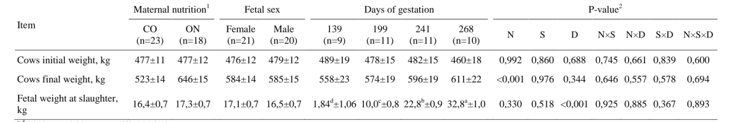
21 treatment, fetal sex and DG were not observed (P≥0.557). Fetal weight (Table 1) was affected by days of gestation (P<0.001) but not by fetal sex. Thus, it can be noted that the results of the effect of maternal nutrition, fetal sex and days of gestation on muscular development of fetus occurred independently of fetal size.
Gene expression of myogenic markers and phenotypic indicators of myogenesis. The mRNA expression for Cadherin-associated protein, β1 ( -catenin) was significantly affected by all fixed effects evaluated (Table 2). The analysis of main effects showed greater expression of -catenin in the skeletal muscle of fetuses from ON-fed cows than in fetuses from CO-fed cows (P=0.035) and in males compared to females (P=0.003). However, interactions between MN and DG showed that -catenin mRNA expression was greater in ON fetuses only at 139 days of pregnancy whereas was similar between fetuses from both MN groups in the subsequent stages of pregnancy. Another significant interaction for -catenin mRNA expression was observed between fetal sex and DG (P=0.054), were gene expression was greater in male fetuses than female fetuses at 139 days of gestation (P<0.10) but no in the subsequent periods (P>0.10). Similar values of mRNA expression for Myogenic differentiation I (MyoD; P = 0.508) and Myogenin (MyoG; P = 0.320) were observed
among ON and CO fetal muscle (Table 2). MyoD was 16% greater expressed in male fetal muscle than in female (P=0.033), whereas MyoG expression was not affected by fetal sex (P=0.120). No interactions among the fixed effects evaluated were observed (P≥0.122) on MyoD and MyoG mRNA expression. The three myogenic markers
22 Crude protein (CP) content of fetal muscle (g/kg) was greater (P=0.051) in fetuses from ON cows compared to those from CO cows and increased (P<0.001) along the gestation (Table 2), being 78% greater at 268 days of gestation compared to 139 days. Fetal sex had no effect (P=0.849) on CP content of fetal muscle. However, different results were observed for the number of myocytes in fetal skeletal muscle (Table 2). The number of muscle cells was in average 10% greater (P=0.009) in males than in females in a same area. An interaction among MN and DG (P=0.091, Table 2) revealed that fetuses from ON cows had greater (P<0.10) number of myocytes at 139 days of gestation (Figure 1) but this phenotypic indicator of myogenesis was similar (P>0.10) between the both MN groups along the subsequent stages of gestation (Table 2).
Gene expression of adipogenic markers and phenotypic quantification of adipogenesis. The mRNA expression of early adipogenic marker zinc finger protein
423 (Zfp423) was affected by interactions between the main effects evaluated.
23 compared to middle gestation (P<0.001, Table 3). The mRNA expression of Peroxisome proliferator activated receptor gamma (PPAR ), another late adipogenic marker, was 26% greater in male than in female fetuses (P=0.002, Table 3). PPAR was
also greater expressed in muscle from ON fetuses compared to CO fetuses at 139 days of gestation (P<0.10) but not different in among the both MN groups in the subsequent stages of gestation (P>0.10).
Although some effects of MN were observed on mRNA expression of adipogenic markers, the phenotypic indicator of adipogenesis evaluated (ether extract content of skeletal muscle) was not affected by MN (P=0.891). Males and females fetuses also presented similar contents of intramuscular fat during pregnancy (P=0.191). However, the fat content in fetal muscle increased about 36% from 139 days of gestation to the subsequent stages (P=0.017, Table 3).
Gene expression of fibrogenic markers and phenotypic quantification of intramuscular collagen deposition. The mRNA expression of Collagen type I alpha 1 (Collagen I) was greater in ON fetuses than in CO fetuses (P=0.091, Table 4) and in male than female fetuses (P=0.095). The expression of Collagen I also decreased from 199 days to the subsequent stages of gestation (P<0.001, Table 4). An interaction between fetal sex and DG was observed on the mRNA expression of Collagen type III alpha 1 (Collagen III, P=0.072) and Fibronectin 1 (FN1, P=0.002). In both cases the
marker was greater expressed in males than in females (P<0.10) only at 139 days of gestation (Table 4), with no differences among fetal sex at the subsequent periods (P>0.10). The mRNA expression of FN1 was greater in ON than in CO fetuses (P=0.027). In other hand, the mRNA expression of transforming growth factor β
24 sex (P=0.100). The expression of TGF- was greater in ON male fetuses than in CO male fetuses (P<0.10), but not in females, which were not affected by the MN (P>0.10). To evaluate the phenotypic fibrogenesis we quantified histochemically the collagen deposition in fetal muscle (Figure 2). The intramuscular collagen deposition was not affected by MN (P=0.907) and fetal sex (P=0.227, Table 4). However, significantly increase was observed due to the increase days of gestation (P<0.001, Figure 2). Total area of collagen increase about five times from 139 to 268 days of gestation (Table 4, Figure 2).
DISCUSSION
All effects observed in this study on fetal skeletal muscle development occurred without fetal weight differences within the same time of gestation (Table 1). These findings agree with previous reports in the literature which have shown that the vast majority of changes in the trajectory of fetal development due to environmental effects occurs without variation of weight at birth (16, 20, 21, 23).
Both maternal nutrient restriction (32) and obesity (33) have been shown to decrease myogenesis in ruminants. In this study, however, we did not find any reduction of myogenesis due to maternal overfeeding. Otherwise, we observed increase in
-Catenin expression and number of myocytes at 139 days of gestation due to maternal overfeeding, despite no differences among MN levels in the subsequent time points of gestation (Table 2). In a previous study with beef cattle breed, the maternal overnutrition tended to decrease gene expression of -Catenin (19), contrary to the
25 CO (P=0.051, Table 2). The differences between ON and CO fetuses for expression of -Catenin and number of myocytes may be explained by the greater availability of nutrients to the gravid uterus in ON cows at the point of greater function of myogenesis (20). However, with the advance of gestation, since the CO feeding level was not restriction, the myogenesis seems to have been compensated in fetuses from CO-fed cows. Evidence of compensatory fetal muscle growth from nutrient restricted and then refed cows has been recent reported (34) and may be related to the effects that were observed in this study.
In general, the mRNA expression of all myogenic markers and the number of myocytes decreases from middle to late gestation (Table 2). These findings corroborate with those observed in beef cattle fetuses (19) and revised by Du et al. (20) based on data from studies with sheep, rodents, and humans. The general report is that myogenesis decreases as gestation progresses where after midgestation (4.5 mo) the formation of new muscle cells is reduced concomitantly with the increase of intramuscular adipogenesis (20). Increase in fat content of the muscle was observed in this study from middle to late gestation (Table 3), signaling increase of intramuscular adipogenesis in the last third of gestation. However, the mRNA expression of adipogenic markers was observed to decrease from middle to late gestation (Table 3), instead of an expected increase. Non-significant increase in mRNA expression trough the gestation of the same markers evaluated in this study was previous reported (19).
26 Likewise, the fat content of skeletal muscle of fetuses did not change as function of MN in this study (Table 3).
Previous studies have suggested that the -Catenin can alters the expression of
other myogenic markers by regulating the expression of paired box 3 (Pax3), which acts upstream of MyoD during skeletal muscle development (35, 36). The -Catenin can also downregulates the myogenesis by regulates the expression of PPAR (37). Although in the current study the results of -Catenin and MyoD expression were correlated (Table 2), there was no signal of PPAR downregulation by -Catenin, since
the pattern of the mRNA expression of both genes were similar (Tables 2 and 3), differently from previous reports when greater expression of PPAR was observed
when expression of -Catenin decreased (19).
27 (Gionbelli et al., unpublished data1). The causes or effects related to these findings are, however, still not well elucidated.
In general, intensification of maternal nutrition altered the gene expression of some myogenic, adipogenic and fibrogenic markers at midgestation (greater in ON than in CO) but some compensatory expression made the effect of MN not significant at late gestation, in accordance to previous literature reports (34). Thus, although previous studies (23) have reported that an increase in the quality of maternal diet from only 60 days during the midgestation increased tenderness, adipose tissue and growth in the offspring, our results suggest that these kind of potential effects does not occurs when cows nutrition is intensified over the maintenance requirements in dual purpose cattle. It can be noted, however, that potential increase in adipogenesis of skeletal muscle has been shown in fetuses from overnourished cows of beef specialized breed (19). What we find is that, although MN intensification over maintenance can alter the trajectory and speed of skeletal muscle development (see interactions between MN and DG in Tables 2, 3 and 4 and Figure 1), the effects that remain until the late gestation are marginal. On the other hand, however, our finds regarding to the differences between males and females during the intrauterine development of skeletal muscle suggest that in fetal programming studies the effect of fetal sex should be carefully controlled and isolated.
MATERIALS AND METHODS
Animal Husbandry. All animal care and handling procedures were approved by Animal Care and Use Committee of the Department of Animal Science of the Universidade Federal de Viçosa, Viçosa, Minas Gerais, Brazil, prior to initiation of the
28 experiment. Fourty-four multiparous non-lactating Holstein × Gyr cows with initial average body weight of 480 ± 10.1 kg and 5 ± 0.5 years old had pregnancy confirmed after an fixed time artificial insemination protocol using semen from a single bull. The general procedures for the reproductive protocol as well the management of the cows during the adaptation period are well described by Rotta et al. (31). Day 0 of pregnancy was considered the day of insemination. At 60 days of gestation the fetal sex diagnose was performed and feeding treatments application started and lasted until the slaughter. Cows were divided at random in two groups with different feeding levels, being the control group (CO, n=24) the feeding restriction to 1.15% of cow body weight per day to maintain the gestation and body weight. Overnourished (ON, n=20) treatment consisted in fed cows ad libitum with the same diet of CO group. The diet was based on corn silage (93%), cotton meal (5%) and mineral + urea mixture (2%). Both groups were fed twice daily (60% in the morning and 40% afternoon).
29 were collected from each fetus. One of the LM samples was placed in sterile tubes containing RNAlater (Qiagen, Hilden, North Rhine, Westphalia, Germany) stored at 4°C overnight and then kept at –80°C before RNA isolation. A small portion of samples was fixed in fresh 10% (wt/vol) formalin in phosphate buffer (pH = 7.4) immediately after slaughter and then processed for histological analysis. A third sample was frozen at -20 ºC for further ether extract and crude protein quantification.
Real-Time Quantitative PCR Analysis. Total RNA (1 μg) was extracted from 0.5 g of powdered tissue samples using Trizol reagent (Invitrogen, Carlsbad, CA), treated with DNase I, Amplification Grade (Invitrogen), and reverse transcribed into cDNA using the GoScript Reverse Transcription System (Promega, Madison, WI). The primer sets used are shown in Table 2. Reverse transcription PCR was performed on a 7300 Real-Times PCR System (Applied Biosystems, Foster City, CA) using SYBR Green RT-PCR kit from Bio-Rad and the following cycle parameters: 95°C for 3 min and 40 cycles at 95°C for 10 s and 60°C for 30 s. The amplification efficiency was 0.90 to 0.99. After amplification, a melting curve (0.01 °C/s) was used to confirm product purity. Results are expressed relative to 18S using the ΔΔCt method (44). To be used as a reference gene, the stability of 18S expression among experimental treatments was determined through statistical analysis by Tukey’s method at α=0.05. No variation of 18S
expression was observed (P=0.845) among experimental treatments.
30 Bothell, WA). A total of 10 images per fetus were taken (2 images per section and 5 sections per fetus) at 400-fold magnification to quantify a total of muscle cells by using ImageJ software (National Institute of Health, Baltimore, MD). Samples were embedded in Paraplast (Sigma-Aldrich, St. Louis, MO) and cut into sections of 5 μm by using a RM2245 microtome (Leica Microsystems Inc.). Sections were rehydrated through incubations on Histochoice (Sigma-Aldrich) and ethanol solutions. After rehydration, sections were stained for 1 h in the picrosirius solution (45). Picrosirius solution was composed of 0.1% (wt/vol) of Direct Red 80 (Sigma-Aldrich) in 1.3% (wt/vol) aqueous picric acid solution. After 1 h incubation, stained sections were washed for 2 min in 0.01 N HCl solution, dehydrated, cleared, and mounted in synthetic resin. Picrosirius stained sections were observed under polarized light by using an Olympus BX53 coupled with a DP21 digital camera (Olympus, Tokyo, Japan). For quantification of picrosirius-stained intramuscular collagen, a total of 10 images per fetus (2 images per section and 5 sections per fetus) were taken into the same background under the polarized light and then analyzed by using ImageJ software. All the images were converted into grayscale and split into red, green, and blue channels. After that, the green channel of all images was thresholded to the same level to highlight the stained collagen. Collagen was quantified as percentage of the total image area. All images were analyzed for collagen quantification at 100-fold magnification.
Chemical Analysis. Ether extract quantification and crude protein content in the fetal skeletal muscle follow the procedures described by Lage et al. (46) for adult animals.
31 Yijkl = µ + MNi + FSj + DGk + (MN×FS)ij + (MN×DG)ik + (FS×DG)jk + (MN×FS×DG)ijk
+ eijkl
where MNi is the ith level of the fixed effect of maternal nutrition, FSj is the jth level of
the fixed effect fetal sex, DGk is the kth level of the fixed effect days of gestation and e
eijkl is the random error associated with Yijkl. Gene expression levels were transformed
using the natural logarithm of the expression values + 1 to achieve normality (47). Outliers were removed to achieve normality using Shapiro-Wilks test at α = 0.10 (48). Least square means were estimated for all effects and compared using Tukey’s method at α = 0.10. All statistical procedures were performed using the MIXED procedure from
SAS (SAS, version 9.2, software; SAS Institute).
REFERENCES
1. Newton P, Agrawal A, & Wollenberg L (2013) Enhancing the sustainability of commodity supply chains in tropical forest and agricultural landscapes. Global Environmental Change 23(6):1761-1772.
2. Nepstad DC, Boyd W, Stickler CM, Bezerra T, & Azevedo AA (2013) Responding to climate change and the global land crisis: REDD+, market transformation and low-emissions rural development.
3. Cohn AS, et al. (2014) Cattle ranching intensification in Brazil can reduce global greenhouse gas emissions by sparing land from deforestation. Proceedings of the National Academy of Sciences 111(20):7236-7241.
4. FAO (2010) United Nations Food and Agriculture Organization (Global Forest Resource Assessment). (Rome, Italy).
5. Bowman MS, et al. (2012) Persistence of cattle ranching in the Brazilian Amazon: A spatial analysis of the rationale for beef production. Land Use Policy 29(3):558-568.
6. Detmann E, Gionbelli MP, & Huhtanen P (2014) A meta-analytical evaluation of the regulation of voluntary intake in cattle fed tropical forage-based diets. J. Anim. Sci.
7. Detmann E, Paulino MF, & Valadares Filho SC (2010) Otimização do uso de recursos forrageiros basais. VII Simpósio de Produção de Gado de Corte, eds Valadares Filho SC, Paulino MF, Paulino PV, & Marcondes MI (Suprema), pp 191-240.
8. Valente É, et al. (2013) Effect of calves’ supplementation on performance, nutritional and behavioral characteristics of their dams. (Translated from English) Tropical Animal Health and Production 45(2):487-495 (in English). 9. Valente E, et al. (2012) Strategies of supplementation of female suckling calves
32 10. Bouman BAM & Nieuwenhuyse A (1999) Exploring options for sustainable beef cattle ranching in the humid tropics: a case study for the Atlantic Zone of Costa Rica. Ag. Syst. 59(2):145-161.
11. Sampaio CB, et al. (2010) Intake and digestibility in cattle fed low-quality tropical forage and supplemented with nitrogenous compounds. (Translated from eng) Trop Anim Health Prod 42(7):1471-1479 (in eng).
12. Gionbelli MP, et al. (2015) Quantitative aspects of growth and development of cow, udder and gravid uterus of Zebu beef cows during pregnancy. PLoS One (in revision - PONE-D-14-27792).
13. Duarte MS, et al. (2013) Effects of maternal nutrition on development of gastrointestinal tract of bovine fetus at different stages of gestation. Livestock Science 153(1):60-65.
14. Hammer CJ, et al. (2011) Effects of maternal selenium supply and plane of nutrition during gestation on passive transfer of immunity and health in neonatal lambs. J. Anim. Sci. 89(11):3690-3698.
15. Meyer AM, et al. (2010) Effects of stage of gestation and nutrient restriction during early to mid-gestation on maternal and fetal visceral organ mass and indices of jejunal growth and vascularity in beef cows. J. Anim. Sci. 88(7):2410-2424.
16. Trahair JF, DeBarro TM, Robinson JS, & Owens JA (1997) Restriction of Nutrition In Utero Selectively Inhibits Gastrointestinal Growth in Fetal Sheep. The Journal of Nutrition 127(4):637-641.
17. Yanusova RD, et al. (2013) Impacts of maternal selenium supply and nutritional plane on visceral tissues and intestinal biology in 180-day-old offspring in sheep. J. Anim. Sci. 91(5):2229-2242.
18. Reed JJ, et al. (2007) Effects of selenium supply and dietary restriction on maternal and fetal body weight, visceral organ mass and cellularity estimates, and jejunal vascularity in pregnant ewe lambs. J. Anim. Sci. 85(10):2721-2733. 19. Duarte MS, et al. (2014) Maternal overnutrition enhances mRNA expression of
adipogenic markers and collagen deposition in skeletal muscle of beef cattle fetuses. J. Anim. Sci.
20. Du M, et al. (2010) Fetal programming of skeletal muscle development in ruminant animals. J. Anim. Sci. 88:E51-E60.
21. Wu G, Bazer FW, Wallace JM, & Spencer TE (2006) BOARD-INVITED REVIEW: Intrauterine growth retardation: Implications for the animal sciences. J. Anim. Sci. 84(9):2316.
22. Vonnahme KA (2007) Nutrition during gestation and fetal programming. Range Beef Cow Symposium XX, (University of Nebraska Lincoln), pp 1-11.
23. Underwood KR, et al. (2010) Nutrition during mid to late gestation affects growth, adipose tissue deposition, and tenderness in cross-bred beef steers. Meat Science 86(3):588-593.
24. Ferrell CL, Garrett WN, & Hinman N (1976) Growth, development and composition of the udder and gravid uterus of beef heifers during pregnancy. J. Anim. Sci. 42(6):1477-1489.
25. Gionbelli MP, et al. (2015) Achieving Body Weight Adjustments for Feeding Status and Pregnant or Non-Pregnant Condition in Beef Cows. PLoS ONE 9(12):e115724.
33 27. Mellado M, Coronel F, Estrada A, & Ríos FG (2011) Lactation performance of Holstein and Holstein x Gyr cattle under intensive condition in a subtropical environment. Tropical and subtropical agroecosystems 14:927-931.
28. Ruas JRM, et al. (2014) Lactation productive characteristics of four genetic groups F1 Holstein x Zebu. R. Bras. Ci. Vet. 21(1).
29. Silva AA, et al. (2011) Quantitative trait loci affecting milk production traits on bovine chromosome 6 in zebuine Gyr breed. J. Dairy. Sci. 94(2):971-980.
30. Santana ML, Jr., et al. (2014) History, structure, and genetic diversity of Brazilian Gir cattle. Livestock Science 163:26-33.
31. Rotta PP, et al. (2015) Effects of day of gestation and feeding regimen in Holstein x Gyr cows: I. Apparent total tract digestibility, nitrogen balance, and fat deposition. J. Dairy. Sci.:(accepted).
32. Zhu MJ, et al. (2006) Maternal nutrient restriction affects properties of skeletal muscle in offspring. The Journal of Physiology 575(Pt 1):241-250.
33. Tong JF, et al. (2009) Maternal obesity downregulates myogenesis and -catenin signaling in fetal skeletal muscle. American Journal of Physiology - Endocrinology And Metabolism 296(4):E917-E924.
34. Gonzalez JM, et al. (2013) Realimentation of nutrient restricted pregnant beef cows supports compensatory fetal muscle growth. J. Anim. Sci. 91(10):4797-4806.
35. Ridgeway AG & Skerjanc IS (2001) Pax3 Is Essential for Skeletal Myogenesis and the Expression of Six1 and Eya2. Journal of Biological Chemistry 276(22):19033-19039.
36. Gustafsson MK, et al. (2002) Myf5 is a direct target of long-range Shh signaling and Gli regulation for muscle specification. Genes & Development 16(1):114-126.
37. Okamura M, et al. (2009) COUP-TFII acts downstream of Wnt/ -catenin signal to silence PPAR gene expression and repress adipogenesis. Proceedings of the National Academy of Sciences of the United States of America 106(14):5819-5824.
38. Hinde K, Carpenter AJ, Clay JS, & Bradford BJ (2014) Holsteins Favor Heifers, Not Bulls: Biased Milk Production Programmed during Pregnancy as a Function of Fetal Sex. PLoS One 9(2):e86169.
39. Trivers RL & Willard DE (1973) Natural Selection of Parental Ability to Vary the Sex Ratio of Offspring. Science 179(4068):90-92.
40. Clark AB (1978) Sex Ratio and Local Resource Competition in a Prosimian Primate. Science 201(4351):163-165.
41. Smith JM (1980) A new theory of sexual investment. (Translated from English) Behav Ecol Sociobiol 7(3):247-251 (in English).
42. Brasil (1997) Regulamento da Inspeção Industrial e Sanitária de Produtos de Origem Animal [Regulation of Industrial and Sanitary Inspection of Animal Products] (Ministério da Agricultura Pecuária e Abastecimento, Brasília, DF, Brazil (In Portuguese)).
43. AVMA (2013) Guidelines for the Euthanasia of Animals: 2013 Edition (American Veterinary Medical Association, Schaumburg, IL, USA) p 102. 44. Bustin S (2002) Quantification of mRNA using real-time reverse transcription
34 45. Junqueira LCU, Bignolas G, & Brentani RR (1979) Picrosirius staining plus polarization microscopy, a specific method for collagen detection in tissue sections. (Translated from English) Histochem J 11(4):447-455 (in English). 46. Lage JF, et al. (2012) Influence of genetic type and level of concentrate in the
finishing diet on carcass and meat quality traits in beef heifers. Meat Science 90(3):770-774.
47. Voge JL, et al. (2004) Quantification of insulin-like growth factor binding protein mRNA using real-time PCR in bovine granulosa and theca cells: effect of estradiol, insulin, and gonadotropins. Domestic Animal Endocrinology 26(3):241-258.
35 TABLES
Table 1. Least square means ± standard errors of the means of the effects of maternal feeding level, fetal sex and days of gestation on initial and final cow live weight and fetal weight
Item
Maternal nutrition1 Fetal sex Days of gestation P-value2
CO (n=23)
ON (n=18)
Female (n=21)
Male (n=20)
139 (n=9)
199 (n=11)
241 (n=11)
268
(n=10) N S D N×S N×D S×D N×S×D Cows initial weight, kg 477±11 477±12 476±12 479±12 489±19 478±15 482±15 460±18 0,992 0,860 0,688 0,745 0,661 0,839 0,600 Cows final weight, kg 523±14 646±15 584±14 585±15 558±23 574±19 596±19 611±22 <0,001 0,976 0,344 0,646 0,557 0,578 0,694 Fetal weight at slaughter,
kg 16,4±0,7 17,3±0,7 17,1±0,7 16,5±0,7 1,84 d
±1,06 10,0c±0,8 22,8b±0,9 32,8a±1,0 0,330 0,518 <0,001 0,925 0,885 0,367 0,893 a-d
Within a variable, means differ (P<0.10) 1CO = control and ON = overnourished cows.
36 Table 2. Least square means ± standard errors for mRNA expression of myogenic markers and phenotypic indicators of myogenesis evaluated on fetal skeletal muscle of Holstein × Gyr cattle according to maternal nutrition, fetal sex and days of gestation
Item
Maternal nutrition1 Fetal sex Days of gestation P-value2
CO (n=23) ON (n=18) Female (n=21) Male (n=20) 139 (n=9) 199 (n=11) 241 (n=11) 268
(n=10) N S D N×S N×D S×D N×S×D mRNA expression of myogenic markers (arbitrary units)
-Catenin 2.25±0.08 2.50±0.08 2.19±0.08 2.56±0.08 2.78±0.13 2.63±0.11 2.16±0.11 1.93±0.11 0.035 0.003 0.002 0.986 <0.001 0.054 0.140 CO - - - - 2.10cd±0.20 2.57bc±0.14 2.22cd±0.14 2.10ef±0.14 - - - - ON - - - - 3.45a±0.18 2.69ab±0.16 2.09de±0.16 1.76f±0.17 - - - - Female - - - - 2.17CD±0.22 2.59BC±0.14 2.06D±0.14 1.93D±0.16 - - - - Male - - - - 3.39A±0.16 2.67B±0.16 2.25CD±0.16 1.92D±0.16 - - - - MyoD 2.61±0.11 2.72±0.13 2.47±0.12 2.87±0.13 3.31a±0.20 3.09a±0.15 2.27b±0.16 2.01c±0.17 0.508 0.033 <0.001 0.856 0.122 0.288 0.444 Myogenin 3.74±0.11 3.92±0.12 3.70±0.11 3.97±0.13 4.32a±0.19 4.67a±0.14 3.63b±0.14 2.72b±0.19 0.320 0.120 <0.001 0.486 0.250 0.254 0.364 Phenotypic indicators of myogenesis
Crude protein content of skeletal muscle (g/kg)
117±2 122±2 120±2 119±2 82.9d±3.6 112c±2 135b±2 148a±2 0.051 0.849 <0.001 0.885 0.458 0.618 0.150
Number of
myocytes (400-fold magnification)
379±9 382±9 362±9 399±9 459±15 458±12 324±12 280±14 0.796 0.009 <0.001 0.555 0.091 0.291 0.247
CO - - - - 427b±22 453ab±16 337c±16 297d±16 - - - - ON - - - - 491a±20 464ab±18 311cd±18 262d±23 - - - - A-DWithin a variable, means differ (P<0.10).
a-fWithin a variable, means differ (P<0.10). 1
CO = control and ON = overnourished cows.
37 Table 3. Least square means ± standard errors for mRNA expression of adipogenic markers and muscle fat content as a phenotypic indicator of adipogenesis evaluated on fetal skeletal muscle of Holstein × Gyr cattle according to maternal nutrition, fetal sex and days of gestation
Item
Maternal nutrition1 Fetal sex Days of gestation P-value2
CO (n=23) ON (n=18) Female (n=21) Male (n=20) 139 (n=9) 199 (n=11) 241 (n=11) 268
(n=10) N S D N×S N×D S×D N×S×D mRNA expression of adipogenic markers (arbitrary units)
Zfp423 2.32±0.10 2.62±0.09 2.20±0.09 2.74±0.11 3.14±0.15 3.02±0.12 2.11±0.14 1.61±0.16 0.049 <0.001 <0.001 0.581 0.001 <0.001 0.307 CO - - - - 2.43c±0.21 3.08b±0.16 2.15cd±0.22 1.62de±0.22 - - - - ON - - - - 3.86a±0.19 2.95b±0.17 2.05cde±0.17 1.59e±0.22 - - - - Female - - - - 2.26C±0.23 2.86B±0.15 2.08CD±0.15 1.57E±0.15 - - - - Male - - - - 4.03A±0.17 3.17B±0.17 2.12CD±0.23 1.64DE±0.27 - - - - C/EBPα 2.93±0.11 2.97±0.11 2.77±0.11 3.13±0.11 3.02ab±0.17 3.39a±0.15 2.56c±0.15 2.82cd±0.15 0.791 0.029 <0.001 0.384 0.377 0.624 0.209 PPAR 2.21±0.11 2.45±0.10 2.07±0.11 2.60±0.11 2.90±0.16 2.82±0.15 2.08±0.16 1.52±0.14 0.128 0.002 <0.001 0.482 0.008 0.190 0.168 CO - - - - 2.32cd±0.23 2.58bc±0.21 2.27cd±0.24 1.66ef±0.19 - - - - ON - - - - 3.48a±0.21 3.06ab±0.21 1.90de±0.21 1.38f±0.21 - - - - Phenotypic indicator of adipogenesis
Fat content of skeletal muscle (g/kg)
6.10±0.26 6.05±0.29 5.81±0.26 6.34±0.29 4.78b±0.52 6.25a±0.34 6.99a±0.34 6.28a±0.35 0.891 0.191 0.017 0.906 0.887 0.968 0.565 A-EWithin a variable, means differ (P<0.10).
a-fWithin a variable, means differ (P<0.10). 1CO = control and ON = overnourished cows. 2
38 Table 4. Least square means ± standard errors for mRNA expression of fibrogenic markers and intramuscular collagen deposition as a phenotypic indicator of fibrogenesis evaluated on fetal skeletal muscle of Holstein × Gyr cattle according to maternal nutrition, fetal sex and days of gestation
Item
Maternal nutrition1 Fetal sex Days of gestation P-value2
CO (n=23) ON (n=18) Female (n=21) Male (n=20) 139 (n=9) 199 (n=11) 241 (n=11) 268
(n=10) N S D N×S N×D S×D N×S×D mRNA expression of fibrogenic markers (arbitrary units)
Collagen I 4.06±0.20 4.56±0.20 4.06±0.20 4.56±0.20 5.15a±0.32 5.51a±0.26 3.76b±0.26 2.84c±0.28 0.091 0.095 <0.001 0.953 0.739 0.130 0.198 Collagen III 3.77±0.15 4.05±0.15 3.72±0.15 4.11±0.15 4.34±0.25 4.95±0.20 3.73±0.20 2.62±0.21 0.216 0.085 <0.001 0.382 0.844 0.072 0.154 Female - - - - 3.58b±0.40 4.97a±0.26 3.65b±0.26 2.67c±0.26 - - - - Male - - - - 5.11a±0.28 4.94a±0.29 3.81b±0.32 2.58c±0.32 - - - - Fibronectin 3.59±0.12 4.02±0.13 3.63±0.12 3.98±0.13 4.55±0.20 4.90±0.16 3.41±0.17 2.36±0.20 0.027 0.069 <0.001 0.806 0.121 0.002 0.201 Female - - - - 3.62c±0.32 4.93b±0.21 3.43c±0.21 2.55d±0.21 - - - - Male - - - - 5.48a±0.23 4.86b±0.24 3.39c±0.26 2.18d±0.33 - - - - TGF- 3.27±0.13 3.22±0.13 3.29±0.11 3.21±0.13 3.43ab±0.18 3.69a±0.18 3.18b±0.16 2.67c±0.18 0.759 0.629 0.004 0.100 0.812 0.624 0.747
Female 3.16ab±0.18 3.38ab±0.18 - - - -
Male 3.02b±0.19 3.41a±0.15 - - - -
Phenotypic indicator of fibrogenesis Intramuscular
collagen deposition (percentage of area at 100-fold
magnification)
2.56±0.09 2.55±0.10 2.64±0.09 2.47±0.10 0.70c±0.15 2.72b±0.13 3.33a±0.13 3.46a±0.14 0.907 0.227 <0.001 0.974 0.203 0.656 0.907
a-dWithin a variable, means differ (P<0.10) 1CO = control and ON = overnourished cows. 2
39 Table 5. Primers for genes analyzed by qRT-PCR
Gene Gene abbreviation UniGene access code Forward sequence Reverse sequence
Collagen type I, alpha 1 COL1A1 NM_001034039.1 CCACCCCAGCCGCAAAGAGT ACGCAGGTGACTGGTGGGATGTC
Collagen type III, alpha 1 COL3A1 NM_001076831.1 GGCCCCCTGGAAAGGACGGA CCCCGCCAGCACCACAACAT
Fibronectin 1 FN1 NM_001163778.1 GCGTGTCACCTGGGCTCCAC CGGTGCCGGGCAGGAGATTT
Transforming growth factor, beta 1 TGF -1 NM_001166068.1 AGCCAGGGGGATGTGCCA TAGCACGCGGGTGACCTCCT
CCAAT enhancer binding protein, alpha C/EBPα NM_176784.2 TGCGCAAGAGCCGGGACAAG ACCAGGGAGCTCTCGGGCAG
Peroxissome proliferator actiated-receptor gamma PPAR NM_001098905.1 TGGAGACCGCCCAGGTTTGC AGCTGGGAGGACTCGGGGTG
Zinc finger protein 423 Zfp423 NM_001101893.1 GGATTCCTCCGTGACAGCA TCGTCCTCATTCCTCTCCTCT
Cadherin-associated protein, beta 1 Β-catenin NM_001076141.1 CCGGCTATTGTAGAAGCTGGTG AAGGCGCTGACTTGGATCTGTC
Myogenic differentiation 1 MYOD NM_001040478.2 TTCCGACGGCATGATGGACTAC TAAGTGCGGTCGTAGCAGTTCC
Myogenin (Myogenic factor 4) MYOG NM_001111325.1 TACAGACGCCCACAATCTGCAC AGCGACATCCTCCACTGTGATG
40 FIGURE LEGENDS
Figure 1. Images of fetal Longissimus dorsi muscle stained with toluidine blue at 400-fold magnification from a CO (A) and ON (B) male fetuses at 139 days of gestation.
41 Figure 1.
42 Figure 2.
B
A
43 CAPÍTULO 2
Desenvolvimento intestinal fetal ao longo da gestação é afetado pelo sexo e nutrição materna em bovinos
RESUMO: Com este estudo objetivou-se avaliar o efeito do nível alimentar materno e sexo fetal sobre o desenvolvimento intestinal de fetos bovinos em diferentes estágios da gestação. Quarenta e quatro vacas Holandês × Gir, multíparas, não lactantes, com peso médio inicial de 480 ± 10,1 kg, foram alimentadas com consumo restrito a 1,15% do peso corporal (CON, n=24) ou à vontade (SUP, n=20), com a mesma dieta, que continha 93% de silagem de milho e 7% de concentrado. Aos 139, 199, 241 e 268 dias de gestação, 11 vacas (6 CON e 5 SUP) foram abatidas para avaliação do desenvolvimento intestinal fetal. O efeito do sexo fetal foi também considerado, perfazendo um arranjo fatorial 2×2×4, com dois níveis alimentares, dois sexos e quatro tempos de gestação. Não foram observados efeitos da nutrição materna (P=0,330) e gênero fetal (P=0,518) sobre o peso fetal. A massa, comprimento e densidade intestinal fetal não foram afetados pela nutrição materna (P≥0,260). Observou-se interação entre a