Agonist and COX Inhibitor on Inflammation and
Microcirculatory Damage in Acute Gastric Lesions
José Roberto Santin
1, Isabel Daufenback Machado
1, Stephen F. P. Rodrigues
1, Simone Teixeira
2, Marcelo
N. Muscará
2, Suely Lins Galdino
3, Ivan da Rocha Pitta
3, Sandra H. P. Farsky
1*1 Laboratory of Experimental Toxicology, Department of Clinical and Toxicological Analyses, School of Pharmaceutical Sciences, University of Sao Paulo, Sao Paulo, Brazil, 2 Department of Pharmacology, Institute of Biomedical Sciences, University of São Paulo, São Paulo, Brazil, 3 Department of Chemistry, Federal University of Pernambuco, Pernabumbuco, Recife, Brazil
Abstract
The present study aimed to show the in vivo mechanisms of action of an indole-thiazolidine molecule peroxisome-proliferator activated receptor pan-agonist (PPAR pan) and cyclooxygenase (COX) inhibitor, LYSO-7, in an ethanol/ HCl-induced (Et/HCl) gastric lesion model. Swiss male mice were treated with vehicle, LYSO-7 or Bezafibrate (p.o.) 1 hour before oral administration of Et/HCl (60%/0.0γM). In another set of assays, animals were injected i.p. with an anti-granulocyte antibody, GW996β or L-NG-nitroarginine methyl ester (L-NAME) before treatment. One hour after Et/HCl administration, neutrophils were quantified in the blood and bone marrow and the gastric microcirculatory network was studied in situ. The gastric tissue was used to quantify the percentage of damaged area, as well as myeloperoxidase (MPO), inducible nitric oxide synthase (iNOS), endothelial nitric oxide synthase (eNOS) protein and PPAR protein and gene expression. Acid secretion was evaluated by the pylorus ligation model. LYSO-7 or Bezafibrate treatment reduced the necrotic area. LYSO-7 treatment enhanced PPAR gene and protein expression in the stomach, and impaired local neutrophil influx and stasis of the microcirculatory network caused by Et/HCl administration. The effect seemed to be due to PPAR agonist activity, as the LYSO-7 effect was abolished in GW996β pre-treated mice. The reversal of microcirculatory stasis, but not neutrophil influx, was mediated by nitric oxide (NO), as L-NAME pre-treatment abolished the LYSO-7-mediated reestablishment of microcirculatory blood flow. This effect may depend on enhanced eNOS protein expression in injured gastric tissue. The pH and concentration of H+ in the stomach were not modified by LYSO-7 treatment. In addition, LYSO-7 may induce less
toxicity, as β8 days of oral treatment did not induce weight loss, as detected in pioglitazone treated mice. Thus, we show that LYSO-7 may be an effective treatment for gastric lesions by controlling neutrophil influx and microcirculatory blood flow mediated by NO.
Citation: Santin JR, Daufenback Machado I, Rodrigues SFP, Teixeira S, Muscará MN, et al. (β01γ) Role of an Indole-Thiazolidine Molecule PPAR Pan-Agonist and COX Inhibitor on Inflammation and Microcirculatory Damage in Acute Gastric Lesions. PLoS ONE 8(10): e76894. doi:10.1γ71/journal.pone. 0076894
Editor: Valli De Re, Centro di Riferimento Oncologico, IRCCS National Cancer Institute, Italy Received May 14, β01γ; Accepted August β7, β01γ; Published October 4, β01γ
Copyright: © β01γ Santin et al. This is an open-access article distributed under the terms of the Creative Commons Attribution License, which permits unrestricted use, distribution, and reproduction in any medium, provided the original author and source are credited.
Funding: The authors thank FAPESP for financial support (Grants No. β010/17175-0 and β011/01848-8). MN Muscará, SL Galdino, IR Pitta and SHP Farsky are fellow of the Conselho Nacional de Pesquisa e Tecnologia (CNPq). The funders had no role in study design, data collection and analysis, decision to publish, or preparation of the manuscript.
Competing interests: The authors have declared that no competing interests exist. * E-mail: sfarsky@usp.br
Introduction
Gastric ulcers, a chronic disease that affects millions of people worldwide, are considered a global health problem and are linked to gastric cancer [1,β]. The genesis of the disease is associated with an imbalance of endogenous protective agents and aggressors to the gastric mucosa. Moreover, exogenous factors, such as stress and the excessive intake of non-steroidal anti-inflammatory drugs (NSAIDs) or alcohol may also cause gastric injury. Experimental models using ethanol/HCl
(Et/HCl) to induce gastric ulcers have been recently employed to study the genesis and progression of gastric lesions and to evaluate therapeutic approaches [γ–7]. Alcohol intake causes mucosal edema, hemorrhage, sub-epithelial cell exfoliation, and infiltration of inflammatory cells. These latter cells release chemical mediators, which contribute to vasoconstriction/ ischemia and stasis in the microcirculation, leading to the death of gastric cells [8–10].
exert both protective and deleterious effects during gastric ulceration [11]. Endothelial nitric oxide synthase (eNOS)-produced NO exerts protective actions by increasing vasodilatation and blood flow, and also reduces endothelial leukocyte interactions in the microcirculation [1β]. Furthermore, expression and activation of inducible nitric oxide synthase (iNOS) by constitutive and recruited cells during gastric ulceration leads to increased NO expression, which enhances tissue injury by evoking inflammatory cell infiltration and edema formation. Thus, the balance between iNOS/eNOS expression and activity in gastric tissue is important for adequate production of NO, which contributes to the maintenance of the structure of gastric tissue in response to aggressive agents [1γ,14].
The main currently available gastric ulcer treatment involves oral administration of histamine receptor antagonists (Hβ-Ras) and/or proton pump inhibitors (PIPs), which act by inhibiting gastric secretion. As gastric ulcers are caused by a complex mechanism, the search for new therapeutic pathways has been the target of recent investigations.
Peroxisome proliferator-activated receptor (PPAR) is a transcription factor of the nuclease hormone receptor superfamily. Three isoforms have been identified, i.e. PPARα, PPAR /δ and PPAR ; they are broadly distributed in the organism and are responsible for regulating important physiological process. PPAR isoforms play key roles in the regulation of adipogenesis, insulin sensitivity and energy homeostasis [15,16]. Evidence has shown that PPAR regulates several actions of immune cells, exerting a marked effect on the control of inflammation. The mechanisms of action are complex and not yet completely understood, as post-transcriptional mechanisms have been proposed for the α and δ isoforms [17,18], and PPAR activation can induce transactivation or transrepression of target genes [19–ββ]. PPAR pan-agonists, which activate all three PPAR isoforms, have been suggested as a potential tool as they amplify the therapeutic action and reduce the adverse effects [βγ,β4].
Recent evidence has demonstrated that simultaneous activation of PPAR isoforms and inhibition of cyclooxygenase-β (COX-β) may be a good approach to treat inflammatory diseases and cancer [β5,β6]. Despite the use of PPAR agonists in a diversity of inflammatory disorders, only little evidence has associated PPAR activation by thiazolidine molecules, such as pioglitazone and rosiglitazone, with the control and healing of gastric tissue damage. Lesions caused by ischemia/reperfusion or NSAID intake were reduced in rats pre-treated with a PPAR agonist, and the beneficial effect was correlated to reduced mRNA levels and protein content of pro-inflammatory cytokines and enzymes, such as COX-β, iNOS, and oxidative enzymes, as well as overexpression of platelet-endothelial cell adhesion molecule (PECAM-1) and heat-shock protein 70 (HSP70) in injured gastric tissue [β7–γ1]. More recently, it has been shown that rosiglitazone prevents indomethacin-induced gastric ulcers in type II diabetic rats [γβ]. Here, we investigated the efficacy and mechanisms of action of an indole-thiazolidine molecule designed to be a PPAR pan-agonist and COX inhibitor, named LYSO-7 [γγ], on Et/HCl-induced gastric lesions in mice. LYSO-7 provided
cytoprotection by impairing neutrophil influx and reestablishing the vascular network. The latter effect was mediated by the in vivo balance of iNOS/eNOS protein expression. To our knowledge, the proposed mechanism of a PPAR pan-agonist molecule has not been previously demonstrated in vivo in an Et/HCl model, and points out the use of PPAR pan-agonists as a possible therapeutic approach for acute gastric lesions.
Materials and Methods
Animals
Male Swiss mice (β0–γ0 g) were provided by the Central Animal House of the School of Pharmaceutical Science and the Chemistry Institute of the University of São Paulo. The animals were housed in standard cages, at room temperature (β5±γ°C), with 1β h dark/1β h light cycles, and supplemented with food and water ad libitum. They were transferred to the laboratory 1β hours prior to the experiments and were given water ad libitum. In all experiments, the animals were kept in cages with raised floors constructed from wide mesh, to prevent coprophagy.
Ethics statement
The project was approved by the Institutional Animal Care and Use Committee (IACUC) at the School of Pharmaceutical Sciences, University of São Paulo (Protocol number: β98 and γ99). All procedures were performed according to the Brazilian Society of Science of Laboratory Animals guidelines for the proper care and use of experimental animals.
Drug treatments
Bezafibrate, pioglitazone (Sigma, St Louis, MO, USA) and LYSO-7 [γγ] were dissolved in water containing 0.5% (v/v) ethanol absolute, and then added to β% carboxymethylcellulose (CMC) in distilled water as the vehicle. In some cases, before the treatments, the mice were additionally treated intraperitoneally with L-NG-nitroarginine methyl ester (L-NAME; Sigma, St Louis, MO, USA), anti-granulocyte antibody (eBioscience, San Diego, CA, USA) or GW996β (Sigma, St Louis, MO, USA).
Gastric lesion protocol
then scanned. The images obtained were analyzed using “EARP” software to measure each lesion.
Determination of gastric secretion
Pylorus ligation is an important procedure that shows possible changes to gastric content parameters, e.g., total acidity and pH [γ6]. The assay employed here was based on a method described by Shay [γ7] with a few modifications. Briefly, the animals were divided into groups (n=5), according to the treatment used, as previously described. After 18 h of fasting, the animals were anesthetized; the abdomen was incised, and the pylorus ligated. Immediately after pylorus ligation, LYSO-7 was administered at a dose of 50 mg/kg. Omeprazole (γ0 mg/kg) was used as the positive control, and the vehicle was administered as the negative control. All the samples were administered intraduodenally. Four hours later, the animals were sacrificed; the abdomen was opened, and another ligature was placed at the esophageal end. The stomachs were removed, and the gastric contents were collected and centrifuged at γ,000 rpm (8,000×g, β5°C, 10 min). The amount of gastric juice acid (in milliliters) and the pH values were determined. The total acid secretion in the gastric juice was determined in the supernatant volume by titration (pH 7.0), using a 0.01 M NaOH solution and phenolphthalein as the indicator.
GW9662 pre-treatment
To evaluate PPAR agonist activity, animals were pre-treated with GW966β, an PPAR antagonist or PBS, γ0 min before treatment with LYSO-7; after 1 hour, the Et/HCl solution was administered. One hour later, the animals were sacrificed, and the stomachs were removed and opened along the greater curvature. The stomachs were gently rinsed with water to remove the gastric contents and blood clots, and then scanned. The images obtained were analyzed using “EARP” software to measure each lesion.
Histological analysis
After macroscopic analysis, a small portion of each stomach was fixed in 4% formalin solution, dehydrated through increasing grades of alcohol and embedded in paraffin. Five-micrometer sections were made using a microtome, and stained with hematoxylin–eosin solution. Tissue preparations were observed and micro-photographed using an Axioplan β Zeiss light microscope. Five different fields of the tissue were evaluated to count the number of leukocytes.
Myeloperoxidase (MPO) assay
The activity of myeloperoxidase (MPO), a hemoprotein located in the azurophilic granules of neutrophils, was used as a biochemical marker for neutrophil infiltration into the studied tissues. MPO activity was measured according to the method originally described by Bradley [γ8] with some modifications, after heating the organ homogenates at 60°C for β h in order to inactivate endogenous catalase. Briefly, after homogenizing the tissue samples in the presence of hexadecyl trimethyl ammonium bromide (HTAB, Sigma, St Louis, MO, USA) in
order to disrupt the granules, the tubes were centrifuged at 10,000 x g for 5 min. MPO activity was analyzed in the supernatants by its capacity to catalyze the oxidation of o-dianisidine dihydrochloride (Sigma, St Louis, MO, USA) in the presence of hydrogen peroxide. Absorbance was monitored at 460 nm (Spectramax Plus γ84, Molecular Devices Inc., Sunnyvale, USA) and the obtained Vmax (maximum speed)
parameter, related to enzyme activity, was determined using a molar extinction coefficient of 11,γ00 M cm-1.
Anti-granulocyte antibody treatment
To determine whether neutrophil depletion encompasses the entire period of neutrophil infiltration, test mice were injected with 50 µL (0.0β5 mg) of rat anti-mouse Ly6G (Gr-1) antibody, clone RB6-8C5 (eBioscience, San Diego, CA, USA), 48 h before the experiment. Previous findings showed that this Ly6G/Ly6C antibody specifically depletes mature granulocytes, especially neutrophils, and does not influence the total monocyte/macrophage count [γ9,40]. Control mice were injected with 50 µL of PBS. Neutrophil depletion was confirmed by optical microscopy.
Intravital microscopy
Animals were anesthetized with ketamine:xylazine (0.β:0.0β g/kg body weight), injected intraperitoneally, and tracheostomized to facilitate respiration. The femoral vein was cannulated to inject the fluorescent agent rhodamine. Leukocytes were labeled with 0.0β% rhodamine immediately before blood vessel visualization. A portion of the stomach was surgically exteriorized to allow observation of the microcirculation. Animals were maintained on a special board thermostatically controlled at γ7°C. The preparation was kept moist and warmed by irrigating the tissue with a warmed (γ7°C) Tyrode’s solution (in mg/mL: 8 NaCl; 0.β KCl; 0.1 MgClβ; 0.058
NaHβPO4.HβO; 1 glucose; 1 NaHCOγ) (pH 7.β–7.4). The rate of
solution flow onto the exposed tissue was controlled (β mL/min) to keep the preparation in continuous contact with solution. Images were obtained by fluorescence microscopy (Carl-Zeiss, Axioplan II) with epi-illumination, captured with a video camera and simultaneously transmitted to a computer. Assays were performed with animals submitted to the Et/HCl-induced ulcer model, pre-treated or not with L-NAME, and treated or not treated with LYSO-7. Five different fields of the microcirculation were evaluated to determine the percentage of blood vessels in stasis.
PPARγ mRNA expression
PPAR mRNA transcripts were quantified in stomach tissue samples by real-time reverse transcription polymerase chain reaction (RT-PCR) analysis as previously reported. The samples were stored at −80°C in RNA-free Eppendorf tubes containing a lysis and nucleic acid purification solution (Lysis Solution βX), homogenized by centrifugation and then digested with proteinase K (β00 µg mL−1 in lysis solution) overnight at
related chemistry. RNA was dried using a UNIVAPO 100H drier (UNIEQUIP). Subsequently, the RNA was dissolved in RNAse-free water (DEPC water) and quantified with a Nanodrop spectrophotometer (ND-4β0, Nanodrop Technology). Nucleic acid quality was assessed by measuring the Aβ60/Aβ80 ratio. Ten microliters of total RNA were used for the RT reaction following the manufacturer’s protocol (High Capacity cDNA Archive kit, Applied Biosystems, USA). The cDNA was then amplified by real-time PCR. The reaction mix (all reagents from Applied Biosystems) contained TaqMan DNA polymerase (TaqMan® Universal PCR Master Mix βX) and primers. Real-time PCR was performed with an ABI PRISM 7000 Sequence Detection System (Applied Biosystems, USA).
iNOS, eNOS and PPARγ protein expression
iNOS, eNOS and PPAR protein expression in the stomach was quantified by Western blot. Briefly, stomach tissue proteins were extracted in Tris buffer (50 mM, pH 7.4) containing leupeptin (10 µg/mL), soybean trypsin inhibitor (10 µg/ml), aprotinin (β µg/ml) and PMSF (1 mM). Homogenized proteins (87.5 µg) were separated by sodium dodecyl sulfate-polyacrylamide gel electrophoresis (SDS–PAGE; 7%) and were electrophoretically transferred to a nitrocellulose membrane. After blocking non-specific sites with 1% casein, membranes were incubated overnight with primary rabbit polyclonal antibodies raised against iNOS (BD, CA, USA), eNOS (BD, CA, USA) and PPAR (Santa Cruz Biotechnology, INC, CA, USA) (500 ng/mL). Membranes were washed with Tris-buffered saline containing 0.1% Tween-β0 and incubated with horseradish peroxidase-conjugated goat anti-rabbit secondary antibody. A chemiluminescent assay (HRP SuperSignalWestPico; Pierce, USA) was used to detect immunoreactive bands. The intensities of the bands were estimated by densitometry analysis and were compared to the intensity of -actin expression.
Effects of LYSO-7 chronic treatment on body weight To evaluate possible adverse effects associated with LYSO-7 administration (50 mg/kg, oral route), the animals were treated daily for β8 days by the oral route. Body weight and food consumption were assessed every two days. Animals treated with pioglitazone (40 mg/kg, oral route) were used as the control.
Statistical analysis
The means and standard error of the mean (SEM) of all data presented here were compared using Student’s t-test or ANOVA. Tukey’s multiple comparisons test and Dunnett’s test was used to determine the significance of differences between values according to the experimental conditions. The statistical software GraphPad Prism® was used for this purpose. p<0.05 was considered significant.
Results
In vivo LYSO-7 treatment protects Et/HCl-induced gastric tissue damage
Oral administration of Et/HCl solution caused lesions in the gastric tissue, which were prevented in a dose-dependent manner by LYSO-7 pre-treatment (Figure 1). It is noteworthy that tissue protection was seen with β5 mg/kg of LYSO-7 and with 50 mg/kg of bezafibrate, a PPAR pan-agonist (Figure 1).
In vivo LYSO-7 treatment enhances PPARγ gene and protein expression in the gastric tissue
Tissue PPAR expression has been highlighted as a mechanism of action of PPAR agonists. Here, we show that this is a mechanism of action of LYSO-7, as levels of PPAR mRNA and protein in the gastric tissue were markedly enhanced after in vivo LYSO-7 treatment (Figure β). In addition, we show that the effect of LYSO-7 in Et/HCl-induced gastric lesions is dependent on its PPAR agonist activity, as the protective effect of LYSO-7 in gastric tissue was reversed in mice pre-treated with GW996β, a recognized antagonist of PPAR (Figure γA and B).
LYSO-7 does not impair acid gastric secretion
Data presented in Table 1 show that the pH and H+
concentration in the stomach after pylorus ligation surgery were γ.β6 and 1γ5.0, respectively. These values were modified by omeprazole treatment, represented by increased and reduced pH and H+ concentration, respectively. On the other hand,
LYSO-7 treatment did not affect gastric secretion parameters.
LYSO-7 inhibits neutrophil migration into Et/HCl-damaged gastric tissue
Although neutrophil participation in Et/HCl-induced gastric damage has been proposed, its actual role in the process has not been previously shown. Here, we show that neutrophil is a hallmark of the lesion, as administration of an anti-granulocyte monoclonal antibody into mice markedly reduced both the number of granulocytes in the blood (Figure 4A) and in the gastric lesion 48 hours later (Figure 4B and C). Moreover, histological images and enhanced MPO activity in the gastric tissue showed that neutrophils rapidly accumulated in the tissue after Et/HCl administration, which were reduced by LYSO-7 treatment (Figure 5A, B and C). LYSO-7 acts by blocking neutrophil trafficking from the bone marrow, as a reduced number of circulating neutrophils and enhanced number of mature granulocytes in the bone marrow were observed in LYSO-7 treated mice (Figure 5 D and E).
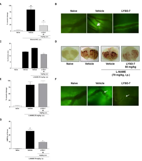
LYSO-7 reverses blood flow stasis in Et/HCl-damaged gastric tissue, mediated by NO
LYSO-7 pre-treatment prohibited vessel stasis (Figure 6 A and
B). of gastric damage induced by different agents. Here, we showNO has been proposed as protective endogenous mediator that pre-treatment with L-NAME, a non-specific inhibitor of NO
Figure 1. Effect of LYSO-7 on Et/HCl-induced gastric tissue damage. Male Swiss mice were treated with CMC (vehicle), LYSO-7 or bezafibrate, p.o., 1 hour before oral administration of Et/HCl solution. Gastric tissue was collected 1 hour after Et/HCl administration. (A) shows the percentage of the lesioned area and (B) shows representative images of the gastric tissue. Results are expressed as mean±SEM of 5 animals in each group. Statistical analysis was performed using ANOVA followed by Dunnett’s test. *P<0.01 vs. vehicle.
doi: 10.1γ71/journal.pone.0076894.g001
synthesis, enhanced gastric lesions caused by Et/HCl administration and partially inhibited the protective effect of LYSO-7 (Figure 6C and D). Furthermore, L-NAME treatment blocked the action of LYSO-7 on gastric microcirculatory blood flow (Figure 6E and F), but did not alter the reduced neutrophil influx into the gastric area caused by LYSO-7 (Figure 6 G).
LYSO-7 treatment inhibits iNOS expression and enhances eNOS protein expression in Et/HCl-damaged gastric tissue
Based on the evidence supporting a role for NO in the protective effects of LYSO-7 in Et/HCl-damaged gastric tissue, and since eNOS/iNOS imbalance is involved in gastric ulcer disease, the actions of LYSO-7 on the protein expression of NOS enzymes were investigated in gastric tissue. The data
presented in Figure 7 show that LYSO-7 inhibited iNOS and enhanced eNOS protein expression.
LYSO-7 treatment does not alter body weight
In order to evaluate a possible adverse effect of LYSO-7, changes in body weight were evaluated for β8 days. The data show that LYSO-7 treatment did not alter the cumulative weight change or food intake. In contrast, pioglitazone treatment evoked significant weight loss, which was not dependent on impaired feed intake (Figure 8).
Figure 3. Role of PPARγ receptor in the protective effect of LYSO-7 on Et/HCl-induced gastric tissue damage. Male Swiss mice were pretreated with GW996β or PBS (i.p.) and treated with CMC (vehicle) or LYSO-7 β0 min later. Et/HCl solution was administered 1 hour after the treatments. Gastric tissue was collected 1 hour later. (A) shows the percentage of the lesioned area; (B) shows representative images of the gastric tissue. Results are expressed as mean±SEM of 5 animals in each group. Statistical analysis was performed using ANOVA followed by Dunnett’s test **P<0.01 vs. vehicle.
doi: 10.1γ71/journal.pone.0076894.g00γ
Table 1. Effects of LYSO-7 and omeprazole treatment on biochemical parameters of gastric juice obtained from mice with pylorus ligation.
Treatment Dose pH [H+] mequiv./L/4h
(mg/kg)
Vehicle - γ.β6 ± 0.1β 1γ5.0 ± 10.41
Omeprazole γ0 4.β4 ± 0.19* 6β.5 ± 5.18*
LYSO-7 50 γ.γβ ± 0.17 117.5 ± 18.γγ
Discussion and Conclusions
The effectiveness of preventive and therapeutic approaches for gastric ulcers has been limited to one pathway, i.e. proton pump inhibition, and the adverse effects of drugs [4β]. Using an acute experimental model of gastric lesions, we show here that
a indole-thiazolidine molecule, a PPAR pan-agonist and COX inhibitor named LYSO-7, does not affect gastric secretion, but causes cytoprotection by inhibiting neutrophil influx into the injured area and by maintaining blood flow in the gastric microcirculatory network. The latter effect is mediated by NO, which seems to be produced by eNOS.
Figure 4. Role of neutrophils in Et/HCl-damaged gastric tissue. Male Swiss mice were pre-treated with PBS or anti-granulocyte antibody (i.p.) and blood was collected β4 or 48 hours later; Et/HCl solution was orally administered a further 48 hours later. Gastric tissue was collected 1 hour after Et/HCl delivery. (A) number of neutrophils in the blood; (B) percentage of the gastric lesion and (C) representative images of gastric tissue. Results are expressed as mean±SEM of 5 animals in each group. Statistical analysis was performed using ANOVA followed by Tukey’s test. *P<0.05 and ***P<0.001 vs. PBS treatment.
The thiazolidine-β,4-dione region of the thiazolidione molecule binds to the retinoid X receptor (RXR) coupled to PPARs to form heterodimeric complexes, which then bind to the peroxisome proliferator response element (PPRE) gene promoter, leading to the regulation of gene transcription [4γ,44]. Although LYSO-7 maintains the thiazolidine-β,4-dione group, it is an indole-substituted properly synthesized to also display inhibitory activity against COX [γγ]. In vitro studies had already shown the PPAR pan-agonist activity of LYSO-7 [γγ], and here we confirm that the activity is maintained in vivo, as levels of PPAR gene and protein expression were enhanced by LYSO-7 treatment. It is noteworthy that the expression of PPAR is a known end-point of PPAR activation [16]; however, the effect of a PPAR pan-agonist on gastric tissue has been shown here for the first time. Furthermore, we have shown that the cytoprotective effect of LYSO-7 is dependent on PPAR , as the in vivo antagonism of the receptor by GW996β abolished the inhibitory action of LYSO-7 in Et/HCl-induced ulcers. These data corroborate the notion that the isoform seems to be the main class of PPAR in gastric tissue [β7–γ1]. It is worth
mentioning that GW996β has been previously used to determine the PPAR agonistic activity of newly synthesized compounds and to clarify the mechanisms of action of PPAR [45–49].
Neutrophil influx has been observed in several models of gastric ulcers, and they have been thought to act as an inducer of the harmful process [50,51]. The participation of neutrophils in acute Et/HCl-induced gastric lesions in mice was shown here, as they rapidly accumulated in the injured tissue and in vivo neutrophil depletion significantly reduced the injured area. Together, these data corroborate the idea that inhibition of neutrophil recruitment may be a target for anti-gastric ulcer therapy [5β,5γ], and that this can be modulated by LYSO-7 treatment.
The role of PPAR activation on neutrophil influx has been shown in different models of inflammation, and the majority of them show an inhibitory effect on the process [19,β1,54]. The mechanisms involve the direct inhibition of leukocyte-endothelial interactions and chemotaxis [55,56] or impaired chemotactic mediator secretion [57–59]. Our data show, for the
Figure 5. Effects of LYSO-7 on neutrophil influx into Et/HCl-damaged gastric tissue. Male Swiss mice were treated with CMC (vehicle) or LYSO-7, p.o., 1 hour before oral administration of Et/HCl solution and gastric tissue, blood and bone marrow perfusate were collected 1 hour later. (A) MPO activity in the gastric tissue; (B) leukocyte numbers in the gastric tissue; (C) representative image of leukocyte infiltration in the gastric tissue; (D) numbers of granulocytes in the bone marrow; (E) number of circulating neutrophils. Results are expressed as mean±SEM of 5 animals in each group. Statistical analysis was performed using ANOVA followed by Tukey’s test. *P<0.05; **P<0.01 vs. vehicle; # P<0.05; # # P<0.01 vs. naïve.
first time, that a PPAR agonist affects the trafficking of neutrophils from the bone marrow, as gastric-injured mice pre-treated with LYSO-7 presented higher and lower numbers of neutrophils in the bone marrow and blood, respectively. Our previous results indicate that LYSO-7 may act directly on the locomotory functions of neutrophils. N-formyl-l-methionyl-l-leucyl-l-phenylalanine (fMLP)-induced leukocyte-endothelial interactions in the mesenteric microcirculation are impaired in
LYSO-7 treated rats, depending on reduced gene and protein expression of the CD6βL and CD18 adhesion molecules by neutrophils (Farsky et al., personal communication). The results obtained in the present study contribute to this evidence, as the inhibitory effect on neutrophil trafficking was not dependent on NO mediation. The reduced neutrophil influx into gastric lesion caused by LYSO-7 was not modified by in vivo L-NAME treatment.
Figure 6. Effects of LYSO-7 on microcirculatory blood flow in Et/HCl-damaged gastric tissue. Male Swiss mice were pre-treated with L-NAME (i.p.) or pre-treated with CMC (vehicle) or LYSO-7, p.o., 1 hour before oral administration of Et/HCl solution. Gastric tissue was surgically exposed 1 hour later after treatments for observation by intravital microscopy. (A) percentage of vessels in stasis; (B) representative images of microcirculatory vessels in the gastric tissue; (C) percentage of lesion area; (D) representative images of gastric tissue; (E) percentage of vessels in stasis; (F) representative images of microcirculatory vessels in the gastric tissue (G) MPO activity in gastric tissue. White arrows indicate vessels in stasis. Results are expressed as mean±SEM of 5 animals in each group. Statistical analysis was performed using ANOVA followed by Tukey’s test. **P<0.01 vs. vehicle; *P<0.05 vs. vehicle + L-NAME and # # P<0.01 vs. naïve.
In contrast, maintenance of the surface mucosal microcirculatory blood flow by LYSO-7 treatment occurred via NO mediation, and seemed to be dependent on reduced and increased protein expression of iNOS and eNOS, respectively. A beneficial role of NO on gastric ulcers has been shown, as the in vivo blockade of both eNOS and iNOS favors the
development of gastric lesions and treatments with NO donors heal lesions [60–6β]. Although it has been shown that enhanced activities of iNOS and eNOS in arthritic rats are responsible for the aggravation of cold-resistant stress-induced gastric lesions [61], most studies have demonstrated that a shift in the eNOS/iNOS balance protects gastric tissue
Figure 7. Effects of LYSO-7 on NOS expression in Et/HCl-damaged gastric tissue. Male Swiss mice were treated with CMC (vehicle) or LYSO-7, p.o., 1 hour before oral administration of Et/HCl solution and gastric tissue was collected 1 hour later. (A) iNOS; (B) eNOS protein expression on gastric tissue (C) representative image of gel electrophoresis. Results are expressed as mean±SEM of 4 animals in each group. Statistical analysis was performed using ANOVA followed by Tukey’s test. *P<0.05 vs. vehicle and # p<0.05 vs. naïve.
[1γ,6β,6γ]. These data are corroborated here, as LYSO-7 promoted a shift in eNOS/iNOS expressed proteins in gastric tissue. It has been shown that PPAR selective agonists favor the shift of the eNOS/iNOS balance in renal tissue after ischemia–reperfusion injury, in endothelial cells after glycation-end product activation, and during vasoconstriction in mesentery vessels [64–67]. Nevertheless, it seems that activation of the three isoforms of PPAR are involved in NO-mediated gastroprotection, as the shift in eNOS/iNOS expression and the reversion of glutathione peroxidase and reductase levels in the damaged stomach were not detected after equivalent treatment with pioglitazone, a selective PPAR agonist (data not shown). Further assays are being carried out to confirm this hypothesis.
The development of PPAR pan-agonists has been focused on the reduction of adverse effects, as they act as partial agonists of two or more isoforms of the receptor [βγ,68]. Although the data are preliminary, LYSO-7 did not modify body weight, which was reduced by equivalent treatment with pioglitazone. In addition, no alterations to biochemical and hematological parameters or on the morphology of the heart,
spleen, liver, lung and kidney were observed (data not shown). Considering that the therapeutic efficacy of PPAR agonists is a worrying challenge [68], additional toxicity and kinetics assays were performed to assure the efficacy of LYSO-7.
In summary, we have shown the in vivo actions of LYSO-7 on the mechanisms of gastric ulcer pathogenesis. We have also demonstrated that inhibition of neutrophil migration and a shift in the eNOS/iNOS balance completely abrogate gastric tissue lesions. Based on the mechanism elucidated here, LYSO-7 may be employed to treat neutrophil-mediated diseases and to reestablish damaged microcirculatory networks.
Author Contributions
Conceived and designed the experiments: SHPF JRS MNM. Performed the experiments: JRS IDM SFPR ST. Analyzed the data: JRS IDM SFPR ST MNM SLG IRP SHPF. Contributed reagents/materials/analysis tools: MNM SLG IRP SHPF. Wrote the manuscript: SHPF JRS.
References
1. Lemos M, Santin JR, Júnior LC, Niero R, Andrade SF (β011) Gastroprotective activity of hydroalcoholic extract obtained from the leaves of Brassica oleracea var. acephala DC in different animal models. J Ethnopharmacol 1γ8: 50γ-507. doi:10.1016/j.jep. β011.09.046. PubMed: β1986ββ9.
β. Sung NY, Choi KS, Park EC, Park K, Lee SY et al. (β007) Smoking, alcohol and gastric cancer risk in Korean men: the National Health
Insurance Corporation Study. Br J Cancer 97: 700-704. doi:10.10γ8/ sj.bjc.660γ89γ. PubMed: 176γ7680.
γ. Li CY, Xu HD, Zhao BT, Chang HI, Rhee HI (β008) Gastroprotective effect of cyaniding γ-glucoside on ethanol-induced gastric lesions in rats. Alcohol 4β: 68γ-687. doi:10.1016/j.alcohol.β008.08.009. PubMed: 190γ8699.
Figure 8. Effects of chronic LYSO-7 treatment on body weight and food intake. Male Swiss mice were treated with CMC (vehicle), pioglitazone or LYSO-7, p.o., once a daily for β8 days. (A) cumulative weight change (%); (B) food intake. Results are expressed as mean±SEM of 5 animals in each group. Statistical analysis was performed using ANOVA followed by Tukey’s test. *P<0.05 vs. vehicle.
4. Alvarez-Suarez JM, Dekanski D, Ristić S, Radonjić NV, Petronijević ND et al. (β011) Strawberry polyphenols attenuate ethanol-induced gastric lesions in rats by activation of antioxidant enzymes and attenuation of MDA increase. PLOS ONE 6: eβ5878. doi:10.1γ71/journal.pone. 00β5878. PubMed: ββ016781.
5. Dembinski A, Warzecha Z, Ceranowicz P, Cieszkowski J, Sendur R et al. (β011) Involvement of Cyclooxygenase-β Activity in Therapeutic Effect of Ghrelin in the Course of Ethanol-Induced Gastric Ulcers in Rats. Gastroenterology 140: S-γ15.
6. Ismail IF, Golbabapour S, Hassandarvish P, Hajrezaie M, Abdul Majid N et al. (β01β) Gastroprotective Activity of Polygonum chinense Aqueous Leaf Extract on Ethanol-Induced Hemorrhagic Mucosal Lesions in Rats. Evid Based Complement Alternat Med, β01β: β01β:40401β. PubMed: βγγ65597
7. Ning JW, Lin GB, Ji F, Xu J, Sharify N (β01β) Preventive effects of geranylgeranylacetone on rat ethanol-induced gastritis. World J Gastroenterol 18: ββ6β-ββ69. doi:10.γ748/wjg.v18.i18.ββ6β. PubMed: ββ611γβ1.
8. Guslandi M (1987) Effects of ethanol on the gastric mucosa. Dig Dis 5: β1-γβ. doi:10.1159/000171159. PubMed: γβ974γβ.
9. Szabo S, Vattay P (1990) Experimental gastric and duodenal ulcers. Gastroenterol Clin North Am 19: 67-85. PubMed: β1841γ1.
10. Medeiros JV, Gadelha GG, Lima SJ, Garcia JA, Soares PM et al. (β008) Role of the NO/cGMP/K(ATP) pathway in the protective effects of sildenafil against ethanol-induced gastric damage in rats. Br J Pharmacol 15γ: 7β1-7β7. doi:10.10γ8/sj.bjp.0707605. PubMed: 18071γ00.
11. Muscará MN, Wallace JL (1999) Nitric Oxide. V. therapeutic potential of nitric oxide donors and inhibitors. Am J Physiol β76: G1γ1γ-G1γ16. PubMed: 10γ6β6γγ.
1β. Kubes P, Suzuki M, Granger DN (1991) Nitric oxide: an endogenous modulator of leukocyte adhesion. Proc Natl Acad Sci U S A 88: 4651-4655. doi:10.107γ/pnas.88.11.4651. PubMed: 1675786. 1γ. Ma L, Wallace JL (β000) Endothelial nitric oxide synthase modulates
gastric ulcer healing in rats. Am J Physiol Gastrointest Liver Physiol β79: Gγ41-Gγ46. PubMed: 1091564γ.
14. Guha P, Dey A, Chatterjee A, Chattopadhyay S, Bandyopadhyay SK (β010) Pro-ulcer effects of resveratrol in mice with indomethacin-induced gastric ulcers are reversed by L-arginine. Br J Pharmacol 159: 7β6-7γ4. doi:10.1111/j.1476-5γ81.β009.0057β.x. PubMed: β0067468. 15. Fajas L, Debril MB, Auwerx J (β001) PPAR gamma: an essential role in
metabolic control. Nutr Metab Cardiovasc Dis 11: 64-69. PubMed: 11γ8γγβ5.
16. Lahiri S, Sen T, Palit G (β009) Involvement of glucocorticoid receptor and peroxisome proliferator activated receptor-gamma in pioglitazone mediated chronic gastric ulcer healing in rats. Eur J Pharmacol 609: 118-1β5. doi:10.1016/j.ejphar.β009.0γ.005. PubMed: 19β81808. 17. Meissner M, Hrgovic I, Doll M, Naidenow J, Reichenbach G et al.
(β010) Peroxisome proliferator-activated receptor {delta} activators induce IL-8 expression in nonstimulated endothelial cells in a transcriptional and posttranscriptional manner. J Biol Chem β85: γγ797-γγ804. doi:10.1074/jbc.M110.1γ794γ. PubMed: β059β0β9. 18. Lee JH, Kim H, Woo JH, Joe EH, Jou I (β01β): 11, β01β) β/MCP-1
expression in IFN- -stimulated astrocytes by increasing MAPK phosphatase-1 mRNA stability. J Neuroinflammation 9: 10.1186/174β-β094-9-γ4. PubMed: ββγγ9770.
19. Long EM, Klimowicz AC, Paula-Neto HA, Millen B, McCafferty DM et al. (β011) A subclass of acylated anti-inflammatory mediators usurp Toll-like receptor β to inhibit neutrophil recruitment through peroxisome proliferator-activated receptor gamma. Proc Natl Acad Sci U S A 108: 16γ57-16γ6β. doi:10.107γ/pnas.110070β108. PubMed: β19γ0915. β0. Yang X, Kume S, Tanaka Y, Isshiki K, Araki S et al. (β011) GW501516,
a PPARδ agonist, ameliorates tubule interstitial inflammation in protein uric kidney disease via inhibition of TAK1-NFκB pathway in mice. PLOS ONE 6: eβ5β71. doi:10.1γ71/journal.pone.00β5β71. PubMed: β1966476.
β1. Farnesi-de-Assunção TS, Alves CF, Carregaro V, de Oliveira JR, da Silva CA et al. (β01β) PPAR- agonists, mainly 15d-PGJ(β), reduce eosinophil recruitment following allergen challenge. Cell Immunol β7γ: βγ-β9. doi:10.1016/j.cellimm.β011.11.010. PubMed: ββ19β475. ββ. Wu L, Yan C, Czader M, Foreman O, Blum JS et al. (β01β) Inhibition of
PPAR in myeloid-lineage cells induces systemic inflammation, immunosuppression, and tumorigenesis. Blood 119: 115-1β6. doi: 10.118β/blood-β011-06-γ6γ09γ. PubMed: ββ05γ106.
βγ. Feldman PL, Lambert MH, Henke BR (β008) PPAR modulators and PPAR pan agonists for metabolic diseases: the next generation of drugs targeting peroxisome proliferator-activated receptors? Curr Top Med Chem 8: 7β8–749. doi:10.β174/15680β6087845γ5084. PubMed: 185γ7685.
β4. Zhang LS, Wang SQ, Xu WR, Wang RL, Wang JF (β01β) Scaffold-based pan-agonist design for the PPARα, PPAR and PPAR receptors. PLOS ONE 7: e4845γ. doi:10.1γ71/journal.pone.004845γ. PubMed: βγ1190β4.
β5. Pancione M, Forte N, Sabatino L, Tomaselli E, Parente D et al. (β009) Reduced beta-catenin and peroxisome proliferator-activated receptor-gamma expression levels are associated with colorectal cancer metastatic progression: correlation with tumor-associated macrophages, cyclooxygenase β, and patient outcome. Hum Pathol 40: 714-7β5. doi:10.1016/j.humpath.β008.08.019. PubMed: 191β1846. β6. Meyer S, Vogt T, Landthaler M, Berand A, Reichle A et al. (β009)
Cyclooxygenase β (COXβ) and Peroxisome Proliferator-Activated Receptor gamma (PPARG) Are Stage-Dependent Prognostic Markers of Malignant Melanoma. PPAR Res, β009: β009:848645. PubMed: 196γ90γβ
β7. Konturek PC, Brzozowski T, Kania J, Konturek SJ, Kwiecien S et al. (β00γ) Pioglitazone, a specific ligand of peroxisome proliferator-activated receptor-gamma, accelerates gastric ulcer healing in rat. Eur J Pharmacol 47β: β1γ-ββ0. doi:10.1016/S0014-β999(0γ)019γβ-0. PubMed: 1β871756.
β8. Takagi T, Naito Y, Ichikawa H, Tomatsuri N, Katada K et al. (β004) A PPAR-gamma ligand, 15-deoxy-Delta1β, 14-prostaglandin. J Volumes β, inhibited gastric mucosal injury induced by ischemia-reperfusion in rats. Redox Rep 9:γ76-981
β9. Villegas I, Martín AR, Toma W, de la Lastra CA (β004) Rosiglitazone, an agonist of peroxisome proliferator-activated receptor gamma, protects against gastric ischemia-reperfusion damage in rats: role of oxygen free radicals generation. Eur J Pharmacol 505: 195-β0γ. doi: 10.1016/j.ejphar.β004.10.0β0. PubMed: 1555615γ.
γ0. Brzozowski T, Konturek PC, Pajdo R, Kwiecień SN, Konturek S et al (β005) Agonist of peroxisome proliferator-activated receptor gamma (PPAR-gamma): a new compound with potent gastroprotective and ulcer healing properties. Inflammopharmacology 1γ: γ17-γγ0. doi: 10.116γ/1568560057744βγ908. PubMed: 16β59750.
γ1. Konturek PC, Brzozowski T, Burnat G, Szlachcic A, Koziel J et al. (β010) Gastric ulcer healing and stress-lesion preventive properties of pioglitazone are attenuated in diabetic rats. J Physiol Pharmacol 61: 4β9-4γ6. PubMed: β0814070.
γβ. Morsy MA, Ashour OM, Fouad AA, Abdel-Gaber SA (β010) Gastroprotective effects of the insulin sensitizers rosiglitazone and metformin against indomethacin-induced gastric ulcers in Type β diabetic rats. Clin Exp Pharmacol Physiol γ7: 17γ-177. doi:10.1111/j. 1440-1681.β009.05β50.x. PubMed: 195668β1.
γγ. Santin JR, Uchôa FD, Lima MD, Rabello MM, Machado ID et al. (β01γ) Chemical synthesis, docking studies and biological effects of a pan peroxisome proliferator-activated receptor agonist and cyclooxygenase inhibitor. Eur J Pharm Sci 48: 689-697. doi:10.1016/j.ejps.β01β.1β.0β9. PubMed: βγγ0599γ.
γ4. Mizui T, Doteuchi M (198γ) Effect of polyamines on acidified ethanol-induced gastric lesion in rats. Jpn J Pharmacol γγ: 9γ9–945. doi: 10.1β54/jjp.γγ.9γ9. PubMed: 6580476.
γ5. Goso Y, Ueno M, Hotta K, Ishihara K (β007) Protective effects of the whisky congeners on ethanol-induced gastric mucosal damage. Alcohol Clin Exp Res γ1: γ90-γ94. doi:10.1111/j.15γ0-0β77.β006.00γ19.x. PubMed: 17β957ββ.
γ6. Lakshmi V, Singh N, Shrivastva S, Mishra SK, Dharmani P et al. (β010) Gedunin and photogedunin of Xylocarpus granatum show significant anti-secretory effects and protect the gastric mucosa of peptic ulcer in rats. Phytomedicine 17: 569–574. doi:10.1016/j.phymed.β009.10.016. PubMed: 1996ββ86.
γ7. Shay H, Komarov SA, Fels SS, Meranze D, Gruenstein M et al. (1945) A simple method for the uniform production of gastric ulceration in the rat. Gastroenterology 5: 4γ–61.
γ8. Bradley PP, Priebat DA, Christensen RD, Rothstein G (198β) Measurement of cutaneous inflammation: estimation of neutrophil content with an enzyme marker. J Invest Dermatol 78: β06-β09. doi: 10.1111/15βγ-1747.ep1β50646β. PubMed: 6β76474.
γ9. Conlan JW, North RJ (1994) Neutrophils are essential for early anti-Listeria defense in the liver, but not in the spleen or peritoneal cavity, as revealed by agranulocyte-depleting monoclonal antibody. J Exp Med 179: β59-β68. doi:10.1084/jem.179.1.β59. PubMed: 8β70870. 40. Daley JM, Thomay AA, Connolly MD, Reichner JS, Albina JE (β008)
Use of Ly6G-specific monoclonal antibody to deplete neutrophils in mice. J Leukoc Biol 8γ: 64-70. PubMed: 1788499γ.
4β. Krznaric Z, Ljubas Kelecic D, Rustemovic N, Vranesic Bender D, Ostojic R et al. (β011) Pharmaceutical principles of acid inhibitors: unmet needs. Dig Dis β9: 469-475. doi:10.1159/000γγ1515. PubMed: ββ09501β.
4γ. Van Beekum O, Fleskens V, Kalkhoven E (β009) Posttranslational modifications of PPAR-gamma: fine-tuning the metabolic master regulator. Obesity 17: β1γ-β19. doi:10.10γ8/oby.β008.47γ. PubMed: 19169ββ1.
44. Dreijerink KM, Varier RA, Van Beekum O, Jeninga EH, Höppener JW et al. (β009) The multiple endocrine neoplasia type 1(MEN1) tumor suppressor regulates peroxisome proliferator-activated receptor gamma-dependent adipocyte differentiation. Mol Cell Biol β9: 5060-5069. doi:10.11β8/MCB.01001-08. PubMed: 1959678γ. 45. Naito Y, Yoshikawa T (β010) PPARgamma as a new therapeutic target
in inflammatory diseases. Nihon Rinsho 68: γ41-γ49. PubMed: β0158107.
46. Takagi T, Naito Y, Ichikawa H, Tomatsuri N, Katada K et al. (β004) A PPAR-gamma ligand, 15-deoxy-Delta1β, 14-prostaglandin. J Volumes β, inhibited gastric mucosal injury induced by ischemia-reperfusion in rats. Redox Rep 9:γ76-981
47. Reddy RC, Narala VR, Keshamouni VG, Milam JE, Newstead MW et al. (β008) Sepsis-induced inhibition of neutrophil chemotaxis is mediated by activation of peroxisome proliferator-activated receptor-{gamma}. Blood 11β: 4β50-4β58. doi:10.118β/blood-β007-1β-1β8967. PubMed: 185γ5β0γ.
48. Lim HA, Lee EK, Kim JM, Park MH, Kim DH et al. (β01β) PPAR activation by baicalin suppresses NF-κB-mediated inflammation in aged rat kidney. Biogerontology 1γ: 1γγ-145. doi:10.1007/ s105ββ-011-9γ61-4. PubMed: ββ0γγ706.
49. Martin HL, Mounsey RB, Mustafa S, Sathe K, Teismann P (β01β) Pharmacological manipulation of peroxisome proliferator-activated receptor (PPAR ) reveals a role for anti-oxidant protection in a model of Parkinson’s disease. Exp Neurol βγ5: 5β8-5γ8. doi:10.1016/ j.expneurol.β01β.0β.017. PubMed: ββ4179β4.
50. Wallace JL, McKnight GW, Bell CJ (1995) Adaptation of rat gastric mucosa to aspirin requires mucosal contact. Am J Physiol β68: G1γ4-G1γ8. PubMed: 7840196.
51. Beserra AM, Calegari PI, Souza Mdo C, Dos Santos RA, Lima JC et al. (β011) Gastroprotective and ulcer-healing mechanisms of ellagic acid in experimental rats. J Agric Food Chem 59: 6957-6965. doi:10.10β1/ jfβ00γβ67. PubMed: β1644797.
5β. Rocha NF, Oliveira GV, Araújo FY, Rios ER, Carvalho AM et al. (β011) (-)-α-Bisabolol-induced gastroprotection is associated with reduction in lipid peroxidation, superoxide dismutase activity and neutrophil migration. Eur J Pharm Sci 44: 455-461. doi:10.1016/j.ejps. β011.08.0β0. PubMed: β19β4γ5γ.
5γ. Bayir Y, Odabasoglu F, Cakir A, Aslan A, Suleyman H et al. (β006) The inhibition of gastric mucosal lesion, oxidative stress and neutrophil-infiltration in rats by the lichen constituent diffractaic acid. Phytomedicine 1γ: 584-590. doi:10.1016/j.phymed.β005.07.00β. PubMed: 169β0514.
54. Celinski K, Dworzanski T, Korolczuk A, Piasecki R, Slomka M et al. (β011) Effects of peroxisome proliferator-activated receptors-gamma ligands on dextran sodium sulphate-induced colitis in rats. J Physiol Pharmacol 6β: γ47-γ56. PubMed: β189γ696.
55. Liang YJ, Jian JH, Liu YC, Juang SJ, Shyu KG et al. (β010) Advanced glycation end products-induced apoptosis attenuated by PPAR delta activation and epigallocatechin gallate through NF-kappaB pathway in
human embryonic kidney cells and human mesangial cells. Diabetes/ Metab Res Rev β6: 406-416. doi:10.100β/dmrr.1100.
56. Imamoto E, Yoshida N, Uchiyama K, Kuroda M, Kokura S et al. (β004) Inhibitory effect of pioglitazone on expression of adhesion molecules on neutrophils and endothelial cells. Biofactors β0: γ7-47. doi:10.100β/biof. 55β0β00104. PubMed: 15096659.
57. Reilly CM, Oates JC, Cook JA, Morrow JD, Halushka PV et al. (β000) Inhibition of mesangial cell nitric oxide in MRL/lpr mice by prostaglandin Jβ and proliferator activation receptor-gamma agonists. J Immunol 164: 1498-1504. PubMed: 10640767.
58. Ricote M, Li AC, Willson TM, Kelly CJ, Glass CK (1998) The peroxisome proliferator-activated receptor-gamma is a negative regulator of macrophage activation. Nature γ91: 79-8β. doi: 10.10γ8/γ4178. PubMed: 94ββ508.
59. Wang AC, Dai X, Luu B, Conrad DJ (β001) Peroxisome proliferator-activated receptor-gamma regulates airway epithelial cell activation. Am J Respir Cell Mol Biol β4: 688-69γ. doi:10.1165/ajrcmb.β4.6.4γ76. PubMed: 114159γγ.
60. El-Demerdash E, El-Mesallamy HO, Abu-Zaid NM, Gad MZ (β010) The potential therapeutic effect of nitric oxide modulators in experimentally-induced gastric ulcers. Drugs Discov Ther 4: β76-β84. PubMed: ββ491β10.
61. Kato S, Ohkawa F, Ito Y, Amagase K, Takeuchi K (β009) Role of endothelial nitric oxide synthase in aggravation of indomethacin-induced gastric damage in adjuvant arthritic rats. J Physiol Pharmacol 60: 147-155. PubMed: β0065509.
6β. Guo JS, Cho CH, Wang JY, Koo MW (β006) Differential effects of selective and non-selective inhibition of nitric oxide synthase on the expression and activity of cyclooxygenase-β during gastric ulcer healing. Eur J Pharmacol 5γ6: γ01-γ08. doi:10.1016/j.ejphar. β005.1β.088. PubMed: 16600β10.
6γ. Guha P, Dey A, Sarkar B, Dhyani MV, Chattopadhyay S et al. (β009) Improved antiulcer and anticancer properties of a trans-resveratrol analog in mice. J Pharmacol Exp Ther γβ8: 8β9-8γ8. doi:10.11β4/jpet. 108.145γγ4. PubMed: 19066γ40.
64. Liang C, Ren Y, Tan H, He Z, Jiang Q et al. (β009) Rosiglitazone via up regulation of Akt/eNOS pathways attenuates dysfunction of endothelial progenitor cells, induced by advanced glycation end products. Br J Pharmacol 158: 1865-187γ. doi:10.1111/j.1476-5γ81.β009.00450.x. PubMed: 19917066.
65. Yuen CY, Wong WT, Tian XY, Wong SL, Lau CW et al. (β011) Telmisartan inhibits vasoconstriction via PPAR -dependent expression and activation of endothelial nitric oxide synthase. Cardiovasc Res 90: 1ββ-1β9. doi:10.109γ/cvr/cvqγ9β. PubMed: β11568β5.
66. Sadaghiani MS, Javadi-Paydar M, Gharedaghi MH, Fard YY, Dehpour AR (β011) Antidepressant-like effect of pioglitazone in the forced swimming test in mice: the role of PPAR-gamma receptor and nitric oxide pathway. Behav Brain Res ββ4: γγ6-γ4γ. doi:10.1016/j.bbr. β011.06.011. PubMed: β1704657.
67. Betz B, Schneider R, Kress T, Schick MA, Wanner C et al. (β01β) Rosiglitazone affects nitric oxide synthases and improves renal outcome in a rat model of severe ischemia/reperfusion injury. PPAR Res: β01β:β19γ19