Arteries from Patients with End-Stage Renal Disease
Leanid Luksha1, Peter Stenvinkel2, Folke Hammarqvist3, Juan Jesu´s Carrero2, Sandra T. Davidge4, Karolina Kublickiene1*
1Division of Obstetrics & Gynecology, Karolinska Institutet, Karolinska University Hospital, Department of Clinical Science, Intervention & Technology, Stockholm, Sweden, 2Division of Renal Medicine, Karolinska Institutet, Karolinska University Hospital, Department of Clinical Science, Intervention & Technology, Stockholm, Sweden, 3Division of Surgery, Karolinska Institutet, Karolinska University Hospital, Department of Clinical Science, Intervention & Technology, Stockholm, Sweden,4Department of Obstetrics and Gynecology, University of Alberta, Edmonton, Alberta, Canada
Abstract
The study focuses on the mechanisms of endothelial dysfunction in the uremic milieu. Subcutaneous resistance arteries from 35 end-stage renal disease (ESRD) patients and 28 matched controls were studied ex-vivo. Basal and
receptor-dependent effects of endothelium-derived factors, expression of endothelial NO synthase (eNOS), prerequisites for myoendothelial gap junctions (MEGJ), and associations between endothelium-dependent responses and plasma levels of endothelial dysfunction markers were assessed. The contribution of endothelium-derived hyperpolarizing factor (EDHF) to endothelium-dependent relaxation was impaired in uremic arteries after stimulation with bradykinin, but not acetylcholine, reflecting the agonist-specific differences. Diminished vasodilator influences of the endothelium on basal tone and enhanced plasma levels of asymmetrical dimethyl L-arginine (ADMA) suggest impairment in NO-mediated regulation of uremic arteries. eNOS expression and contribution of MEGJs to EDHF type responses were unaltered. Plasma levels of ADMA were negatively associated with endothelium-dependent responses in uremic arteries. Preserved responses of smooth muscle to pinacidil and NO-donor indicate alterations within the endothelium and tolerance of vasodilator mechanisms to the uremic retention products at the level of smooth muscle. We conclude that both EDHF and NO pathways that control resistance artery tone are impaired in the uremic milieu. For the first time, we validate the alterations in EDHF type responses linked to kinin receptors in ESRD patients. The association between plasma ADMA concentrations and endothelial function in uremic resistance vasculature may have diagnostic and future therapeutic implications.
Citation:Luksha L, Stenvinkel P, Hammarqvist F, Carrero JJ, Davidge ST, et al. (2012) Mechanisms of Endothelial Dysfunction in Resistance Arteries from Patients with End-Stage Renal Disease. PLoS ONE 7(4): e36056. doi:10.1371/journal.pone.0036056
Editor:Marcelo G. Bonini, University of Illinois at Chicago, United States of America
ReceivedSeptember 27, 2011;AcceptedMarch 29, 2012;PublishedApril 26, 2012
Copyright:ß2012 Luksha et al. This is an open-access article distributed under the terms of the Creative Commons Attribution License, which permits unrestricted use, distribution, and reproduction in any medium, provided the original author and source are credited.
Funding:Financial support was provided by grants from the Swedish Society of Medicine, Swedish Heart and Lung Foundation, Center for Gender Medicine at Karolinska Institutet, Loo and Hans Ostermans Foundation, the Swedish Kidney Association and the Karolinska Institute Research Funds. L. Luksha is supported by PostDoc research grants from AFA insurance and Department of Clinical Science, Intervention & Technology (CLINTEC) at Karolinska Institutet, Sweden. S. Davidge is supported by Canadian Institute for Health Research. The funders had no role in study design, data collection and analysis, decision to publish, or preparation of the manuscript.
Competing Interests:The authors have read the journal’s policy and have the following conflicts: The study was partly funded by AFA Insurance. This does not alter the authors’ adherence to all the PLoS ONE policies on sharing data and materials, as detailed online in the guide for authors.
* E-mail: [email protected]
Introduction
Adverse cardiovascular events are common complications of end-stage renal disease (ESRD) and these patients are more likely to die from cardiovascular disease (CVD) than from kidney failure [1]. Although the underlying mechanisms that predispose ESRD patients to higher risk of CVD are incompletely understood, morphological and functional abnormalities of the endothelium may play an important role [2]. Endothelial dysfunction is considered an early marker of CVD [3], which facilitates the progress of atherosclerosis [4] and contributes to the development of hypertension through the enhancement of vascular resistance [5]. Thus, studies aimed to at investigating the mechanisms of endothelial dysfunction in ESRD are of importance and may provide a means to ameliorate cardiovascular complications and introduce novel treatment strategies.
Our current knowledge of endothelial dysfunction in ESRD is mainly based on findings from animal models [6], circulating plasma markers [7] andin-vivoassessments in the human forearm
to impairment in production and/or bioavailability of NO, but EDHF type responses may also be affected by uremic milieu. To test this hypothesis we isolated arteries from subcutaneous fat to segregate pharmacologically the relative impairments in NO and EDHF type responses that confer endothelial dysfunction in ESRD. Targeted pathways of endothelial dysfunction were assessed using basal and receptor-dependent stimulation of endothelium-derived vasodilators, expression of endothelial NO synthase (eNOS), prerequisites for MEGJ, and associations between endothelium-dependent responses and plasma levels of endothelial dysfunction surrogate markers.
Results
Participants
Age, gender, and smoking status were similar between the groups. The body mass index was lower in ESRD patients vs.
controls. Plasma levels of asymmetrical dimethyl L-arginine (ADMA), soluble vascular cell adhesion molecule-1 (sVCAM-1), interleukin-6, pentraxin-3, high sensitivity C-reactive protein (hsCRP) and lipoprotein(a) and triglycerides were elevated in ESRD. No differences in blood pressure or total cholesterol were observed between the groups (Table 1).
Vascular function
In total, 84 subcutaneous arteries with internal diameter of 20966mm were dissected from 35 ESRD patients and 71 arteries
with internal diameter of 22467mm were dissected from 28 controls (P.0.05). There was no difference in the magnitude of pre-constriction 3mmol/L norepinephrine between the groups
(2.560.2 mN/mm2ESRDvs.2.760.2 mN/mm2controls). Endothelium-dependent relaxation
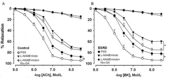
In controls, ACh and bradykinin (BK) caused relaxation of arteries with similar magnitude (Figure 1). However, arteries were more sensitive to BK vs.ACh (pEC50, here and in the following text: BK 7.960.1vs.ACh 7.760.1,P= 0.01). NOS/cyclooxygen-ase (COX) inhibition reduced endothelium-dependent relaxation (Figure 1) and the sensitivity to agonists became similar (ACh: 7.260.1vs.BK: 7.360.1,P= 0.5).
Relaxation and sensitivity to both agonists was attenuated in ESRD vs. controls (Figure 1). In contrast to the controls, the sensitivities of ESRD arteries in PSS were similar between the agonists (ACh: 7.360.1 vs. BK: 7.460.1,P= 0.2).
After NOS/COX inhibition relaxation and sensitivity to ACh and BK were attenuated in ESRD (Figure 1). The concentration-response curves after NOS/COX inhibition were shifted to the right in ESRD vs. controls (Figure 1). However, the maximal
EDHF type relaxation was reduced in ESRD vs. controls in response to BK but not to ACh (BK:P= 0.003; ACh:P= 0.09). Moreover, the relative contribution of EDHF was reduced in ESRDvs.controls in response to BK but not to ACh (Figure 2).
An inhibitor of gap junctions (18-a-glycyrrhetinic acid, 18-aGA) markedly reduced EDHF type relaxation in response to both agonists (Figure 1). There was no difference in residual relaxation after incubation with 18-aGA along with NOS inhibitor,Nv -nitro-L-arginine-methyl ester (L-NAME) and COX inhibitor, indo-methacin (Indo) between ESRD vs. controls (Figure 1). The relative contribution of MEGJs to EDHF type responses was similar between ESRD and controls independently of the agonist used (ACh, 1mmol/l: 8664 ESRD (n = 16) vs. 8066 controls (n = 12),P= 0.7; BK, 1mmol/l: 8565 ESRD (n = 13) vs. 8763 controls (n = 8),P= 0.9).
In order to eliminate the possible interference of co-morbidities, the responses to agonists before and after NOS/COX inhibition in ESRD patients without diabetes mellitus (DM) and CVD were compared with those of controls. In response to BK we observed similar results as above (Figure 1B). In contrast to the whole ESRD group, in arteries from ESRD without DM and CVD, ACh-induced relaxation was reduced in PSS as compared to controls but similar after NOS/COX inhibition (ACh after NOS/COX inhibition: 760.1 ESRD without DM and CVD (n = 18) vs.
7.260.1 controls (n = 23),P= 0.1).
S-albumin (g/L) 34.963.4* 38.463.4
S-Creatinine (mmol/L) 620 (249–1069)* 78 (55–100)
Interleukin-6 (pg/ml) 5.0 (1.9–16.8)* 1.5 (0.4–17.1)
hsC-reactive protein (mg/L) 1.9 (0.2–24.9)* 1.4 (0.4–13.9)
Pentraxin-3 (ng/ml) 1.2 (0.5–8.3)* 0.6 (0.1–2.3)
Fibrinogen (g/L) 4.861.3* 3.261.3
Glomerular filtration rate (ml/min) 1263* 8963
Asymmetric Dimethylarginine (mmol/L) 0.660.1* 0.560.1
Soluble VCAM-1 (ng/ml) 1295 (637–1980)* 588 (368–830)
Soluble ICAM-1 (ng/ml) 223 (138–404) 231 (161–363)
Diabetes mellitus n, (%) 10 (29) 0
CVD, n, (%) 13 (37) 0
Antihypertensive treatment, n, (%) 33 (94) 0
Statin treatment, n, (%) 15 (43) 0
*,P,0.05.
Transmission electron microscopy (TEM)
Analysis of TEM images focused on morphological prerequisites for the gap junctions between EC and smooth muscle cells (SMC) in arteries from ESRD patients and controls (n = 3). The main criteria for identification of MEGJs was the presence of the characteristic pentalaminar membrane structure at points of cell to cell contact, where the central region had a higher electron opacity than the inner parts and distance between the EC and SMC plasma membranes was around 3.5 nm [20]. TEM images showed the presence of long protrusions (up to 4.5mm) from both ECs
(Figure 3A) and SMCs (Figure 3B) penetrating the internal elastic lamina and forming close contacts with each other (Figure 3C and 3D). Although the observed EC-SMC contacts did not fulfill all criteria for the characteristic pentalaminar structures, they could be considered as prerequisites for MEGJs.
Endothelium-independent relaxation
Endothelium-independent relaxation to NO donor sodium nitroprusside (SNP) was similar between the groups (pEC50:
6.060.1 ESRD (n = 17)vs.6.260.2 controls (n = 13),P= 0.6). In ESRD patients pinacidil-induced responses were blunted as compared to controls (6.160.1 ESRD vs 5.760.1 controls,
P= 0.01, Figure 4). NOS/COX inhibition attenuated the response in controls but not in ESRD (6.160.1 PSS vs 5.860.1 L-Figure 1. Concentration response curves to acetylcholine (ACh, A) and bradykinin (BK, B).Responses in physiological salt solution (PSS) and after incubation withNv
-nitro-L-arginine methyl ester plus indomethacin alone (L-NAME+Indo) or together with 18a-glycyrrhetinic acid (L-NAME+Indo+18a-GA) in arteries from ESRD patients (n = 32 for ACh and n = 22 for BK) and controls (n = 23 for ACh and n = 17 for BK). * ESRDvs. controls,P,0.05.
doi:10.1371/journal.pone.0036056.g001
Figure 2. The relative contribution of endothelium-derived hyperpolarizing factor (EDHF). Contribution of EDHF in arteries from ESRD patients and controls in response to acetylcholine (ACh) and bradykinin (BK). * ESRDvs.controls,P,0.05.
doi:10.1371/journal.pone.0036056.g002
Figure 3. Transmission electron images of arteries from ESRD patients.The lower magnification pictures (A,B) show an overview of the vascular wall with endothelium (End) and smooth muscle (SM) being separated by the internal elastic lamina (IEL). The areas denoted by the boxes are magnified and show the sites of intercellular contacts that could be considered as prerequisites of myoendothelial gap junctions (C, D). The width of the gap is,20 nm (C, arrow),,11 nm (D, arrow). Bar: (A) 2mm; (B) 3mm; (C) 0.1mm; (D) 0,2mm.
Name+Indo, P= 0.03 controls; 5.760.1 vs 5.660.1, P= 0.3 ESRD, respectively, Figure 4). There was no difference between ESRD and controls in their responses to pinacidil after NOS/ COX inhibition (Figure 4).
Influence of the endothelium-derived factors on basal tone and expression of eNOS
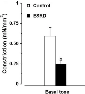
NOS/COX inhibitors induced constriction of arteries from both ESRD and control groups. This constriction was reduced in ESRDvs.controls (Figure 5). Exclusion of the patients with DM and CVD from ESRD group did not change the outcome (Figure 5; 0.2860.1 mN/mm2 ESRD without DM and CVD (n = 17)vs.0.5960.1 mN/mm2controls (n = 26),P= 0.02). There was no difference in eNOS expression in ESRD vs. controls (Figure 6).
Associations between endothelium-dependent responses and plasma markers of endothelial dysfunction
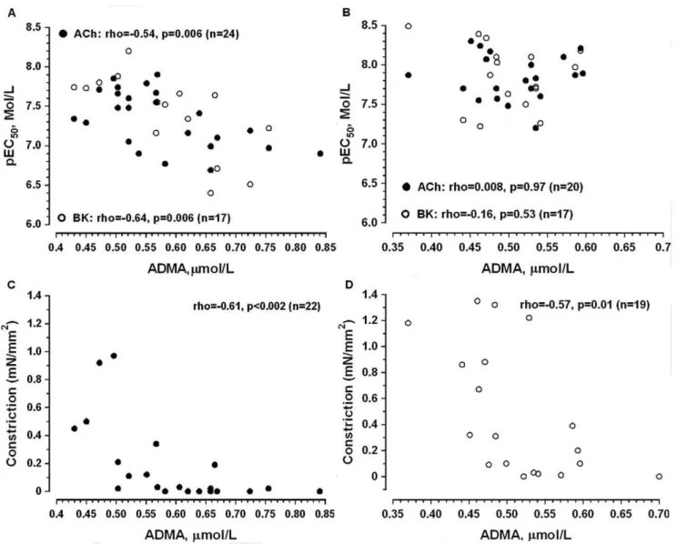
The sensitivity of arteries to ACh and BK was negatively associated with plasma levels of ADMA in ESRD (Figure 7A) but not in controls (Figure 7B). There was no association between ADMA and vascular sensitivity to the endothelium-dependent agonists after NOS/COX inhibition in both groups. Similarly, sensitivity to SNP was not associated with ADMA levels (data not shown).
In contrast to the agonists-induced relaxation, constriction in response to NOS/COX inhibition was negatively associated with plasma levels of ADMA in both ESRD and controls (Figure 7C and 7D).
No relation was found between resistance artery function and other surrogate markers of endothelial dysfunction (soluble intercellular adhesion molecule-1 (sICAM-1) and sVCAM-1) in ESRD and controls (data not shown).
Discussion
In this ex-vivo study of human uremic resistance arteries we describe, for the first time, the relative role of endothelium-derived factors, agonist-specific differences and associations between endothelial function and surrogate plasma markers of endothelial dysfunction. We show that reduced EDHF type responses contribute markedly to endothelial dysfunction in ESRD. Impaired EDHF type responses in ESRD were detected with endothelium-dependent agonist BK but not ACh. Thus, we suggest that changes in signal transduction from endothelial receptors towards generation and/or transformation of hyperpo-larization to the smooth muscle are differently affected by uremic toxins with a predominant impact on those mediated by kinin receptors. Diminished vasodilator influence of the endothelium on basal tone of SMCs along with enhanced plasma levels of ADMA indicates an impairment in NO-mediated control of arterial tone in ESRD. While the eNOS expression and the contribution of Figure 4. Concentration response curves to pinacidil.Responses
in arteries from ESRD patients (n = 10) and controls (n = 7) in physiological salt solution (PSS) and after incubation withNv
-nitro-L
-arginine methyl ester plus indomethacin (L-NAME+Indo). * ESRD vs. controls, P,0.05; # before vs. after incubation with L-NAME+Indo, P,0.05.
doi:10.1371/journal.pone.0036056.g004
Figure 5. Contractile response to NOS/COX inhibitors of arteries from controls (n = 26)vs.ESRD patients with (n = 32). *,P,0.05 ESRDvs.controls.
doi:10.1371/journal.pone.0036056.g005
MEGJs to EDHF type responses appeared to be unaltered in uremic arteries, the upstream machinery of both endothelial pathways (i.e. NO and EDHF) were impaired. Since relaxation in response to NO donor or hyperpolarizing agent pinacidil (after NOS/COX inhibition) were similar between the groups, we confirm that the endothelium is the main target of uremic environment, whereas functional capacity of the vascular smooth muscle appeared to be rather tolerant. In accordance with our previous study [11] we corroborate a central role of the uremic milieu in the genesis of endothelial dysfunction. The present study also shows that among measured plasma markers of endothelial dysfunction only ADMA was strongly associated with the magnitude of endothelial dysfunction in uremic resistance arteries. Thus, our findings provide novel insights into the mechanisms of endothelial dysfunction in resistance circulation of ESRD patients. The pattern of impairment of EDHF type responses after BK but not ACh stimulation in uremic arteries emphasizes the agonist-specific mechanisms of endothelial dysfunction in this toxic milieu. We speculate that conventional and/or disease-specific risk factors may differently affect kinin and muscarinic receptors and/or their
regulatory pathways. For example, endothelial dysfunction in atherosclerosis appears to be receptor-specific, involving the muscarinic receptors with relative sparing of the kinin receptor pathways. Abnormal reactivity of epicardial coronary arteries during physiologic stress is better represented by BK and not by ACh responses [21]. Moreover, differences exist between BK- and ACh- induced relaxation of the mesenteric arteries from spontaneously hypertensive rats at different ages, suggesting a more detrimental effect of increased blood pressure on BK-induced vasorelaxation [22], while selective impairement of endothelium-dependent relaxation to ACh but not BK is observed in isolated small omental arteries from women with preeclampsia [23]. Thus, prior conclusions based only on the vascular effects of one agonist (i.e. ACh) should be considered with caution. In contrast to previous studies, in which only ACh was tested [9–10], our more comprehensive analysis of pathways involved in endothelial dysfunction of resistance arteries in ESRD patients revealed an impairment of EDHF contribution coupled with stimulation of kinin receptors.
Figure 7. Spearman rank correlation between plasma levels of asymmetrical dimethyl L-arginine (ADMA, mmol/L) and artery sensitivity to endothelium-dependent vasodilators (pEC50, A, B) or vasoconstriction in response to NOS/COX inhibition
alterations of BK-induced EDHF type responses in this patient group.
Despite the fact that EDHF normally does not act through the KATP-channels [13], pinacidil-induced responses allowed us to assess the general mechanism of relaxation induced by hyperpo-larization due to outward K+
currents at the level of the smooth muscle [26]. Moreover, an animal study of renal failure suggested that alterations in smooth muscle K+
-channels could be involved in reduced endothelium-dependent hyperpolarization [16]. In our study, relaxation to pinacidil was reduced in ESRDvs.controls but this difference disappeared after NOS/COX inhibition, which opposes the findings in the animal study [16]. Most likely basal NO had a potentiating effect on pinacidil-induced relaxation in controls but not in ESRD patients. While in general relaxation induced by KATP-channels openers has yet been considered endothelium-independent [27], the potentiating effects of endo-thelium-derived factors has been reported before [25,28].
Impaired endothelial influence on pinacidil-induced responses may further support our data about reduced basal release of endothelium-derived factors in ESRD. Indeed, NOS/COX inhibitors induced smaller constriction in uremic vs. control arteries. As basal vascular tone is to a large extend NO-dependent [29], our data implies a reduction in basal production of NO in ESRD. In contrast, a previous study reported increased basal NO production in the forearm of hemodialysis patients [30]. The inconsistent results may be caused by different methodology, and selection of patients. Recently, we demonstrated the lack of NO contribution to shear stress responses in subcutaneous uremic arteries [11]. In the current study, differences in sensitivity between BK and ACh, depending from NOS/COX inhibition in controls but not in ESRD, indicated on distinct NO contribution to agonist-induced relaxation between the two groups. Moreover, the negative correlation between serum ADMA levels and relaxation to ACh and BK in ESRD but not in controls further supports the impaired contribution of NO to agonists-induced responses in uremia.
Multiple mechanisms may lead to NO deficiency in renal failure [12,31]. A decreased bioavailability of NO due to increased pro-oxidative environment has been suggested [11]. Reduced expression of eNOS has been linked to a decreased NO production in an animal model of kidney failure [32]. Studies on EC cultures have shown that erythrocytes [33] or sera fractions enriched with advanced glycation end products [34] from uremic patients may directly affect expression and activity of eNOS. However, we failed to find any difference in eNOS protein expression between uremic and control arteries. Moreover, an elevated vascular expression but unchanged activity of eNOS was demonstrated in radial arteries of ESRD patients [35].
Neverthe-ADMA on endothelial superoxide generation may be due to the activation of other enzymatic sources of superoxide radicals such as NADPH oxidases. ADMA may also stimulate renin angiotensin system, and ADMA induced impairement of NO-mediated function due to increased superoxide production has been shown to occur via activation of of Ang II-NADPH oxidase pathway in isolated small vessels from rats [38]. In animal models of experimental diabetic nephropathy, AngII induced activation of NADPH oxidase and eNOS uncoupling serves as the major source of superoxide, and the blockade of AngII signaling ameliorates eNOS uncoupling by increased tetrahydrobiopterin levels with following restoration of NO bioavailability and improved glomerular hemodynamics [39–40].
Although, an association between elevated ADMA levels and endothelium-dependent dilatation in the forearm was previously reported [41], we are the first to show an association between ADMA and endothelium-dependent relaxation in uremic resis-tance vasculature. The inverse correlation between ADMA and changes in basal tone after NOS/COX inhibition in both experimental groups support in-vivo results demonstrating that ADMA increases vascular resistance in ESRD patients [42] and in healthy humans [43]. Taken together our data endorse the proposal that elevated ADMA may act as a potential mechanism behind the impaired NO-dependent control of uremic resistance artery tone.
On the other hand, adhesion molecules sICAM-1 and sVCAM-1, two purported biomarkers of endothelial dysfunction, did not correlate with changes in basal tone nor with responses to endothelium-dependent agonists. As ADMA is a potentially modifiable risk factor, future interventional studies primarily focusing on acute and long term L-arginine supplementation [44] or regulation of dimethylarginine dimethylaminohydrolase activity that confers the intracellular ADMA concentrations [45] are of interest.
vasculature, our findings may have diagnostic and future therapeutic implications.
Materials and Methods
Participants
The study was approved by the Ethical Committee at Karolinska University Hospital and conducted according to the principles expressed in the Declaration of Helsinki. All participants involved in the research gave written informed consent prior to enrollment.
Subcutaneous fat biopsies were obtained from 35 ESRD patients at the time of peritoneal dialysis catheter insertion. Only patients starting dialysis treatment were included. Exclusion criteria were acute infection, vasculitis or liver disease at the time of evaluation. Control tissue was obtained from 28 age-matched volunteers without renal, mental or diabetic disease who underwent hernia repair (n = 17) or laparoscopic cholecystectomy (n = 11).
Baseline Laboratory and Clinical Assessments
Clinical history of CVD or DM was obtained from medical records. CVD was defined as the presence of ischemic cardiac disease, peripheral vascular disease and/or cerebrovascular disease. Ongoing medication was collected from medical charts. Glomerular filtration rate was estimated by the mean of creatinine- and urea clearances in ESRD patients, whereas cystatin-C estimated glomerular filtration rate in the controls. Fasting venous blood samples were taken. Plasma and serum were stored at 270uC pending further analyses. Serum interleukin-6 was measured on an ImmuliteH analyzer (Siemens Medical Solution Diagnostic, Los Angeles, CA, USA). Serum concentra-tions of albumin, creatinine, lipids and hsCRP were measured routinely. ADMA was assessed in serum by ELISA assays (DLD Diagnostika GMBH, Germany). Concentrations of pentraxin-3, sICAM-1 and sVCAM-1 were measured in serum (ELISA assays from R&D systems, USA).
Vascular function
Arteries were isolated and mounted on two stainless steel wires (25mm in diameter) in the organ baths of a four-channel wire
myograph (model 610, Danish Myo Technology; Aarhus, Den-mark) as described previously [25]. Arteries collected from patients with ESRD we refer as ‘‘uremic arteries’’.
Once a sustained, steady contraction to norepinephrine (3mmol/L) was attained, the concentration-response curves to
the endothelium-dependent vasodilators ACh and BK (1 nmol/L to 3mmol/L) or SNP (10 nmol/L to 100mmol/L), NO-donor and an opener of ATP-sensitive K+-channels (K
ATP-channels), pinaci-dil (10 nmol/L to 100mmol/L), were obtained. Arteries were then
incubated for 20 min with NOS inhibitor (300mmol/L) and COX inhibitor, Indo (10mmol/L). Subsequently, arteries were pre-constricted again and second concentration-response curve for endothelium-dependent agonists was obtained. The term ‘‘EDHF’’ used in this study refers to the L-NAME+ Indo-insensitive component of endothelium-dependent vasodilatation. The level of increased resting tone of the arteries after incubation with L-NAME+Indo was considered as an index of vasoactive properties of the endothelium, reflecting a basal release of endothelium-derived vasoactive factors. To evaluate the contribu-tion of gap junccontribu-tions in EDHF type responses, the concentracontribu-tion- concentration-response curves to ACh and BK were constructed after 15 min co-incubation with 18-aGA (100mmol/L) in the presence of
L-NAME+Indo.
Fluorescence immunohistochemistry
Freshly isolated arteries where cryopreserved in optimal cutting temperature compound on dry ice. Transverse 8mm cryosections
were prepared and mounted onto slides, air-dried, and stored at 280uC. For immunostaining, cryosections were incubated for 1.5 hr at room temperature with the mouse polyclonal anti-eNOS antibody (1:250, BD Biosciences 610296). Incubation with the secondary goat anti-mouse antibody (Invitrogen, Alexa fluor 488 A11001) was done for 1 hr in the dark. Glass coverslips were mounted with Vectashield H-1200 Mounting Kit (Vector Laboratories). Stained sections were examined immediately under fluorescence microscope. All images presented are in (6100) magnification.
Transmission electron microscopy
Artery segments were fixed as described previously [24–25]. Serial transverse, ultra-thin sections (approximately 50–80 nm) were cut. The series consisted of 3–5 sections. For each artery such series were repeated three times after an interval of 10mm.
Sections were examined in a Tecnai 10 transmission electron microscope at 80 kV and digital images were captured.
Chemicals
The composition of PSS was (in mmol/L): NaCl 119, KCl 4.7, CaCl2 2.5, MgSO4 1.17, NaHCO3 25, KH2PO4 1.18, EDTA 0.026, and glucose 5.5. The chemicals were obtained from Sigma, St. Louis. To prepare stock solution, the substances were dissolved in distilled water. Indo and pinacidil were dissolved in ethanol and 18a-GA was dissolved in DMSO. Pilot studies showed that the solvents used had no effect upon vascular responses at their final concentrations.
Data analysis
In figures, results are expressed as mean 6 SEM. In tables, normally distributed variables are expressed as mean6SD, and non-normally distributed variables as medians and interquartile ranges. Baseline characteristics of the patients and arteries used and staining were analysed by conventional parametric and non-parametric methods. The isometric force developed by artery segment during application of vasoactive compounds was calculated using Myodata (Danish Myo Technology, Denmark) and expressed as mN/mm2. Relaxation was expressed as a percentage of the pre-constriction. In order to visualize the relative contribution of EDHF or MEGJs, a percentage of the relaxation was calculated after pre-incubation with L-NAME+Indo or L-NAME+Indo+18a-GA and related to the full response in PSS or after pre-incubation with L-NAME+Indo. Negative log concen-tration (in mol/l) required to achieve 50% of the maximum response (pEC50) was calculated by nonlinear regression analysis (BioDataFit 1.02). ANOVA was used to compare concentration-response curves before and after incubation with different inhibitors. Spearman’s rank correlation was used to determine the associations between artery sensitivity (pEC50) to endothelium-dependent vasodilators and plasma markers of endothelial dysfunction. Significance was taken at the 5% level for all comparisons. All statistical analyses were performed with STA-TISTICA (v.10.0, StatSoft, Uppsala, Sweden).
Acknowledgments
Factors and Hypertension: Possible Roles in Pathogenesis and as Treatment Targets. Curr Hypertens Rep 12: 267–275.
6. Vettoretti S, Ochodnicky P, Buikema H, Henning RH, Kluppel CA, et al. (2006) Altered myogenic constriction and endothelium-derived hyperpolarizing factor-mediated relaxation in small mesenteric arteries of hypertensive subtotally nephrectomized rats. J Hypertens 24: 2215–2223.
7. Malyszko JS, Malyszko J, Hryszko T, Kozminski P, Pawlak K, et al. (2006) Markers of endothelial damage in patients on hemodialysis and hemodiafiltra-tion. J Nephrol 19: 150–154.
8. Yilmaz MI, Saglam M, Carrero JJ, Qureshi AR, Caglar K, et al. (2008) Serum visfatin concentration and endothelial dysfunction in chronic kidney disease. Nephrol Dial Transplant 23: 959–965.
9. Morris S, McMurray J, Spiers A, Jardine AG (2001) Impaired endothelial function in isolated human uremic resistance arteries. Kidney Int 60: 1077–1082.
10. Passauer J, Pistrosch F, La¨ssig G, Herbrig K, Bu¨ssemaker E, et al. (2005) Nitric oxide- and EDHF-mediated arteriolar tone in uremia is unaffected by selective inhibition of vascular cytochrome P450 2C9. Kidney Int 67: 1907–1912. 11. Luksha N, Luksha L, Carrero JJ, Hammarqvist F, Stenvinkel P, et al. (2011)
Impaired resistance artery function in patients with end-stage renal disease. Clin Sci (Lond) 120: 525–536.
12. Baylis C (2008) Nitric oxide deficiency in chronic kidney disease. Am J Physiol Renal Physiol 294: F1–9.
13. Luksha L, Agewall S, Kublickiene K (2009) Endothelium-derived hyperpolar-izing factor in vascular physiology and cardiovascular disease. Atherosclerosis 202: 330–344.
14. Benchetrit S, Green J, Katz D, Bernheim J, Rathaus M (2003) Early endothelial dysfunction following renal mass reduction in rats. Eur J Clin Invest 33: 26–33. 15. Kohler R, Eichler I, Schonfelder H, Grgic I, Heinau P, et al. (2005) Impaired EDHF-mediated vasodilation and function of endothelial Ca2+-activated K+ -channels in uremic rats. Kidney Int 67: 2280–2287.
16. Kalliovalkama J, Jolma P, Tolvanen J-P, Ka¨ho¨nen M, Hutri-Ka¨ho¨nen N, et al. (1999) Potassium channel-mediated vasorelaxation is impaired in experimental renal failure. Am J Physiol Heart Circ Physiol 277: H1622–H1629. 17. Kimura K, Nishio I (1999) Impaired endothelium-dependent relaxation in
mesenteric arteries of reduced renal mass hypertensive rats. Scand J Clin Lab Invest 59: 199–204.
18. Gschwend S, Haug MB, Nierhaus M, Schulz A, Vetter R, et al. (2009) Short-term treatment with a beta-blocker with vasodilative capacities improves intrarenal endothelial function in experimental renal failure. Life Sci 85: 431–437.
19. Passauer J, Pistrosch F, Bu¨ssemaker E, La¨ssig G, Herbrig K, et al. (2005) Reduced Agonist-Induced Endothelium-Dependent Vasodilation in Uremia Is Attributable to an Impairment of Vascular Nitric Oxide. J Am Soc Nephrol 16: 959–965.
20. Sokoya EM, Burns AR, Marrelli SP, Chen J (2007) Myoendothelial gap junction frequency does not account for sex differences in EDHF responses in rat MCA. Microvascular Research 74: 39–44.
21. Prasad A, Husain S, Schenke W, Mincemoyer R, Epstein N, et al. (2000) Contribution of bradykinin receptor dysfunction to abnormal coronary vasomotion in humans. J Am Coll Cardiol 36: 1467–1473.
22. Wirth KJ, Linz W, Wiemer G, Scho¨lkens BA (1996) Differences in acetylcholine-and bradykinin-induced vasorelaxation of the mesenteric vascular bed in spontaneously hypertensive rats of different ages. Naunyn Schmiedebergs Arch Pharmacol 354: 38–43.
23. Pascoal IF, Lindheimer MD, Nalbantian-Brandt C, Umans JG (1998) Preeclampsia selectively impairs endothelium-dependent relaxation and leads to oscillatory activity in small omental arteries. J Clin Invest 101: 464–470. 24. Luksha L, Luksha N, Kublickas M, Nisell H, Kublickiene K (2010) Diverse
Mechanisms of Endothelium-Derived Hyperpolarizing Factor-Mediated Dila-tation in Small Myometrial Arteries in Normal Human Pregnancy and Preeclampsia. Biol Reprod 83: 728–735.
differentially modulates vasodilations by pinacidil and levcromakalim in goat coronary artery. Eur J Pharmacol 348: 11–23.
29. Rees DD, Palmer RM, Moncada S (1989) Role of endothelium-derived nitric oxide in the regulation of blood pressure. Proc Natl Acad Sci USA 86: 3375–3378.
30. Passauer J, Bussemaker E, Range U, Plug M, Gross P (2000) Evidence In Vivo Showing Increase of Baseline Nitric Oxide Generation and Impairment of Endothelium-Dependent Vasodilation in Normotensive Patients on Chronic Hemodialysis. J Am Soc Nephrol 11: 1726–1734.
31. Kao M, Ang D, Pall A, Struthers AD (2009) Oxidative stress in renal dysfunction: mechanisms, clinical sequelae and therapeutic options. J Hum Hypertens 24: 1–8.
32. Kim SW, Lee JU, Paek YW, Kang DG, Choi KC (2000) Decreased nitric oxide synthesis in rats with chronic renal failure. J Korean Med Sci 15: 425–430. 33. Bonomini M, Pandolfi A, Pietro ND, Sirolli V, Giardinelli A, et al. (2005)
Adherence of uremic erythrocytes to vascular endothelium decreases endothelial nitric oxide synthase expression. Kidney Int 67: 1899–1906.
34. Linden E, Cai W, He JC, Xue C, Li Z, et al. (2008) Endothelial Dysfunction in Patients with Chronic Kidney Disease Results from Advanced Glycation End Products-Mediated Inhibition of Endothelial Nitric Oxide Synthase through RAGE Activation. Clin J Am Soc Nephrol 3: 691–698.
35. Go´mez-Ferna´ndez P, Pe´rez-Requena J, Sa´nchez-Margalet V, Esteban J, Murillo-Carretero M, et al. (2005) Vascular damage in chronic renal failure. The increase of vascular nitrotyrosine and cytochines accumulation is accompanied by an increase of endothelial nitric oxide synthase expression. Nefrologia 25: 155–162.
36. Billecke SS, D’Alecy LG, Platel R, Whitesall SE, Jamerson KA, et al. (2009) Blood content of asymmetric dimethylarginine: new insights into its dysregu-lation in renal disease. Nephrol Dial Transplant 24: 489–496.
37. Antoniades C, Shirodaria C, Leeson P, Antonopoulos A, Warrick N, et al. (2009) Association of plasma asymmetrical dimethylarginine (ADMA) with elevated vascular superoxide production and endothelial nitric oxide synthase uncou-pling: implications for endothelial function in human atherosclerosis. European Heart Journal 30: 1142–1150.
38. Veresh Z, Racz A, Lotz G, Koller A (2008) ADMA Impairs Nitric Oxide-Mediated Arteriolar Function Due to Increased Superoxide Production by Angiotensin II-NAD(P)H Oxidase Pathway. Hypertension 52: 960–966. 39. Satoh M, Fujimoto S, Arakawa S, Yada T, Namikoshi T, et al. (2008)
Angiotensin II type 1 receptor blocker ameliorates uncoupled endothelial nitric oxide synthase in rats with experimental diabetic nephropathy. Nephrology Dialysis Transplantation 23: 3806–3813.
40. Satoh M, Fujimoto S, Haruna Y, Arakawa S, Horike H, et al. (2005) NAD(P)H oxidase and uncoupled nitric oxide synthase are major sources of glomerular superoxide in rats with experimental diabetic nephropathy. American Journal of Physiology - Renal Physiology 288: F1144–F1152.
41. Yilmaz MI, Saglam M, Caglar K, Cakir E, Ozgurtas T, et al. (2005) Endothelial Functions Improve with Decrease in Asymmetric Dimethylarginine Levels after Renal Transplantation. Transplantation 80: 1660–1666.
42. Mittermayer F, Schaller G, Pleiner J, Vychytil A, Sunder-Plassmann G, et al. (2005) Asymmetrical Dimethylarginine Plasma Concentrations Are Related to Basal Nitric Oxide Release but Not Endothelium-Dependent Vasodilation of Resistance Arteries in Peritoneal Dialysis Patients. J Am Soc Nephrol 16: 1832–1838.
43. Achan V, Broadhead M, Malaki M, Whitley G, Leiper J, et al. (2003) Asymmetric Dimethylarginine Causes Hypertension and Cardiac Dysfunction in Humans and Is Actively Metabolized by Dimethylarginine Dimethylaminohy-drolase. Arterioscler Thromb Vasc Biol 23: 1455–1459.