Autores
Guilherme Albertoni1 Nestor Schor1
1 Federal University of São
Paulo (UNIFESP).
Data de submissão: 07/02/2014. Data de aprovação: 17/03/2014.
Correspondência para:
Nestor Schor.
Federal University of São Paulo (UNIFESP).
Rua Botucatu, nº 740. São Paulo, SP, Brazil. CEP: 04023-900. E-mail: [email protected] Tel.: 55 (11) 5904-1699. Fax: 55 (11) 59041684. This work was supported by grants from Research supported by CNPq, FINEP, FAPESP, CAPES, and Fundação Oswaldo Ramos (FOR). The funders had no role in study design, data collection and analysis, decision to publish, or preparation of the manuscript.
Resveratrol (RESV) is a polyphenolic compound found in various plants, including grapes, berries and peanuts, and its processed foods as red wine. RESV possesses a variety of bioactivities, including antioxidant, anti-inflammatory, cardioprotective, antidiabetic, anticancer, chemopreventive, neuroprotective, renal lipotoxicity preventative, and renal protective effects. Numerous studies have demonstrated that polyphenols promote cardiovascular health. Furthermore, RESV can ameliorate several types of renal injury in animal models, including diabetic nephropathy, hyperuricemic, drug-induced injury, aldosterone-induced injury, ischemia-reperfusion injury, sepsis-related injury, and endothelial dysfunction. In addition, RESV can prevent the increase in vasoconstrictors, such as angiotensin II (AII) and endothelin-1 (ET-1), as well as intracellular calcium, in mesangial cells. Together, these findings suggest a potential role for RESV as a supplemental therapy for the prevention of renal injury.
A
BSTRACTResveratrol (RESV) é um composto fenólico encontrado em várias plantas, como a uva e amendoim, e seus produtos derivados, como o vinho tinto. RESV possui uma variedade de bioatividades, incluindo antioxidantes, anti-inflamatória, cardio-protetoras, antidiabetes, anticancerígeno, quimiopreventivo, neuroprotetor, lipotoxi-cidade renal, e efeitos protetores renais. Numerosos estudos demonstraram que os polifenois promovem a saúde cardio-vascular e podem reparar vários tipos de lesões renais em modelos animais, incluindo a nefropatia diabética, hiperuricemia, lesão induzida por droga, lesão induzida pela aldosterona, lesão de isquemia-reperfusão, lesões relacionadas com sepsis, e disfunção endotelial. Além disso, RESV pode prevenir o aumento de vasoconstritores, tais como angiotensina II (AII) e endotelina-1 (ET-1), bem como o cálcio intracelular, em células mesangiais. Em conjunto, estes resultados sugerem um importante papel para o RESV como uma terapia complementar na prevenção de lesões renais.
R
ESUMOResveratrol plays important role in protective mechanisms
in renal disease - mini-review
Resveratrol desempenha importante papel no mecanismo de proteção
na doença renal - mini-revisão
Keywords: angiotensin II; endothelin-1; polyphenols; protective agents.
Palavras-chave: angiotensina II; endote-lina-1; polifenóis; substâncias protetoras.
DOI: 10.5935/0101-2800.20150015
I
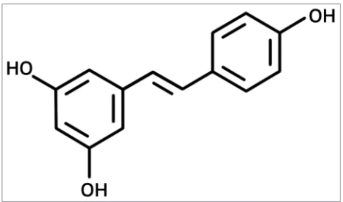
NTRODUCTIONResveratrol, trans-3,5,4′ -trihydroxydro-xystilbene (RESV) (Figure 1), is a polyphenolic phytoalexin of natural oc-currence in many plants and its processed products, such as grapes, berries, red wi-ne, and peanut,1 that presents numerous
health benefits. RESV is one of the most important natural stilbenes and has been extensively studied. It has been shown to possess health-promoting properties, su-ch as antioxidation, anti-inflammation,
cardioprotection, antidiabetes, anticancer, chemoprevention, and neuroprotection.2-6
Several studies performed in recent years have reported the potential health bene-fits of RESV in cardiovascular and renal disease.
RESV is a potent antioxidative agent that can act as a reactive oxygen species (ROS) scavenger and iron chelator.7 In addition,
the NAD+-dependent deacetylase, silent mating type
information regulation 2 homolog surtuin 1 (SIRT1). This protein has been implicated in calorie-restricted lifespan extension and delayed onset of age-related diseases. Furthermore, SIRT1 may regulate multiple cellular functions, including apoptosis, mitochondrial biogenesis, inflammation, glucose/lipid metabolism, autophagy, and adaptations to cellular stress, through the deacetylation of target proteins.7
An excess of ROS is involved in a variety of diseases and in the aging process, which implicate numerous cellular response pathways.8,9 Oxidative stress is
induced by an imbalance between ROS production and antioxidant defenses; therefore, exogenous antioxidants or the modulation of antioxidant enzymes can be expected to reduce oxidative stress. Previous studies have shown that RESV can directly scavenge ROS.10 In addition to scavenging ROS, exogenously
administered RESV modulates the expression and activity of antioxidant enzymes, such as superoxide dismutase (SOD), glutathione peroxidase (GPx), and catalase, either through transcriptional regulation via nuclear factor E2-related factor 2 (Nrf2), activator protein (AP) 1, forkhead box protein O (FOXO), or through enzymatic modifications.11
P
ROTECTIVEMECHANISMOFRESVINRENALDISEASESSILENTMATINGTYPEINFORMATIONREGULATION 2 HOMOLOG
(SIRT1)
Aging is an inevitable process that affects all organs; age-related disruption of cellular homeostasis results in reduced responsiveness to physiological stress and organ dysfunction. Seven mammalian sirtuins exist, and SIRT1 is the sirtuin most closely related to Sir2.12 SIRT1 deacetylates several substrates and
is an important regulator of a wide variety of cellular processes, including stress responses, cell survival,
mitochondrial biogenesis, and metabolism in response to cellular energy and the redox status.12,13 RESV
has been shown to activate SIRT1 through multiple mechanisms.
Park et al.14 demonstrated that RESV activates
SIRT1 through the activation of AMP-activated protein kinase (AMPK). This was achieved via inhibition of phosphodiesterase 4 (PDE 4) and elevation of cyclic adenosine monophosphate (cAMP) in cells, thereby providing a new mechanism to explain SIRT1 activation by RESV.14 A direct link between SIRT1 and
the metabolic benefits of RESV were demonstrated in a more recent study by Price et al.15
SIRT1, P53, AND CISPLATIN
Cisplatin is a chemotherapeutic agent widely used for the treatment of malignant tumors in solid organs. However, a fundamental dose-limiting factor of cisplatin treatment is nephrotoxicity. Direct DNA damage, inflammatory injury, and oxidative stress have been recognized as the mechanisms by which cisplatin induces renal injury.16
In particular, cisplatin-induced apoptotic cell death after DNA damage is the major mechanism for cytotoxicity in renal tubule cells.16 In response to DNA
damage, p53 can induce cell cycle arrest and apoptosis; p53-induced apoptosis affects transcriptional activity and members of the Bcl-2 family in mitochondria.17
In kidney disease, p53 is involved in the apoptotic process observed in ischemic injury and aristocolic acid-induced nephrotoxicity.18
Furthermore, it has been demonstrated that downregulation of p53 by small interference RNA is an effective way of preventing or treating cisplatin-induced nephrotoxicity.19 Activation of p53 is
regulated by posttranslational modifications of p53, such as ubiquitination, phosphorylation, and acetylation.20 Notably, acetylation of p53 affects its
affinity to bind DNA.21
Kim et al.16 demonstrated that activation of SIRT1
by RESV reduces cisplatin-mediated p53 acetyla-tion and ameliorates cisplatin-induced kidney injury through inhibition of the apoptotic pathway. SIRT1 protein expression was decreased by cisplatin in mou-se proximal tubular cells and the SIRT1 activator, RESV, reduced cisplatin-induced p53 acetylation and apoptosis. Through in vivo experimentation, the au-thors revealed that SIRT1 activation by RESV decrea-sed cisplatin-induced apoptosis in the kidney.16
Stiaccini et al.22 were able to demonstrate that
RESV is a strong SIRT1 activator with high activity on SIRT1 protein expression. In a recent study published by Schirmer et al.23 SIRT1 mRNA levels
were not changed in zebrafish exposed to 20-200 µM doses of RESV for 30 and 60 min. However,
in vitro studies using rat cells24 and human visceral
adipocytes25 showed that longer incubation times
were required to observe changes in SIRT1 mRNA and protein expression. Indeed, in the in vitro study by Stiaccini et al.,22 cells were incubated for 24 h with
RESV 200 nM in order to measure increases in SIRT1 expression. However, RESV was a weak activator of the cell signaling system as it caused increases in SIRT1 expression.22
SIRT1, SMAD3, AND 5/6 NEPHRECTOMIZED
It is well documented that Smad3 phosphorylation is a key signaling mechanism underlying fibrogenesis in response to fibrogenic mediators, such as TGF-β, angiotensin II (AII), and advanced glycation end products.26 The evidence to suggest that Smad3
acetylation is also an important signaling pathway leading to ECM production, includes data from experiments using a rodent model of CKD and cultured cells treated with TGF-β1. Significantly elevated levels of Smad acetylation were observed in rats with 5/6 nephrectomy and following TGF-β1 treatment in cultured cells. Furthermore, RESV significantly reduced Smad3 acetylation levels in the remnant kidney of 5/6 nephrectomized rodents and in cultured cells subjected to TGF-β1 treatment. Knocking down SIRT1 in cultured cells increased acetylation levels of Smad3 and attenuated the effect of RESV on Smad3 acetylation.26
RESV has been shown to protect the remnant kidney of 5/6 nephrectomized rats, a rodent model of chronic kidney disease (CKD).27 In the same
model, RESV treatment significantly attenuated the decline of glomerular filtration rates (GFR). In cultured mesangial cells, RESV reduced extracellular matrix (ECM) protein expression induced by tumor growth factor β1 (TGF-β1), and its effects were dependent on SIRT1. SIRT1 inhibits TGF-β1 signaling by deacetylating Smad3. The loss of the allele for SIRT1 aggravates kidney damage in 5/6 nephrectomized mice. Furthermore, knocking down SIRT1 enhances the effects of TGF-β1 on the ECM, and markedly suppresses
the protective effects of RESV. This study provides strong evidence that SIRT1 protects the kidney in a rodent model of CKD through inhibition of TGF-β1 signaling by deacetylating Smad3, and reducing kidney fibrosis.27 In summary, RESV
treatment significantly attenuates renal damage in nephrectomized rats. The renal protective effects of RESV are associated with SIRT1 activation, and a reduction in Smad3 acetylation and TGF-β1 signaling.27
R
ESV AS AN ANTIOXIDANT-
FORKHEADBOX PROTEINO1 (F
OXO1)
ANDSUPEROXIDEDISMUTASE(SOD)
RESV regulates the expression of target genes of FOXO, and may regulate cell survival and/ or apoptosis through global modulation of gene expression via deacetylation of FOXO transcription factors.28 SIRT1 plays an intermediary role in the
action of RESV on FoxO1-mediated gene expression. The dephosphorylated form of FoxO1, which is distributed in the nucleus, is deacetylated by SIRT1, and upregulates the expression of gluconeogenic genes.29
FoxO1 belongs to a family of transcription factors that includes FoxO3a, FoxO4, and FoxO6 in mammals. These proteins play important roles in aging, cell metabolism, insulin resistance, and resistance to oxidative stress.30 Recently, it was demonstrated in
a rat model of diabetes that hyperglycemia induces FoxO1 phosphorylation and suppresses expression of FoxO1 in the kidney. Furthermore, H2O2 negatively regulated FoxO1 by PI3 kinase/AKT-dependent phosphorylation, and FoxO1 dowregulated the expression of catalase in mesangial cells.31
In a recent study by Kitada et al.32 the authors
demonstrated that RESV ameliorated renal injury and enhanced mitochondrial biogenesis with manganese superoxide dismutase (Mn-SOD) dysfunction in the kidney of db/db mice. This was achieved through improvements in the oxidative stress status in the kidney by ROS scavenger activity, normalization of Mn-SOD dysfunction, and partial rescue of glucose-lipid metabolism.32
Subauste & Burant33 reported that excessive
FOXO1, SOD AND DIABETIC NEPHROPATHY
Oxidative stress has emerged as a critical pathogenic factor in the development DN. ROS are thought to play multiple roles in the pathogenesis and progression of DN since ROS production in the kidney is high in the presence of diabetes and DN.34
In a rat model of diabetes, RESV protects the kidney from oxidative stress induced by elevated expression of fibronectin and collagen IV. Under stress conditions, high levels of ROS have been shown to inhibit phosphorylation and acetylation of FoxO1 proteins, resulting in enhanced FoxO1-DNA binding activity.35
FoxO1 subsequently controls ROS levels by transcriptional regulation of a multilayered system.35 Suppressed FoxO1 mRNA levels and
elevated phosphor-FoxO1 levels correlated with the downregulation of catalase mRNA in the kidney of diabetic rats.35 RESV has been shown to increase both
FoxO and SIRT1 levels in multiple cell types,29,36,37
and this was associated with increased longevity and defense against oxidative stress.37
Oxidative stress has been implicated in the pathogenesis of diabetic nephropathy (DN).35
However, the mechanisms involved in ROS generation in diabetes have yet to be elucidated. Nicotinamide adenine dinucleotide phosphate (NADPH) oxidase38
and endothelial nitric oxide synthase (eNOS) uncoupling38 in diabetic glomeruli have been shown
to be major sources of ROS production in a rat model of DN.38
Wu et al.35 observed that malondialdehyde (MDA),
a product of lipid peroxidation, is a sensitive indicator of ROS levels. In that study, the authors observed that increased ROS levels and decreased SOD activity correlated with increased levels of fibronectin and collagen. These data suggest that enhanced oxidative stress increases expression of fibronectin and collagen IV. These findings also indicate that overproduction of ROS in diabetes is associated with the progression of DN, and that antioxidants may provide a useful treatment.35
E
FFECTS OF RESV ON NITRIC OXIDE(NO)
LIPOPOLYSACCHARIDES (LPS) AND RESV
The effect of LPS is accompanied by an elevation of NO in plasma and organs, which was not observed in the presence of RESV. There is much debate on this topic in the literature; some studies demonstrate the
lack of NO involvement in the mechanism of action of RESV,39 whereas others indicate the effects of RESV
are mediated through NO.40 Regardless, the data
support the use of RESV as therapeutic treatment of induced sepsis and endotoxemia-induced death.41
Subacute treatment (7 days) with RESV was effective at preventing lipopolysaccharide (LPS)-induced lethality in mice.42 In prior studies, RESV
attenuated LPS-induced renotoxicity,43 neurotoxicity44
as well as acute phase response in rats.45
Sebai et al.43 reported a clear reduction in
LPS-induced oxidative stress and lethality after subacute pretreatment with RESV, whereas acute RESV treatment administered 12 and 24 h before intoxication failed to reduce the lethality of LPS in mice.
HYPERTENSION, ENDOTHELIALDYSFUNCTION, AND RESV
Early treatment with RESV attenuates the development of hypertension and prevents endothelial dysfunction in spontaneously hypertensive rats (SHR). The mechanisms involved appear to be threefold: 1) attenuation of vascular oxidative stress resulting in increased NO bioavailability, 2) prevention of eNOS uncoupling possibly via inhibition of tetrahydrobiopterin (BH4) oxidation by free radicals, and 3) increased expression of important proteins involved in the NO pathway, namely eNOS and soluble guanylyl cyclase (sGC).46
Endothelial dysfunction is a hallmark of hypertension.47 Impairment of NO synthesis
and/or bioavailability causes endothelial dysfunction. Oxidative stress, particularly induced by superoxide, scavenges NO by forming highly reactive peroxynitrite radicals.48,49 Oxidative stress has also been shown to
uncouple eNOS resulting in impaired endothelium-dependent relaxations.50 Uncoupled eNOS generates
ROS instead of NO, thereby reducing NO production and increasing oxidative stress.51 In summary, oxidative
stress can contribute to endothelial dysfunction by scavenging NO and uncoupling eNOS.
Numerous studies have shown that RESV significantly alters the NO response and increases endothelium dependent vasorelaxation.52 RESV also
increases expression of eNOS in cell culture studies.53
not conclusive as most report no change in blood pressure,54 and only one study in female rats reported
a decrease in blood pressure.55
Bhatt et al.46 demonstrated that RESV significantly
attenuates the rise in blood pressure observed in SHR. Consequently, several studies have investigated the effects of RESV and other red wine polyphenols on endothelial function.54-56 No change in blood pressure
was observed following chronic RESV treatment in SHR in studies by Thandapilly et al.54 It is interesting
to note that in both studies, RESV was administered to adult SHR with established hypertension. In another study, RESV treatment normalized endothelial function and significantly lowered blood pressure in SHR.46 Thus, it appears that the beneficial effects of
RESV treatment on blood pressure may be related to events occurring prior to increases in blood pressure.
Protein nitrosylation is an indicator of peroxynitrite formation in vascular tissue. SHR had significantly elevated nitrotyrosine levels in the aortic homogenate as compared to Wistar Kyoto rats (WKY). Furthermore, elevated nitrotyrosine levels were normalized by RESV treatment. Thus, RESV prevents NO scavenging and increases its biological availability in SHR by lowering oxidative stress.46
In addition to scavenging NO, another important mechanism by which oxidative stress can contribute to endothelial dysfunction is by uncoupling the eNOS enzyme. The physiological consequences of eNOS uncoupling are particularly harmful, since it reduces the production of NO and results in formation of superoxides, thereby further increasing oxidative stress.56 Superoxide production is sensitive to the
eNOS inhibitor, l-NNA, suggesting that eNOS is most
likely uncoupled in SHR. Interestingly, treatment with RESV normalizes superoxide production, which is suggestive of a novel role for RESV in preventing eNOS uncoupling.51
It is well recognized that oxidation of the cofactor tetrahydrobiopterin (BH4) to BH2 provides an important contribution to eNOS uncoupling. Interestingly, BH4 supplementation abolished elevated superoxide production in SHR. This finding indicates that vascular oxidative stress contributes to endothelial dysfunction and hypertension by uncoupling eNOS, and possibly by oxidation of BH4. It has been reported that BH4 supplementation starting at an early age (5-16 weeks of age) suppressed the development of hypertension in SHR.46 Furthermore,
upregulated eNOS protein expression was observed in SHR as well as WKY rats receiving RESV, which is suggestive of transcriptional upregulation. Together, these data suggest that eNOS uncoupling plays an important role in endothelial dysfunction. RESV prevents eNOS uncoupling and rescues endothelial function in SHR.46
The effect of RESV in the presence of sodium nitroprusside (SNP) was investigated in SHR and WKY rats. SNP-induced vasorelaxation was similar in both groups. RESV significantly increased relaxation in response to higher doses of SNP in SHR.46 The proximal
mediator for NO-induced vasorelaxation is soluble guanylyl cyclase (sGC), and its β1 subunit is responsible for the responsiveness of sGC to NO.57 Basal expression
of sGC was higher in SHR as compared to WKY rats, which could be explained by a compensatory increase in response to reduced NO bioavailability. SHR and WKY rats treated with RESV demonstrated a greater expression of the sGC β1 subuni.46
E
FFECTS OF RESV ON RENAL ORGANIC ION TRANSPORTERSURICACID (UA) AND RESV
Hyperuricemia, as a metabolic disorder, is usually associated with gout, kidney disease, hypertension, cardiovascular disease, inflammation, diabetes, and metabolic syndrome.58 Reabsorption and secretion of
uric acid (UA) are controlled by specific organic anion transport proteins in renal apical and basolateral membranes. Urate transporter 1 (URAT1) and glucose transporter 9 (GLUT9) mediate urate reabsorption from the lumen of kidney tubules into the blood, and maintain blood urate homeostasis.59 Human
ATP-binding cassette, subfamily G, 2 (ABCG2) is located in the brush border membranes of renal proximal tubules to control urate secretion, and its gene mutation in Xenopus oocytes results in a reduction of the rate of urate transport.60 ABCDG2 is associated
with hyperuricemia and gout in Caucasian, Han Chinese, Japanese, and African-American subject.61
Uricosuric agents lower urate levels by regulating renal URAT1, GLUT9, and OAT1.59 Therefore,
these renal urate transport-related proteins present important targets for the prevention and treatment of hyperuricemia and gout.62 Renal organic cation/
of renal OCTs and OCTNs impair kidney organic cation balance and induce renal solute toxicity.63,64
Downregulation of renal mOCT1, mOCT2, mOCTN1, and mOCTN2 has been demonstrated in hyperuricemic mice with renal injury.65
Uromodulin (UMOD), the most abundant protein in normal urine, is associated with hyperuricemia and kidney disease.66 UMOD-deficient mice have reduced
creatinine clearance and upregulated expression of major distal electrolyte transporters.67 UMOD is a
useful marker for renal dysfunction in hyperuricemia associated with abnormalities in renal organic ion transporters.67
In oxonate-induced hyperuricemic mice, RESV reduced serum urate levels and enhanced urate excretion. The antihyperuricemic effects of RESV were related to the regulation of renal mURAT1, mGLUT9, mABCG2, and mOAT1.68 Moreover, improvements
of renal function, as well as upregulation of renal mOCT1, mOCT2, mOCTN1, and mOCTN2 protein levels, contributed to the renoprotective effects of RESV.68
Serum urate level is most often linked to renal urate excretion. Renal urate transport becomes increasingly relevant in blood urate homeostasis. RESV reduces serum urate levels by downregulating mGLUT9 expression. As a consequence, this inhibits urate reabsorption, downregulates mABCG2, and upregulates mOAT1 expression to increase urate secretion in the kidney of hyperuricemic mice.68
Therefore, it has been suggested that RESV exhibits antihyperuricemic effects through the regulation of different renal urate transport-related proteins to enhance renal urate excretion in hyperuricemic mice.52
Hyperuricemia is one of several well-described risk factors contributing to kidney function disorders. Creatinine, a substrate of OCT1 and OCT2 in renal proximal tubules, is also considered a biomarker of renal dysfunction.64 Consistent with the amelioration
of kidney dysfunction, renoprotective effects of RESV may be mediated by increased renal mOCT1 expression in hyperuricemic mice.68,69
E
FFECTS OF RESV ON ANGIOTENSINII (AII)
AND ENDOTHELIN-1 (ET-1)
SYSTEMANGIOTENSIN II (AII), ENDOTHELIN-1 (ET-1) AND RESV
There is an increasing body of evidence that implicates the renin-angiotensin system (RAS) in the pathogenesis of chronic vascular disease. AII is an important
component of RAS and a vasoactive peptide.70 It
appears from the literature that AII is able to turn on
the synthesis of ET-1 in several vascular cell types, including cultured vascular smooth muscle cells.71
ET-1 was shown to mediate the growth-promoting effect of AII, and thus plays an important role in cardiovascular disease and vascular remodeling.72
AII has also been shown to stimulate membrane-bound NADPH oxidase, which generates oxygen species in vascular smooth cells.73 Previous reports
indicate that ROS mediate ET-1 gene induction within cardiac fibroblasts, vascular endothelial cells, and smooth muscle cells.73
Zhang et al.71 demonstrated that RESV exerts an
antioxidant-like inhibitory effect on smooth muscle cellular proliferation and ET-1 gene expression induced by AII. In addition, RESV suppresses the extracellular signal-regulated kinase (ERK) pathway, reduces AII-induced cell proliferation, and reduces ET-1 gene expression. It is plausible that the AII-activated signaling pathway consists of a number of redox-sensitive steps, and that RESV treatment modulates the redox state of the cell through its antioxidants properties. In summary, RESV inhibits AII-induced ROS formation, ERK phosphorylation, ET-1 gene expression, and cell proliferation in vascular smooth muscle cells.71
Albertoni et al.73 demonstrated that 24-h UA
treatment in mesangial cells stimulated ET-1, AII, and the renin-angiotensin system. In further experiments by the same group (article in press) UA induced an increase in pre-proET-1 (ppET-1) mRNA expression and peptide synthesis, angiotensinogen (AGT) mRNA expression, and AII peptide production after 6 and 12 h. Furthermore, the study demonstrated that RESV reduced UA-induced ppET-1 gene expression and the production of AII and ET-1 in mesangial cells, suggesting that RESV is able to minimize the impact of these hormones on glomerular function (article in press).
In mesangial cells, UA induces an increase in intracellular Ca2+ concentration ([Ca2+]
i).
73 This
increase in [Ca2+]
i is inhibited by RESV, providing the
first direct evidence that UA induces an increase in [Ca2+]
i that is suppressed by RESV (article in press).
The main novel finding of this study is that UA-induced increases in AII and [Ca2+]
i in smooth muscle
C
ONCLUSIONSRESV exerts protective effects against acute and chronic kidney injury through various mechanisms. RESV activates SIRT1 through multiple mechanisms, such as activation of AMPK via the inhibition of PDE 4 and the elevation of cAMP, downregulation of p53 by siRNA. SIRT1 subsequently inhibits TGF-β1 sig-naling by deacetylating Smad3.
RESV ameliorates renal injury and enhances mitochondrial biogenesis with Mn-SOD. Furthermore, RESV regulates the expression of FOXO target genes and may regulate cell survival and/or apoptosis through global modulation of gene expression via deacetylation of FOXO transcription factors. RESV has been shown to protect the kidney of diabetic rats from oxidative stress induced by increased expression of fibronectin and collagen IV. Additional benefits of RESV include attenuation of LPS-induced renotoxicity, neurotoxicity, and acute phase response in rat. RESV significantly alters NO response and increases endothelium-dependent vasorelaxation. It also reduces serum urate levels and enhances urate excretion in hyperuricemia. Finally, RESV prevents some of the effects of hyperuricemia on glomerular function that lead to glomerulosclerosis. Taking this into consideration, RESV may provide a useful supplemental treatment for preventing renal injury.
R
EFERENCES1. Bertelli AA, Das DK. Grapes, wines, resveratrol, and heart health. J Cardiovasc Pharmacol 2009;54:468-76. DOI: http:// dx.doi.org/10.1097/FJC.0b013e3181bfaff3
2. Timmers S, Konings E, Bilet L, Houtkooper RH, van de Weijer T, Goossens GH, et al. Calorie restriction-like effects of 30 days of resveratrol supplementation on energy metabolism and me-tabolic profile in obese humans. Cell Metab 2011;14:612-22. DOI: http://dx.doi.org/10.1016/j.cmet.2011.10.002
3. Brasnyó P, Molnár GA, Mohás M, Markó L, Laczy B, Cseh J, et al. Resveratrol improves insulin sensitivity, reduces oxi-dative stress and activates the Akt pathway in type 2 diabetic patients. Br J Nutr 2011;106:383-9. PMID: 21385509 DOI: http://dx.doi.org/10.1017/S0007114511000316
4. Magyar K, Halmosi R, Palfi A, Feher G, Czopf L, Fulop A, et al. Cardioprotection by resveratrol: A human clinical trial in patients with stable coronary artery disease. Clin Hemorheol Microcirc 2012;50:179-87.
5. Kondratyuk TP, Park EJ, Marler LE, Ahn S, Yuan Y, Choi Y, et al. Resveratrol derivatives as promising chemopreventive agents with improved potency and selectivity. Mol Nutr Food Res 2011;55:1249-65. PMID: 21714126 DOI: http://dx.doi. org/10.1002/mnfr.201100122
6. Catalgol B, Batirel S, Taga Y, Ozer NK. Resveratrol: French paradox revisited. Front Pharmacol 2012;3:141. DOI: http:// dx.doi.org/10.3389/fphar.2012.00141
7. Kitada M, Koya D. Renal protective effects of resveratrol. Oxid Med Cell Longev 2013;2013:568093.
8. Palsamy P, Subramanian S. Resveratrol protects diabetic kidney by attenuating hyperglycemia-mediated oxidative stress and renal inflammatory cytokines via Nrf2-Keap1 signaling. Bio-chim Biophys Acta 2011;1812:719-31. PMID: 21439372 DOI: http://dx.doi.org/10.1016/j.bbadis.2011.03.008
9. Zhang L, Pang S, Deng B, Qian L, Chen J, Zou J, et al. High glucose induces renal mesangial cell proliferation and
fibro-nectin expression through JNK/NF-κB/NADPH oxidase/ROS
pathway, which is inhibited by resveratrol. Int J Biochem Cell Biol 2012;44:629-38. DOI: http://dx.doi.org/10.1016/j.bio-cel.2012.01.001
10. Holthoff JH, Woodling KA, Doerge DR, Burns ST, Hinson JA, Mayeux PR. Resveratrol, a dietary polyphenolic phytoalexin, is a functional scavenger of peroxynitrite. Biochem Pharma-col 2010;80:1260-5. PMID: 20599800 DOI: http://dx.doi. org/10.1016/j.bcp.2010.06.027
11. Kitada M, Kume S, Imaizumi N, Koya D. Resveratrol impro-ves oxidative stress and protects against diabetic nephropathy through normalization of Mn-SOD dysfunction in AMPK/ SIRT1-independent pathway. Diabetes 2011;60:634-43. DOI: http://dx.doi.org/10.2337/db10-0386
12. Guarente L. Franklin H. Epstein Lecture: Sirtuins, aging, and medicine. N Engl J Med 2011;364:2235-44. DOI: http://dx.doi. org/10.1056/NEJMra1100831
13. Kitada M, Kume S, Kanasaki K, Takeda-Watanabe A,
Koya D. Sirtuins as possible drug targets in type 2 diabe-tes. Curr Drug Targets 2013;14:622-36. DOI: http://dx.doi. org/10.2174/1389450111314060002
14. Park SJ, Ahmad F, Philp A, Baar K, Williams T, Luo H, et al. Resveratrol ameliorates aging-related metabolic phenotypes by inhibiting cAMP phosphodiesterases. Cell 2012;148:421-33. PMID: 22304913 DOI: http://dx.doi.org/10.1016/j. cell.2012.01.017
15. Price NL, Gomes AP, Ling AJ, Duarte FV, Martin-Montalvo A, North BJ, et al. SIRT1 is required for AMPK activation and the beneficial effects of resveratrol on mitochondrial function. Cell Metab 2012;15:675-90. DOI: http://dx.doi.org/10.1016/j. cmet.2012.04.003
16. Kim DH, Jung YJ, Lee JE, Lee AS, Kang KP, Lee S, et al. SIRT1 activation by resveratrol ameliorates cisplatin-induced renal injury through deacetylation of p53. Am J Physiol Renal Phy-siol 2011;301:F427-35. PMID: 21593185 DOI: http://dx.doi. org/10.1152/ajprenal.00258.2010
17. Mihara M, Erster S, Zaika A, Petrenko O, Chittenden T, Pancoska P, et al. p53 has a direct apoptogenic role at the mitochondria. Mol Cell 2003;11:577-90. DOI: http://dx.doi. org/10.1016/S1097-2765(03)00050-9
18. Zhou L, Fu P, Huang XR, Liu F, Lai KN, Lan HY. Activation of p53 promotes renal injury in acute aristolochic acid nephro-pathy. J Am Soc Nephrol 2010;21:31-41. DOI: http://dx.doi. org/10.1681/ASN.2008111133
19. Molitoris BA, Dagher PC, Sandoval RM, Campos SB, Ashush H, Fridman E, et al. siRNA targeted to p53 attenuates ische-mic and cisplatin-induced acute kidney injury. J Am Soc Ne-phrol 2009;20:1754-64. DOI: http://dx.doi.org/10.1681/ ASN.2008111204
20. Brooks CL, Gu W. Ubiquitination, phosphorylation and ace-tylation: the molecular basis for p53 regulation. Curr Opin Cell Biol 2003;15:164-71. DOI: http://dx.doi.org/10.1016/S0955-0674(03)00003-6
21. Luo J, Li M, Tang Y, Laszkowska M, Roeder RG, Gu W. Ace-tylation of p53 augments its site-specific DNA binding both in vitro and in vivo. Proc Natl Acad Sci U S A 2004;101:2259-64. PMID: 14982997 DOI: http://dx.doi.org/10.1073/ pnas.0308762101
23. Schirmer H, Pereira TC, Rico EP, Rosemberg DB, Bonan CD, Bogo MR, et al. Modulatory effect of resveratrol on SIRT1,
SIRT3, SIRT4, PGC1α and NAMPT gene expression profiles in
wild-type adult zebrafish liver. Mol Biol Rep 2012;39:3281-9. DOI: http://dx.doi.org/10.1007/s11033-011-1096-4
24. Morita Y, Wada-Hiraike O, Yano T, Shirane A, Hirano M, Hiraike H, et al. Resveratrol promotes expression of SIRT1 and StAR in rat ovarian granulosa cells: an implicative role of SIRT1 in the ovary. Reprod Biol Endocrinol 2012;10:14. DOI: http://dx.doi.org/10.1186/1477-7827-10-14
25. Costa Cdos S, Rohden F, Hammes TO, Margis R, Bortolotto JW, Padoin AV, et al. Resveratrol upregulated SIRT1, FOXO1,
and adiponectin and downregulated PPARγ1-3 mRNA
expres-sion in human visceral adipocytes. Obes Surg 2011;21:356-61. DOI: http://dx.doi.org/10.1007/s11695-010-0251-7
26. Chung AC, Zhang H, Kong YZ, Tan JJ, Huang XR, Kopp JB, et al. Advanced glycation end-products induce tubular CTGF via TGF-beta-independent Smad3 signaling. J Am Soc Nephrol 2010;21:249-60. DOI: http://dx.doi.org/10.1681/ ASN.2009010018
27. Huang XZ, Wen D, Zhang M, Xie Q, Ma L, Guan Y, et al. Sirt1 activation ameliorates renal fibrosis by inhibiting the
TGF-β/Smad3 pathway. J Cell Biochem 2014;115:996-1005.
DOI: http://dx.doi.org/10.1002/jcb.24748
28. Chen Q, Ganapathy S, Singh KP, Shankar S, Srivastava RK. Resveratrol induces growth arrest and apoptosis through ac-tivation of FOXO transcription factors in prostate cancer ce-lls. PLoS One 2010;5:e15288. DOI: http://dx.doi.org/10.1371/ journal.pone.0015288
29. Park JM, Kim TH, Bae JS, Kim MY, Kim KS, Ahn YH. Role of resveratrol in FOXO1-mediated gluconeogenic gene expression in the liver. Biochem Biophys Res Commun 2010;403:329-34. PMID: 21078299 DOI: http://dx.doi.org/10.1016/j. bbrc.2010.11.028
30. Dröge W. Free radicals in the physiological control of cell func-tion. Physiol Rev 2002;82:47-95. PMID: 11773609
31. Venkatesan B, Mahimainathan L, Das F, Ghosh-Choudhury N, Ghosh Choudhury G. Downregulation of catalase by reactive oxygen species via PI 3 kinase/Akt signaling in mesangial cells. J Cell Physiol 2007;211:457-67. PMID: 17186497 DOI: http:// dx.doi.org/10.1002/jcp.20953
32. Kitada M, Kume S, Imaizumi N, Koya D. Resveratrol impro-ves oxidative stress and protects against diabetic nephropathy through normalization of Mn-SOD dysfunction in AMPK/ SIRT1-independent pathway. Diabetes 2011;60:634-43. DOI: http://dx.doi.org/10.2337/db10-0386
33. Subauste AR, Burant CF. Role of FoxO1 in FFA-induced oxidative stress in adipocytes. Am J Physiol Endocrinol Metab 2007;293:E159-64. DOI: http://dx.doi.org/10.1152/ajpendo.00629.2006
34. Forbes JM, Coughlan MT, Cooper ME. Oxidative stress as a major culprit in kidney disease in diabetes. Diabetes 2008;57:1446-54. PMID: 18511445 DOI: http://dx.doi. org/10.2337/db08-0057
35. Wu L, Zhang Y, Ma X, Zhang N, Qin G. The effect of resve-ratrol on FoxO1 expression in kidneys of diabetic nephropa-thy rats. Mol Biol Rep 2012;39:9085-93. DOI: http://dx.doi. org/10.1007/s11033-012-1780-z
36. Howitz KT, Bitterman KJ, Cohen HY, Lamming DW, Lavu S, et al. Small molecule activators of sirtuins extend Saccha-romyces cerevisiae lifespan. Nature 2003;425:191-6. PMID: 12939617 DOI: http://dx.doi.org/10.1038/nature01960 37. Brunet A, Sweeney LB, Sturgill JF, Chua KF, Greer PL, Lin Y,
et al. Stress-dependent regulation of FOXO transcription fac-tors by the SIRT1 deacetylase. Science 2004;303:2011-5. PMID: 14976264 DOI: http://dx.doi.org/10.1126/science.1094637 38. Satoh M, Fujimoto S, Haruna Y, Arakawa S, Horike H, Komai
N, et al. NAD(P)H oxidase and uncoupled nitric oxide syn-thase are major sources of glomerular superoxide in rats with experimental diabetic nephropathy. Am J Physiol Renal Phy-siol 2005;288:F1144-52. PMID: 15687247 DOI: http://dx.doi. org/10.1152/ajprenal.00221.2004
39. Mokni M, Limam F, Elkahoui S, Amri M, Aouani E. Strong cardioprotective effect of resveratrol, a red wine polyphenol, on isolated rat hearts after ischemia/reperfusion injury. Arch Biochem Biophys 2007;457:1-6. PMID: 17125727 DOI: http:// dx.doi.org/10.1016/j.abb.2006.10.015
40. Tsai SK, Hung LM, Fu YT, Cheng H, Nien MW, Liu HY, et al. Resveratrol neuroprotective effects during focal cerebral ischemia injury via nitric oxide mechanism in rats. J Vasc Surg 2007;46:346-53. DOI: http://dx.doi.org/10.1016/j.jvs.2007.04.044
41. Hobbs AJ, Higgs A, Moncada S. Inhibition of nitric oxide synthase as a potential therapeutic target. Annu Rev Pharma-col ToxiPharma-col 1999;39:191-220. PMID: 10331082 DOI: http:// dx.doi.org/10.1146/annurev.pharmtox.39.1.191
42. Sebai H, Sani M, Ghanem-Boughanmi N, Aouani E. Preven-tion of lipopolysaccharide-induced mouse lethality by resvera-trol. Food Chem Toxicol 2010;48:1543-9. DOI: http://dx.doi. org/10.1016/j.fct.2010.03.022
43. Sebai H, Ben-Attia M, Sani M, Aouani E, Ghanem-Boughanmi N. Protective effect of resveratrol on acute endotoxemia-in-duced nephrotoxicity in rat through nitric oxide independent mechanism. Free Radic Res 2008;42:913-20. PMID: 19031312 DOI: http://dx.doi.org/10.1080/10715760802555577 44. Sebai H, Gadacha W, Sani M, Aouani E,
Ghanem-Bou-ghanmi N, Ben-Attia M. Protective effect of resveratrol against lipopolysaccharide-induced oxidative stress in rat brain. Brain Inj 2009;23:1089-94. DOI: http://dx.doi. org/10.3109/02699050903379370
45. Sebai H, Ben-Attia M, Sani M, Aouani E, Ghanem-Boughanmi N. Protective effect of resveratrol in endotoxemia-induced acu-te phase response in rats. Arch Toxicol 2009;83:335-40. DOI: http://dx.doi.org/10.1007/s00204-008-0348-0
46. Bhatt SR, Lokhandwala MF, Banday AA. Resveratrol prevents endothelial nitric oxide synthase uncoupling and attenuates de-velopment of hypertension in spontaneously hypertensive rats. Eur J Pharmacol 2011;667:258-64. PMID: 21640096 DOI: http://dx.doi.org/10.1016/j.ejphar.2011.05.026
47. Puddu P, Puddu GM, Zaca F, Muscari A. Endothelial dysfunc-tion in hypertension. Acta Cardiol 2000;55:221-32. PMID: 11041120 DOI: http://dx.doi.org/10.2143/AC.55.4.2005744 48. Escobales N, Crespo MJ. Oxidative-nitrosative stress in
hyper-tension. Curr Vasc Pharmacol 2005;3:231-46. DOI: http:// dx.doi.org/10.2174/1570161054368643
49. Pryor WA, Squadrito GL. The chemistry of peroxynitrite: a product from the reaction of nitric oxide with superoxide. Am J Physiol 1995;268:L699-722. PMID: 7762673 DOI: http:// dx.doi.org/10.1006/abbi.1995.1435
50. Landmesser U, Dikalov S, Price SR, McCann L, Fukai T, Holland SM, et al. Oxidation of tetrahydrobiopterin leads to uncoupling of endothelial cell nitric oxide synthase in hyperten-sion. J Clin Invest 2003;111:1201-9. PMID: 12697739 DOI: http://dx.doi.org/10.1172/JCI200314172
51. Münzel T, Daiber A, Ullrich V, Mülsch A. Vascular consequen-ces of endothelial nitric oxide synthase uncoupling for the ac-tivity and expression of the soluble guanylyl cyclase and the cGMP-dependent protein kinase. Arterioscler Thromb Vasc Biol 2005;25:1551-7. DOI: http://dx.doi.org/10.1161/01. ATV.0000168896.64927.bb
52. Naderali EK, Doyle PJ, Williams G. Resveratrol induces vaso-relaxation of mesenteric and uterine arteries from female gui-nea-pigs. Clin Sci (Lond) 2000;98:537-43. DOI: http://dx.doi. org/10.1042/CS19990303
53. Wallerath T, Deckert G, Ternes T, Anderson H, Li H, Witte K, et al. Resveratrol, a polyphenolic phytoalexin present in red wine, enhances expression and activity of endothelial nitric oxide synthase. Circulation 2002;106:1652-8. PMID: 12270858 DOI: http://dx.doi.org/10.1161/01.CIR.0000029925.18593.5C 54. Thandapilly SJ, Wojciechowski P, Behbahani J, Louis XL, Yu
55. López-Sepúlveda R, Jiménez R, Romero M, Zarzuelo MJ, Sán-chez M, Gómez-Guzmán M, et al. Wine polyphenols improve endothelial function in large vessels of female spontaneously hypertensive rats. Hypertension 2008;51:1088-95. DOI: http:// dx.doi.org/10.1161/HYPERTENSIONAHA.107.107672 56. Rivera L, Morón R, Zarzuelo A, Galisteo M. Long-term
res-veratrol administration reduces metabolic disturbances and lowers blood pressure in obese Zucker rats. Biochem Phar-macol 2009;77:1053-63. DOI: http://dx.doi.org/10.1016/j. bcp.2008.11.027
57. Lucas KA, Pitari GM, Kazerounian S, Ruiz-Stewart I, Park J, Schulz S, et al. Guanylyl cyclases and signaling by cyclic GMP. Pharmacol Rev 2000;52:375-414. PMID: 10977868
58. Bhole V, Choi JW, Kim SW, de Vera M, Choi H. Serum uric acid levels and the risk of type 2 diabetes: a prospective study. Am J Med 2010;123:957-61. DOI: http://dx.doi.org/10.1016/j. amjmed.2010.03.027
59. Preitner F, Bonny O, Laverrière A, Rotman S, Firsov D, Da Cos-ta A, et al. Glut9 is a major regulator of urate homeosCos-tasis and its genetic inactivation induces hyperuricosuria and urate ne-phropathy. Proc Natl Acad Sci U S A 2009;106:15501-6. DOI: http://dx.doi.org/10.1073/pnas.0904411106
60. Woodward OM, Köttgen A, Coresh J, Boerwinkle E, Guggino WB, Köttgen M. Identification of a urate transporter, ABCG2, with a common functional polymorphism causing gout. Proc Natl Acad Sci U S A 2009;106:10338-42. PMID: 19506252 DOI: http://dx.doi.org/10.1073/pnas.0901249106
61. Wang B, Meng D, Wang J, Liu S, Zhou S, Miao Z, et al. Ge-netic association of polymorphism rs1333049 with gout. Rheumatology (Oxford) 2011;50:1559-61. DOI: http://dx.doi. org/10.1093/rheumatology/ker135
62. Saito H. Pathophysiological regulation of renal SLC22A orga-nic ion transporters in acute kidney injury: pharmacological and toxicological implications. Pharmacol Ther 2010;125:79-91. PMID: 19837111 DOI: http://dx.doi.org/10.1016/j.phar-mthera.2009.09.008
63. Glube N, Closs E, Langguth P. OCTN2-mediated carniti-ne uptake in a carniti-newly discovered human proximal tubule cell line (Caki-1). Mol Pharm 2007;4:160-8. DOI: http://dx.doi. org/10.1021/mp060073a
64. Grover B, Buckley D, Buckley AR, Cacini W. Reduced expression of organic cation transporters rOCT1 and rOCT2 in experimen-tal diabetes. J Pharmacol Exp Ther 2004;308:949-56. PMID: 14718608 DOI: http://dx.doi.org/10.1124/jpet.103.058388
65. Wang CP, Wang Y, Wang X, Zhang X, Ye JF, Hu LS, et al. Mulberroside a possesses potent uricosuric and ne-phroprotective effects in hyperuricemic mice. Planta Med 2011;77:786-94. PMID: 21154198 DOI: http://dx.doi. org/10.1055/s-0030-1250599
66. Dahan K, Devuyst O, Smaers M, Vertommen D, Loute G, Poux JM, et al. A cluster of mutations in the UMOD gene causes familial juvenile hyperuricemic nephropathy with abnormal expression of uromodulin. J Am Soc Ne-phrol 2003;14:2883-93. DOI: http://dx.doi.org/10.1097/01. ASN.0000092147.83480.B5
67. Bachmann S, Mutig K, Bates J, Welker P, Geist B, et al. Re-nal effects of Tamm-Horsfall protein (uromodulin) deficien-cy in mice. Am J Physiol Renal Physiol 2005;288:F559-67. PMID: 15522986 DOI: http://dx.doi.org/10.1152/ajpre-nal.00143.2004
68. Shi YW, Wang CP, Liu L, Liu YL, Wang X, Hong Y, et al. Antihyperuricemic and nephroprotective effects of resve-ratrol and its analogues in hyperuricemic mice. Mol Nutr Food Res 2012;56:1433-44. DOI: http://dx.doi.org/10.1002/ mnfr.201100828
69. Vaughan D. Pharmacology of ACE inhibitors versus AT1 blo-ckers. Can J Cardiol 2000;16:36E-40E.
70. Chao HH, Juan SH, Liu JC, Yang HY, Yang E, Cheng TH, et al. Resveratrol inhibits angiotensin II-induced endothe-lin-1 gene expression and subsequent proliferation in rat aortic smooth muscle cells. Eur J Pharmacol 2005;515:1-9. PMID: 15878161 DOI: http://dx.doi.org/10.1016/j. ejphar.2005.03.035
71. Zhang X, Wang Y, Yang W, Hou X, Zou J, Cao K. Resve-ratrol inhibits angiotensin II-induced ERK1/2 activation by downregulating quinone reductase 2 in rat vascular smooth muscle cells. J Biomed Res 2012;26:103-9. DOI: http://dx.doi. org/10.1016/S1674-8301(12)60019-0
72. Kim CS, Choi JS, Park JW, Bae EH, Ma SK, Lee J, et al. Al-tered regulation of nitric oxide and natriuretic peptide system in cisplatin-induced nephropathy. Regul Pept 2012;174:65-70. DOI: http://dx.doi.org/10.1016/j.regpep.2011.12.001