Preparation and photocatalytic activity of the layered titanates
Marija Milanović
*, Ivan Stijepović, Ljubica M. Nikolić
Department of Materials Engineering, Faculty of Technology, University of Novi Sad, Serbia Received 15 April 2010; received in revised form 23 June 2010; accepted 28 June 2010
Abstract
Titanate structures were synthesized in highly alkaline solution using hydrothermal procedure. As-prepared powders were characterized by X-ray diffraction (XRD), Fourier transform infrared spectroscopy (FT-IR) and transmission electron microscopy (TEM). A speciic surface area of the powders was measured by BET method. Results conirmed formation of layered trititanates, already after one hour of hydrothermal synthesis. To examine the photocatalytic activity of the as-prepared layered titanates, methylene blue (MB) was employed as a target compound in response to visible light at ambient temperature. It was observed that the speciic surface area, size distribution and crystallinity are important factors to get high photocatalytic activity for the decomposition of MB.
Keywords: layered titanates, hydrothermal synthesis, photocatalytic activity
I. Introduction
During the past decade, a great research interest has been directed to the development of one-dimensional (1D) nanostructured powders based on oxide ceramics. Compared with bulk materials, one-dimensional nano-scale materials, with their large speciic surface areas and possible quantum coninement effects, exhibit dis-tinct electronic, optical, chemical and thermal proper-ties [1–3]. In many cases, 1D nanostructures are supe-rior to their counterparts with larger dimensions. Major advantages of these materials are their extraordinary lengths, lexibility and structure that can allow them to be physically manipulated into various shapes accord-ing to the design requirements.
Among the large family of 1D nanomaterials, titan-ates are of particulate interest. Depending on the syn-thesis method as well as on the conditions within the method, it is possible to obtain different morphology of titanate structures, such as nanotubes, nanowires, nano-ibers, nanorods, nanobelts etc. Titanate based 1D ma-terials are one of the most promising semiconducting ceramic materials because of their wide range of poten-tial applications in nanoelectronics, in optical and sen -sor devices, in solar cells, pigments, biomedicine etc [1,4–8]. In addition, the high morphological and
struc-tural speciicity of 1D titanates (phase composition, high aspect ratio and large speciic surface area) may enhance their photocatalytic activity, leading to a high-er potential of applications in environmental puriica-tion [9–13]. Beside the growing interest in obtaining the one dimensional titanates, the exact mechanism of their formation is still a controversial topic discussed wide-ly in contemporary literature [14–21]. Also, the clarii-cation of the individual stages that precede the forma-tion of the 1D structures, is one of the opened quesforma-tions; even the composition of these materials is still a subject of debate [1, 22–24].
In this work, layered titanates were synthesized by a hydrothermal method. The main subject of this work was to investigate the photocatalytic activity of the lay-ered structures under irradiation by visible light. These structures can be understood as an early stage in the for-mation of the one dimensional titanates.
II. Experimental
The titanate based structures were synthesized through a simple hydrothermal procedure, reported pre-viously [25]. The commercial TiO2 powder (Degus-sa P25) was dispersed in an aqueous solution of 10M NaOH and stirred for some time. The hydrothermal re-action was carried out at 150°C for a different time (1, 3, 6 and 10 hours). After hydrothermal treatment the pre-cipitates were washed with distilled water and absolute
* Corresponding author: tel: +381 21 485 3750,
ethanol until the neutral pH was reached and separated from the solution by centrifugation. The formed titan-ates were dried at 120°C for one day. The samples were named Tm-x, where x stands for the time (in hours) of the hydrothermal reaction. The sample notations of the obtained titanates are shown in Table 1, together with the values of speciic surface areas.
Table 1. Sample notations and speciic surface areas of
titanate samples
Sample notation
Reaction time
[h] area, SSpeciic surface BET [m2/g]
Tm-1 1 355
Tm-3 3 219
Tm-6 6 322
Tm-10 10 249
Photocatalytic activity of the prepared powders was estimated from the change in the concentration of meth-ylene blue (MB) under visible light irradiation. In sev-eral glasses containing 0.01 g of titanate powder, a 10 ml of aqueous MB solution (100 ppm) was added and maintained in the dark for 1 hour with stirring. After completion of the adsorption of MB, the solution was kept under irradiation of visible light for a different time (10, 20, 30, 40, 50 minutes). Samples were centrifuged in order to separate the titanates from the solution. The concentration change of MB was then determined by measuring the maximum absorbance at λ = 665 nm as a function of irradiation time using a spectrophotometer. Concentration of methylene blue solution was calculat-ed with the absorbency of the methylene blue solution separated from sample. The adsorption degree of titan-ate powders to methylene blue solution was valued as c/co, where c is the concentration of methylene blue
so-lution separated from adsorpted sample and the co is the
concentration of the original methylene blue solution. Structural changes caused by the hydrothermal con-version of the starting P25 powder to titanate structures were studied by FT-IR spectroscopy and electron mi-croscopy. Phase composition of the samples was deter-mined by X-ray powder diffraction (XRD). X-ray dif-fraction patterns of the titanates were collected on a Siemens D500 instrument using Ni-iltered Cu-Kα radi-ation of wavelength 1.5418 Å. The XRD data were re-corded with a step of 0.02°/sec. Microstructure and mor-phology of the samples were analyzed using a Hitachi H9000-NA transmission electron microscope (TEM) operating at 200 KV, capable of performing the select-ed area electron diffraction (SAED). Fourier-transform infrared spectroscopy measurements were performed using a Nicolet-Nexsus 670 FT-IR in the range 400– 4000 cm-1. Speciic surface area of the powders was es-timated by low temperature nitrogen adsorption mea-surements performed on a Quantachrom Autosorb-3B instrument applying BET method.
III. Results and discussion
Typical XRD patterns of one-dimensional titanates are shown in Fig. 1. Titanate structures are present al-ready after one hour of hydrothermal synthesis, which is an unusually short time. From the Fig. 1, it can be iden-tiied four characteristic diffraction peaks at 2θ ~ 9.5, 24.44, 28.37 and 48.32o, related to the Na
2Ti3O7 type of titanates, (ICDD card 31-1329), in accordance with the literature [24,26]. These peaks could be ascribed to the interlayer spacing typical for one-dimensional titan-ate structures [24–28]. The intensity of the peak around 9.5° is increasing with the reaction time, indicating the rise in crystallinity.
Figure 1. XRD patterns of titanate structures prepared under hydrothermal reaction
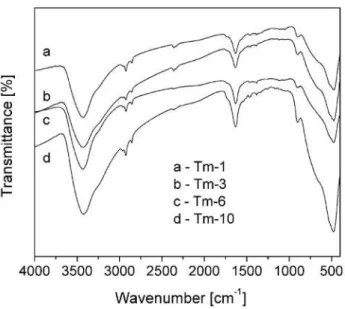
The FT-IR spectra of the prepared powders, Fig. 2, are in accordance with the XRD results. The peak at around 900 cm-1 is typical for one-dimensional
struc-tures [3,25]. This peak corresponds to the vibration of Ti-O nonbridging oxygen bonds, and could imply on the formation of Ti-O-Na bonds. The characteristic bands in the range 450–700 cm-1 are due to the different
types of Ti-O vibrations, and they are related with tet-rahedrally and octahedrally coordinated titanium ions [29]. As previously reported [25], those peaks are miss-ing in the startmiss-ing powder and it can be assumed that observed vibrations are the result of the braking of the Ti-O bonds and formation of titanate layers by the in-tercalation of Na+ and OH– ions into the structure. Two prominent absorption bands at 3400 and 1600 cm-1 in all
TEM images of titanate structures, shown in Fig. 3, are in good agreement with previously presented re-sults. Formation of lamellar titanate sheets is obvious already after 1 hour of hydrothermal synthesis, Fig. 3a. Complete transformation of P25 nanoparticles into titanate nanosheets indicates that the rate of hydrother-mal reaction is very high in the early stages of lay-er formation. As the reaction proceeds, formed nano-sheets grow together with a tendency to curl at their edges, leading to the formation of nanotubes. Hence, similarly to our indings reported earlier [30], obtained results implied that the most probable formation mech-anism of titanate nanotubes is a transformation of ti-tania nanoparticles into titanate nanosheets due to the attack of sodium hydroxide during the hydrothermal reaction. These results have shown that the formation of one dimensional titanate structures (layered titan-ates) is possible after unusually short time, but
com-plete transformation into the nanotubes requires a lon-ger time for the synthesis than 10 hours tested in this work.
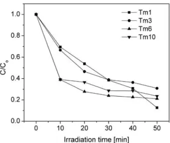
Eficiency of the formed layered titanates in water puriication is shown in Fig. 4. Photocatalytic activity of the prepared powders was estimated from the change in the concentration of methylene blue (MB) under visi-ble light irradiation. It can be seen that obtained layered titanates have shown high eficiency in the photocata-lytic degradation of MB. It was particularly noteworthy that the process showed an extraordinary fast initial rate of adsorption, which can be veriied by the fact that the amount of adsorbed MB onto Tm-6 and Tm-10 samples within 10 minutes achieved almost 60% in respect to the initial concentration. With increasing the time of ir-radiation, the concentration of MB decreases, indicating the rise of titanates activity. These changes are clear-ly noticeable from the transition of the solution colour from dark blue to light blue shades with increasing time of excitation, Fig. 5. The remarkable photocatalytic ac-tivity of the layered titanates can be attributed to their defective structure and high speciic surface area. The BET speciic surface area, Table 1, increases following the order: Tm-3 < Tm-10 < Tm-6 < Tm-1, which does not correlate with the cristallinity of the powders, but is probably caused by the different porous nanostructure in the investigated samples. Further analyses regarding the pore shape and size distribution are needed to ex-plain this behaviour. With respect to the starting powder (SBET(P25) = 80 m2/g), the high speciic surface area of as-synthesized layered titanates made it possible to ad-sorb pollutant molecules into the surface of the pores, thus favouring the photocatalytic degradation mediated with these structures. It can be seen that after comple-tion of the MB absorpcomple-tion, the most effective titanates in MB degradation are those with the highest specif-ic areas.
Figure 3. TEM images of one dimensional layered titanate structures: a) Tm-1, b) Tm-3 and c) Tm-10
a) b) c)
IV. Conclusions
We have successfully synthesized layered titanates through a simple hydrothermal procedure. Layered ti-tanates can be understood as an early stage in the forma-tion of the titanate nanotubes. The XRD patterns have shown that the particulate structure of the starting pow-der is completely altered already after one hour of hy-drothermal treatment at 150°C and layered titanates have started to appear. The structure of the obtained ti-tanates corresponds to the sodium titi-tanates. The cris-tallinity of the powders increases with the time of hy-drothermal reaction. As-prepared layered titanates have very high values of speciic surface area, up to 355 m2/g in contrast with 80 m2/g of the starting powder, P25 Degussa. The obtained layered titanate structures have shown the high eficiency in photocatalytic deg-radation of MB, already after 10 minutes under visi-ble light excitation. Remarkavisi-ble photocatalytic activity of the layered titanates can be attributed to their defec-tive structure and high speciic surface area. Addition-al investigations regarding the pore size distribution are
needed in order to explain the inluence of the titanates pore structure on their photocatalytic eficiency.
Acknowledgment: The authors gratefully
acknowl-edge the Ministry of Science and Technological Devel-opment Republic of Serbia for the inancial support of this work Project 142059 and COST 539
References
H.-H. Ou, S.-L. Lo, “Review of titania nanotubes syn-1.
thesized via the hydrothermal treatment: Fabrication, modiication and application”, Sep. Purif. Technol.,
58 (2007) 179–191.
E.I. Kapinus, T.A. Khalyavka, V.V. Shimanovskaya, 2.
T.I. Viktorova and V.V. Strelko, “Photocatalytic ac-tivity of spectro-pure titanium dioxide: Effects of crystalline structure, speciic surface area and sorp-tion properties”, Inter. J. Photoen., 5 [3] (2003) 159– 166.
J.A. Toledo-Antonio, S. Capula, M.A. Cortes-Ja-3.
come, C. Angeles-Chavez, E. Lopez-Salinas, G. Fer-rat, J. Navarrete, J. Escobar, “Low-tempareature FTIR study of CO adsorption on titania nanotubes”, J. Phys. Chem. C, 111 (2007) 10799–10805.
M.R. Hoffmann, S.T. Martin, W. Choi, D.W. Bahne-4.
mann, “Environmental applications of semiconductor photocatalysis”, Chem. Rev., 95 (1995) 69–96.
W. Wang, H. Lin, J. Li, N. Wang, “Formation of tita-5.
nia nanoarrays by hydrothermal reaction and their ap-plication in photovoltaic cells”, J. Am. Ceram. Soc.,
91 [2] (2008) 628–631.
M.’Ou Li, X. Xiao, R. Liu, “Synthesis and bioactivity 6.
of highly ordered TiO2 nanotube arrays”, Appl. Surf. Sci., 255 [2] (2008) 365–367.
A. Corma, “From microporous to mesoporous molec-7.
ular sieve materials and their use in catalysis”, Chem.
Rev., 97 (1997) 2373–2420, and the references therein.
F. Dong, W. Zhao, Z. Wu, “Characterization and pho-8.
tocatalytic activities of C, N and S co-doped TiO2 with
1D nanostructure prepared by the nano-coninement effect”, Nanotechnol., 19 (2008) 365607.
L. Xionga, Y. Yang, J. Maia, W. Suna, C. Zhang, D. 9.
Wei, Q. Chen, J. Ni, “Adsorption behavior of methyl-ene blue onto titanate nanotubes”, Chem. Eng. J., 156
(2010) 313–320.
P. Wena, H. Itoh, W. Tang, Q. Feng, “Transformation 10.
of layered titanate nanosheets into nanostructured po-rous titanium dioxide in polycation solution”, Micro-porous MesoMicro-porous Mater., 116 (2008) 147–156.
C.-W. Peng, M. Richard-Plouet, T.-Y. Ke, C.-Y. Lee, 11.
H.-T. Chiu, C. Marhic, E. Puzenat, F. Lemoigno, L. Brohan, “Chimie douce route to sodium hydroxo ti-tanate nanowires with modulated structure and con-version to highly photoactive titanium dioxides”, Chem. Mater., 20 (2008) 7228–7236.
Z.-J. Chen, B.-Z. Lin, B.-H. Xu, X.-L. Li, Q.-Q. Wang, 12.
K.-Z. Zhang, M.-C. Zhu, “Preparation and characteri-zation of mesoporous TiO2-pillared titanate photocat-Figure 4. Photocatalytic degradation of MB onto layered
titanate structures under visible light excitation
Figure 5. Change of the solution color inluenced by MB
alyst”, J. Porous Mater., DOI 10.1007/s10934-010-9369-1.
S.-Y. Kim, T.-H. Lim, T.-S. Chang, C.-H. Shin, “Pho-13.
tocatalysis of methylene blue on titanium dioxide na-noparticles synthesized by modiied sol-hydrothermal process of TiCl4”, Catal. Lett., 117 (2007) 3–4.
N. Viriya-empikul, N. Sano, T. Charinapanitkul, T. 14.
Kikuchi. W. Tanthapanichakoon, “A step towards length control of titanate nanotubes using hydrother-mal reaction with sonication pretreatment”, Nanote-chnol., 19 (2008) 036501.
Z. Y. Yuan, B. L. Su, “Titanium oxide nanotubes, na-15.
noibers and nanowires”, Colloids Surf., A241 (2004) 173–183.
A. Thorne, A. Kruth, D. Tunstall, T. S. J. Irvine, W. 16.
Zhou, “Formation, Structure, and stability of titan-ate nanotubes and their proton conductivity”, J. Phys. Chem. B, 109 (2005) 5439–5444.
Q. Chen, G.H. Du, S. Zhang, L.-M. Peng, “The struc-17.
ture of trititanate nanotubes”, Acta Crystallogr. B, 58
(2002) 587–593.
Y. Yang, X. Wang, L. Li, “Crystallization and phase 18.
transition of titanium oxide nanotube arrays”, J. Am.
Ceram. Soc., 91 [2] (2008) 632–635.
B. Poudel, W.Z. Wang, C. Dames, J.Y. Huang, S. 19.
Kunwar, D.Z. Wang, D. Banerjee, G. Chen, Z.F. Ren, “Formation of crystallized titania nanotubes and their transformation into nanowires”, Nanotechnol., 16
(2005) 1935–1940.
E. Horvth, A. Kukovecz, Z. Knya, I. Kiricsi, “Hydro-20.
thermal conversion of self-assembled titanate nano-tubes into nanowires in a revolving autoclave”, Chem.
Mater., 19 4 (2007) 927–931.
A. Kukovecz, M. Hodos, E. Horvth, G. Radnczi, Z. 21.
Knya, I. Kiricsi, “Oriented crystal growth model ex-plains the formation of titanate nanotubes”, J. Phys. Chem. B, 109 38 (2005) 17781–17783.
C.-C. Tsai, H. Teng, “Structural features of nanotubes 22.
synthesized from NaOH treatment on TiO2 with
dif-ferent post-treatments”, Chem. Mater., 18 (2006) 367–373.
S. XuChun, E. Yang, Z. YiFan, “Synthesis of M
23. xH
y-Ti3O7 nanotubes by simple ion-exchanged process and
their adsorption property”, Chin. Sci. Bull., 52 [18]
(2007) 2491–2495.
M. Qamar, C.R. Yoon, H.J. Oh, N.H. Lee, K. Park, 24.
D.H. Kim, K.S. Lee, W.J. Lee, S.J. Kim, “Preparation and photocatalytic activity of nanotubes obtained from titanium dioxide”, Catal. Today, 131 (2008) 3–14.
Lj. M. Nikolic, M. Maletin, P. Ferreira, P. M. Vilar-25.
inho, “Synthesis and characterization of one-dimen-sional titanate structure”, Process. Appl. Ceram., 2
(2008) 109–114.
A-L. Sauvet, S. Baliteau, C. Lopez, P. Fabry, “Synthe-26.
sis and characterization of sodium titanates Na2Ti3O7
and Na2Ti6O13”, J. Solid State Chem., 177 (2004)
4508–4515.
D.V. Bavkin, V.N. Parmon, A.A. Lapkin, F.C. Walsh, 27.
“The effect of hydrothermal conditions on the mesopo-rous structure of TiO2 nanotubes”, J. Mater. Chem., 14
(2004) 3370–3377.
J. Yu, M. Zhou, “Effects of calcination temperature on 28.
microstructures and photocatalytic activity of titanate nanotube ilms prepared by an EPD method”, Nanote-chnol., 19 (2008) 045606.
E. Morgado Jr., M.A.S. de Abreu, G.T. Moure, B.A. 29.
Marinkovic, P.M. Jardim, A.S. Araujo, “Characteri-zation of nanostructured titanates obtained by alkali treatment of TiO2-anatases with distinct crystal sizes”,
Chem. Mater., 19 [4] (2007) 665–676.
Lj.M. Nikolić, M. Milanović, S. Nedić, K. Gianna-30.