marinum
Ting-Shu Wu1,2, Cheng-Hsun Chiu3, Chih-Hsun Yang4, Hsieh-Shong Leu1, Ching-Tai Huang1, Yi-Chieh Chen5, Tsu-Lan Wu6,7, Pi-Yueh Chang6,7,8, Lin-Hui Su6,7, An-Jing Kuo6,7, Ju-Hsin Chia6,7,8, Chia-Chen Lu9, Hsin-Chih Lai7,8,10*
1Division of Infectious Diseases, Department of Internal Medicine, Chang Gung Memorial Hospital, Chang Gung University College of Medicine, Taoyuan, Taiwan, 2Graduate Institute of Clinical Medical Sciences, Chang Gung University, Taoyuan, Taiwan,3Division of Infectious Diseases, Department of Pediatrics, Chang Gung Children’s Hospital, Chang Gung Memorial Hospital, Chang Gung University College of Medicine, Taoyuan, Taiwan,4Department of Dermatology, Chang Gung Memorial Hospital, Chang Gung, Taoyuan, Taiwan,5Department of Plastic and Reconstructive Surgery, Chang Gung Memorial Hospital, Chang Gung University College of Medicine, Taoyuan, Taiwan,6Department of Laboratory Medicine, Chang Gung Memorial Hospital, Chang Gung University College of Medicine, Taoyuan, Taiwan,7Department of Medical Biotechnology and Laboratory Sciences, Chang Gung University, Taoyuan, Taiwan,8Graduate Institute of Medical Biotechnology and Laboratory Sciences, Chang Gung University, Taoyuan, Taiwan,9Department of Respiratory Therapy, Fu-Jen Catholic University, New Taipei, Taiwan,10Research Center for Pathogenic Bacteria, Chang Gung University, Taoyuan, Taiwan
Abstract
Introduction:Mycobacterium marinumcauses skin and soft tissue, bone and joint, and rare disseminated infections. In this study, we aimed to investigate the relationship between treatment outcome and antimicrobial susceptibility patterns. A total of 27 patients withM. marinuminfections were enrolled.
Methods:Data on clinical characteristics and therapeutic methods were collected and analyzed. We also determined the minimum inhibitory concentrations of 7 antibiotics against 30 isolates from these patients.
Results:Twenty-seven patients received antimycobacterial agents with or without surgical debridement. Eighteen patients were cured, 8 failed to respond to treatment, and one was lost to follow-up. The duration of clarithromycin (147 vs. 28;
p= 0.0297), and rifampicin (201 vs. 91;p= 0.0266) treatment in the cured patients was longer than that in the others. Surgical debridement was performed in 10 out of the 18 cured patients, and in 1 of another group (p= 0.0417). All the 30 isolates were susceptible to clarithromycin, amikacin, and linezolid; 29 (96.7%) were susceptible to ethambutol; 28 (93.3%) were susceptible to sulfamethoxazole; and 26 (86.7%) were susceptible to rifampicin. However, only 1 (3.3%) isolate was susceptible to doxycycline.
Discussion: Early diagnosis of the infection and appropriate antimicrobial therapy with surgical debridement are the mainstays of successful treatment. Clarithromycin and rifampin are supposed to be more effective agents.
Citation:Wu T-S, Chiu C-H, Yang C-H, Leu H-S, Huang C-T, et al. (2012) Fish Tank Granuloma Caused byMycobacterium marinum. PLoS ONE 7(7): e41296. doi:10.1371/journal.pone.0041296
Editor:David M. Ojcius, University of California Merced, United States of America
ReceivedApril 10, 2012;AcceptedJune 19, 2012;PublishedJuly 20, 2012
Copyright:ß2012 Wu et al. This is an open-access article distributed under the terms of the Creative Commons Attribution License, which permits unrestricted use, distribution, and reproduction in any medium, provided the original author and source are credited.
Funding: This work was supported by the National Science Council (grant NSC99-2321-B-002-022-MY3), and Chang Gung Memorial Hospital (grant CMRPG381391). The funders had no role in study design, data collection and analysis, decision to publish or preparation of the manuscript.
Competing Interests:The authors have declared that no competing interests exist.
* E-mail: hclai@mail.cgu.edu.tw
Introduction
Mycobacterium marinum is a Runyon group I, slow-growing mycobacterium with an optimal growth temperature of 30uC [1]. It most frequently causes skin and soft tissue infections in the extremities [2]. Patients typically show clusters of nodules, ulcers, or verrucous plaques that may centripetally spread from the arms or legs in a sporotrichoid pattern; however, pulmonary infections, osteomyelitis, arthritis, and disseminated diseases are encountered to a lesser extent [3–6]. Many factors play important roles in causingM. marinuminfections. These include prescription of local or systemic steroids or immunosuppressive agents, and structural lung diseases. Among these, the primary risk factor is exposure to
aquatic environments or marine animals. Thus, M. marinum infection is also known as ‘‘fish tank granuloma’’ [7].
Methods
Ethics statement
The study was approved by the Institutional Review Board of the Chang Gung Memorial Hospital Linkou Medical Center in December, 2009. Patients were requested to give written informed consent to store and use the data. No linkage of these data with other sources was done. No patient identifiers were included in the dataset used for this analysis. All bacterial strains were obtained from the Bacteria Bank, Department of Laboratory Medicine, Chang Gung Memorial Hospital.
Clinical review, case definition, and classification of outcomes
A subject was defined as a patient if he/she had a culture-positiveM. marinuminfection. We studied the records of 27 patients who were enrolled for treatment at at the Chang Gung Memorial Hospital, Taiwan between January 1, 1999 and December 31, 2010. We retrospectively analyze the patients’ medical charts for demographic data, contact history, infection sites, comorbidities, histological results, antimycobacterial regimens, surgical history, and outcomes. Among the 27 patients, 24 had either skin or soft-tissue infections, 1 had arthritis, 1 had a corneal infection, and 1 had a pulmonary infection. Twenty-six patients received anti-mycobacterial therapy, and 1 patient received steroid inhalation therapy, because this patient was treated as hypersensitivity pneumonitis; 10 of these 27 patients underwent at least 1 episode of surgical debridement. The antimycobacterial regimens included the following: RIF 600 mg/day (for pediatric patients, 10–15 mg/ kg), EMB 1200 mg/day (for pediatric patients, 15–25 mg/kg), CLR 500 mg twice a day, DOX 100 mg twice a day, co-trimoxazole (sulfamethoxazole-trimethoprim) (SXT) 800/160 mg twice a day, and amikacin (AMK) 7.5 mg/kg twice a day. The clinical outcomes were classified as follows: (i) successful: remission of lesions without any sequelae; (ii) unsuccessful: persistent symptoms and signs of infection, relapse, or recurrence of infection within 6 months after completion of therapy, or who was lost to follow-up.
Bacterial strains and chemicals
A total of 30M. marinumisolates were collected. We isolated 3 strains from 1 patient, 2 strains from another, and 1 strain each from the remaining 25 patients. Bacteria were identified by traditional culture and biochemical methods [13]. Thehsp65gene polymorphism analysis was performed to confirm the identifica-tion ofM. marinum, as described by Telenti et al. [14]. All bacterial isolates were routinely maintained in skim milk collection tubes containing 50% glycerol and were refrigerated at270uC before subcultivation. The reference strain M. marinumATCC 927 was purchased from the American Type Cell Collection organization.
Acid-fast staining (AFS)
We followed the procedure described by Kent and Kubica [15]. Briefly, the specimens were stained with carbolfuchsin, decolorized by 3% acid-alcohol, and finally counter-stained with methylene blue. Results of the AFS smears were reported as follows: 1+, if 1– 9 acid-fast bacilli (AFB) were observed per 100 oil power fields; 2+, if 1–9 AFB were observed per 10 oil power fields; 3+, if 1–9 AFB were observed per oil power field; and 4+, if $10 AFB were observed per oil power field.
Drug susceptibility test
The minimal inhibitory concentrations (MIC) of several antibiotics against M. marinum were studied by a microdilution assay, according to the methods described by Wallace and Brown et al. [16,17]. In brief, the drugs were 2-fold serially diluted in 7H9 broth with OADC enrichment. Each well of the 96-well plate contained 1024
L drug-containing broth. The final bacterial inocula had concentrations between 106 and 107CFU/L. A 1025L bacterial suspension in Middlebrook solution that con-tained between 10 and 102CFU was dispensed into each well, including the negative control wells. The following antibiotics were tested: RIF, EMB, CLR, AMK, sulfamethoxazole (SMX), line-zolid (LZD), and DOX. CLR was provided by the Pharmaceutical Products Division, Abbott Laboratories, North Chicago, Ill., USA, and LZD was provided by Pfizer Inc., New York, N.Y., USA. The other antimicrobials were purchased from Sigma Chemical Co., St. Louis, Mo., USA. The MIC was determined as the antibiotic concentration of the last well that showed no microbial growth from a series of dilutions of the antibiotic in a 96-well plate. The exception was SMX, for which the end-point was the well with approximately 80% growth inhibition compared with the growth in the negative control well. Susceptibility and resistance break-points of M. marinum strains were determined according to the Clinical and Laboratory Standards Institute (CLSI) standards [18]. Drugs susceptibility was defined at the following concentrations: RIF#1 mg/L, EMB#5 mg/L, CLR#16 mg/L, AMK#32 mg/ L, SMX#32 mg/L, and DOX#4 mg/L. The breakpoint of LZD against mycobacteria was proposed as#8 mg/L, as described by Rodrı´guez et al. [19].
Statistical analysis
Statistical analyses were performed using STATA version 11 (STATA Corporation, College Station, TX, USA). The Mann– Whitney U test was used for inter-group comparisons of continuous variables. Categorical variables were compared using thex2test or Fisher’s exact test as appropriate. A p-value of,0.05 was considered statistically significant.
Results
Demogaphic data and clinical characteristics
One patient had a right corneal lesion after a hook injury when finshing shrimps, and another patient had a rare lung infection during combination ribavirin and pegylated interferon therapy for her chronic C hepatitis.
These patients did not consult a doctor until the symptoms worsened; the duration from the onset of symptoms to visiting a doctor varied from 15 days to as long as 3 years. The most common presentations were tender, erythematous nodules or plaques (18/27). Seven of the 27 patients presented with ulcerative wounds with a purulent discharge (figure 1). One of these patients had corneal involvement and complained of right-eye pain and photophobia, and another patient had dyspnea.
Acid-fast staining and histopathological findings Of the 27 acid-fast stained pus and tissue specimens, 9 (33.3%) were positive. Of these 5, 3, and 1 specimens scored 1+, 2+, 3+, respectively; 3 specimens had no AFS documentation. Samples from 23 patients were subjected to both histopathological examinations and bacterial culturing, while samples from 4 other patients underwent only culture. The most commonly documen-ted pathological condition was suppurative granulomatous in-flammation (8/23), followed by granulomatous inin-flammation (7/ 23), caseating granulomatous inflammation (2/23), and necrotiz-ing granulomatous inflammation (2/23). Four specimens were reported to indicate chronic inflammation, rheumatoid nodule, focal alveolar damage, and corneal ulcer.
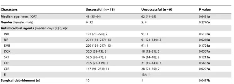
Table 1.Demographic data and treatment ofMycobacterium marinuminfected patients.
Characters Successful (n = 18) Unsuccessful (n = 9) P value
Median age[years (IQR)] 48 (35–64) 62 (41–65) 0.6431a
Gender[female: male] 6: 12 5: 4 0.2770a
Antimicrobial agents[median days (IQR); n]c
INH 191 (73–226); 7 91; 1 0.5102a
RIF 201 (154–247); 13 91 (21–134); 5 0.0266a
EMB 220 (154–247); 13 91; 1 0.1724a
DOX 50.5 (28–73); 3 18 (12–21); 5 0.0507a
SXT 52.5 (28–77); 2 16 (14–18); 2 0.1213a
CIP 70.5 (22–119); 2 21 (15–143); 3 0.5637a
CLR 147 (91–281); 11 28 (21–35); 2 0.0297a
E 134; 1
Surgical debridement[n] 10 1 0.0417b
IQR: inter-quartiles ranges, INH: isoniazid, RIF: rifampicin, EMB: ethambutol, DOX: doxycycline, SXT: sulfamethoxazole-trimethoprim, CIP: ciprofloxacin, CLR: clarithromycin, E: erythromycin.
a) by Mann-Whitney
Utest. b) by Fisher’s exact test.
c) patients received one or more than one agent.
doi:10.1371/journal.pone.0041296.t001
Figure 1. Cutaneous manifestations ofMycobacterium marinuminfections.Footnote:M. marinumskin infections presented with (A) a solitary, or (B) multiple violaceous plaques with hyperkeratotic surface on limbs. The disease also presented as (C) a warty, verrucous plaque with an irregular border on hand, or (D) erythematous swelling of finger with pus discharge.
Antimicrobial susceptibilities
The quality control strain (M. marinum ATCC 927) was susceptible to all 7 drugs. The results of in vitro susceptibility testing indicated thatM. marinumisolates were basically sensitive to the drugs used in the test, with a sensitivity rate ranging between 86.7% (RIF) to 100% (CLR, AMK, and LZD); the only exception was DOX, to which only 1 (3.3%) isolate was sensitive (table 2).
Treatment and outcome
Among these 27 patients, 25 patients gotM. marinumskin and soft tissue infections, one had pulmonary infection, and the last had corneal infection. After medical treatment and/or surgical debridement, 18 were cured without any sequelae, 7 remained draining and unhealed wounds, 1 had complication of hypersen-sitivity pneumonitis, and one had right eye blindness.
For antimicrobial therapy, the median (IQR) duration of prescription (in days) for each antibiotic in the successful versus unsuccessful groups was as follows: CLR, 147 (91–281) vs. 28 (21– 35) (p= 0.0297); and RIF, 201 (154–247) vs. 91 (21–134) (p= 0.0266). These figures indicate that the duration of pre-scription of CLR, and RIF was longer in the successful group than in the unsuccessful group. On the other hand, the median (IQR) duration of prescription for DOX was 50.5 (28–73) vs. 18 (12–21) (p= 0.0507) in the successful versus unsuccessful group , re-spectively. This indicated there is a tendency to treatment failure for prescription of doxycycline. Furthermore, the median (IQR) duration of prescription of the following antibiotics in the successful versus unsuccessful groups was as follows: isoniazid (INH), 191 (73–226) vs. 91 (p= 0.5102); EMB, 220 (154–247) vs. 91 (p= 0.1724); SXT, 52.5 (28–77) vs. 16 (14–18) (p= 0.1213); ciprofloxacin (CIP), 70.5 (22–119) vs. 21 (15–143) (p= 0.5637); and erythromycin, 134, respectively. Besides, 10 patients in the successful group had received surgical debridement; whereas, only 1 patient in the unsuccessful group had received surgical debridement (Fisher’s exact test;p= 0.0417).
Eight patients received DOX treatment; of these, only 3 (37.5%) had successful results (table 3). In addition to the DOX therapy, 2 of these 3 patients further received adjuvant surgical debridement of the lesions, and the third patient received a combination of RIF and EMB treatment for 73 days, and consecutive prescription with CLR, RIF, and EMB for 147 days. Of the 5 patients who underwent unsuccessful DOX treatment, 1 received only local CIP treatment for corneal infection with sequela of right eye blindness, 1 had an overly short CLR and CIP treatment duration (21 days) with persistent nodules on her right hand,1 had an overly short RIF (21 days) and SXT (14 days) treatment duration with
persistent plaques on his right hand, 1 further received SXT, CLR, CIP, and RIF, but had persistent nodules and relapsed 4 months after discontinuing the antibiotics, and the fifth unsuccess-ful patient became lost to follow-up.
Discussion
Efficiently treatment of M. marinum infection remains a chal-lenge. One of the reasons that make diagnosis difficult is the patients’ lack of awareness coupled with the fact thatM. marinum infections progress slowly, with a median (IQR) progression time of 3 (1–6) months. As the patients did not consult the doctor until the clinical symptoms worsened, it was difficult to identify the relationship between M. marinum skin infections and previous trauma history. Besides, cutaneous lesions are generally non-specific and are often initially misdiagnosed as pyoderma, furunculosis, or even sporotrichosis [20–22]. Regarding the clinical treatment, M. marinum infections are usually treated empirically with antituberculosis agents, and antibiotic-suscepti-bility testing is not routinely performed in many clinical laboratories. These issues make the treatment ofM. marinumeven more difficult. The results of this study presented the antibiotic susceptibility pattern of M. marinum and characterized the re-lationship between antimicrobial therapy, surgery, and treatment outcome inM. marinuminfections in Taiwan. These results may thus be used as a reference for clinical treatment ofM. marinum infections in other countries.
The CLSI report does not recommend routine susceptibility testing of this species. However, the test may be necessary for some patients whose samples are still culture positive after receiving several months of unsuccessful therapy [23]. The results ofin vitro susceptibility in this study showed a high susceptibility rate (.90%) for CLR, LZD, AMK, or SMX, which was consistent with effective treatment results. It is suggested that each single agent can be considered for treating in superficialM. marinuminfections. From the viewpoint of pharmacokinetics, LZD could be a good alternative oral agent, although there have been no clinical trials till date to support this hypothesis [24]. Despite some previous reports showing successful treatment ofM. marinuminfection with DOX or minocycline [25,26], the effectiveness of DOX treatment was still controversial due to several reports of treatment failure [11,26–28]. Our results showed that M. marinum strains showed only 3.3% (1/30) sensitivity to DOX. This may reflect the high rate of treatment failure encountered in our patients treated with DOX. The controversy over DOX therapy may be related to the regional drug resistance pattern of M. marinum. Thus, it is suggested that drug susceptibility testing ofM. marinumshould be
Table 2.In vitrosusceptibilities of 30M. marinumisolates.
Antimicrobial agents MIC 50 MIC 90 Range Modal MIC
Geometric mean
MIC 95% CI Sensitive rate (%)
Rifampicin 1 2 0.125–4 1 0.7 0.5–1 86.7
Ethambutol 0.25 4 0.25–16 0.5 1 0.6–1.5 96.7
Clarithromycin 4 6 0.125–8 4 3 2.2–4.1 100
Amikacin 2 8 0.125–16 8 2.3 1.4–3.9 100
Sulfamethoxazole 4 32 0.5–64 4 4.7 2.7–8.1 93.3
Linezolid 2 4 0.5–4 2 1.7 1.4–2.2 100
Doxycycline 16 32 4–32 16 14.9 12.0–18.6 3.3
performed in patients whose treatment has failed and also in regions where the drug-resistance rate is high.
The potential reasons for the treatment failure/difficulty inM. marinum infections include delayed diagnosis and ineffective regimens in antimicrobial therapy. To date, there have been no comparative trials for treatment regimens inM. marinumskin and soft-tissue infections. A general approach is to treat patients with 2 active agents for 1–2 months after resolution of symptoms; the total treatment time is typically 3–4 months [9]. Furthermore, the duration of antimicrobial therapy in pulmonary or disseminated infections is also not standardized. Although, at present, there is no evidence indicating the superiority of combination therapy over monotherapy, there is a tendency to prescribe CLR plus EMB, or RIF plus EMB to treat a deep structure infection [9,29]. In deep structure infections, especially on the hands, early diagnosis and appropriate therapy may play a key role in preventing the loss of normal function [30]. In addition, our results indicated surgical debridement may contribute to favorable treatment outcome.
AmongM. marinuminfections, a rare case involving a pulmonary infection was observed. The patient was a middle-aged woman and received interferon and ribavirin therapy for chronic hepatitis C. She then acquired interstitial pneumonitis where the M. marinumstrain was isolated. This strain was sensitive to all tested antibiotics except DOX. Due to the delayed diagnosis and the lack
of antimicrobial therapy, she developed sequelae such as pulmonary fibrosis and rheumatoid arthritis-like disorders. In 2005, Lai et al. also reported a case of pulmonary infection in an immunocompetent patient [6]. The 2 cases indicatedM. marinum causes superficial infections, in addition to disseminated or even pulmonary infections. It would be worth monitoring and in-vestigating this phenomenon closely for a longer duration.
In conclusion, optimal antimicrobial therapy consisting of CLR and/or RIF plus ethambutol, and surgical debridement may have a high treatment success rate inM. marinuminfection. In contrast, DOX prescription is not suitable for this purpose.
Acknowledgments
We wish to acknowledge Hui-Chin Liao for assistance with the laboratory work. We thank all of our colleagues at Chang Gung Memorial Hospital for the collection of clinical samples. Also, we thank Professor Chee-Jen Chang for the biostatistics consultation.
Author Contributions
Conceived and designed the experiments: TSW HCL. Performed the experiments: TSW HCL. Analyzed the data: CHC CHY HSL CTH YCC. Contributed reagents/materials/analysis tools: TLW PYC LHS AJK JHC CCL. Wrote the paper: TSW HCL CHC CHY.
References
1. Runyon EH (1981) Mycobacteria: an overview. Rev Infect Dis 3: 819–821. 2. Collins CH, Grange JM, Noble WC, Yates MD (1985)Mycobacterium marinum
infections in man. J Hyg (Lond) 94: 135–149.
3. Sivan M, Bose D, Athanasou N, McNally M (2008) Mycobacterium marinum
osteomyelitis of a long bone. Joint Bone Spine 75: 600–602.
4. Patel S, Duke O, Harland C (1995) Septic arthritis due toMycobacterium marinum. J Rheumatol 22: 1607–1608.
5. Holmes GF, Harrington SM, Romagnoli MJ, Merz WG (1999) Recurrent, disseminatedMycobacterium marinuminfection caused by the same genotypically
defined strain in an immunocompromised patient. J Clin Microbiol 37: 3059– 3061.
6. Lai CC, Lee LN, Chang YL, Lee YC, Ding LW, et al. (2005) Pulmonary infection due toMycobacterium marinumin an immunocompetent patient. Clin Infect Dis 40: 206–208.
7. Swift S, Cohen H (1962) Granulomas of the skin due toMycobacterium balneiafter abrasions from a fish tank. N Engl J Med 267: 1244–1246.
Table 3.Clincal manifestations ofM. marinuminfected patients who received doxycycline therapy.
Case Age Gender
Infection
source Site of lesions Histology
Antibiotics
(days) Surgery Outcome
1 41 M Shrimp sting Cornea Ulcer Topical DOX (12),
topical CIP (15)
Keratectomy Failed
2 50 F Trauma Right hand Suppurative
granulomatous inflammation
DOX (7), CLR (21), CIP (21)
None Failed
3 65 M Fish tank Right hand Suppurative
granulomatous inflammation
DOX (21), RIF (21), SXT (14) None Failed
4 69 F Fish tank Right hand Granulomatous
inflammation
DOX (18), SXT (18), CLR (35), CIP (143), RIF (187)
None Relapse
5 31 F ND Right hand Suppurative
granulomatous inflammation
DOX (21) None Lost to
follow-up
6 39 M ND Right
forearm
Suppurative granulomatous inflammation
DOX (ND) Debridement Successful
7 41 M Fish tank Right hand Suppurative
granulomatous inflammation
DOX (28) Debridement Successful
8 51 M Fish Left hand ND DOX (73), RIF
(220), EMB (220), CLR (147)
None Successful
8. Lewis FM, Marsh BJ, von Reyn CF (2003) Fish tank exposure and cutaneous infections due toMycobacterium marinum: tuberculin skin testing, treatment, and prevention. Clin Infect Dis 37: 390–397.
9. Aubry A, Chosidow O, Caumes E, Robert J, Cambau E (2002) Sixty-three cases ofMycobacterium marinuminfection: clinical features, treatment, and antibiotic susceptibility of causative isolates. Arch Intern Med 162: 1746–1752. 10. Wu TS, Chiu CH, Su LH, Chia JH, Lee MH, et al. (2002)Mycobacterium marinum
infection in Taiwan. J Microbiol Immunol Infect 35: 42–46.
11. Ljungberg B, Christensson B, Grubb R (1987) Failure of doxycycline treatment in aquarium-associatedMycobacterium marinuminfections. Scand J Infect Dis 19: 539–543.
12. Pang HN, Lee JY, Puhaindran ME, Tan SH, Tan AB, et al. (2007)Mycobacterium marinumas a cause of chronic granulomatous tenosynovitis in the hand. J Infect 54: 584–588.
13. Vincent V, Brown-Elliott BA, Jost KC Jr, Wallace RJ Jr (2003)Mycobacterium: Phenotypic and Genotypic Identification. In: Murray PR, Baron EJ, Jorgensen JH, Pfaller MA, Yolken RH, editors. Manual of Clinical Microbiology. 8th ed. Washington, D.C., USA: American Society for Microbiology Press. pp. 560– 584.
14. Telenti A, Marchesi F, Balz M, Bally F, Bo¨ttger EC, et al. (1993) Rapid identification of mycobacteria to the species level by polymerase chain reaction and restriction enzyme analysis. J Clin Microbiol 31: 175–178.
15. Kent PT, Kubica GP (1985) Public health mycobacteriology: a guide for the level III laboratory . U.S. Department of Health and Human Services, Centers for Disease Control, Atlanta, Ga.
16. Wallace RJ Jr, Nash DR, Steele LC, Steingrube V (1986) Susceptibility testing of slowly growing mycobacteria by a microdilution MIC method with 7H9 broth. J Clin Microbiol 24: 976–981.
17. Brown BA, Wallace RJ Jr, Onyi GO (1992) Activities of clarithromycin against eight slowly growing species of nontuberculous mycobacteria, determined by using a broth microdilution MIC system. Antimicrob Agents Chemother 36: 1987–1990.
18. NCCLS (2003) Susceptibility Testing of Mycobacteria, Nocardiae, and Other Aerobic Actinomycetes: Approved Standard. Document M24-A. Wayne, PA: NCCLS.
19. Rodrı´guez JC, Ruiz M, Lo´pez M, Royo G (2002) In vitro activity of moxifloxacin, levofloxacin, gatifloxacin and linezolid against Mycobacterium tuberculosis. Int J Antimicrob Agents 20: 464–467.
20. Madan V, Lear JT (2007) Sporotrichoid streptococcal pyoderma. J Eur Acad Dermatol Venereol 21: 572–573.
21. Feddersen A, Kunkel J, Jonas D, Engel V, Bhakdi S, et al. (1996) Infection of the upper extremity byMycobacterium marinumin a 3-year-old boy - diagnosis by 16S-rDNA analysis. Infection 24: 47–48.
22. Adams RM, Remington JS, Steinberg J, Seibert JS (1970) Tropical fish aquariums. A source ofMycobacterium marinuminfections resembling sporotricho-sis. JAMA 211: 457–461.
23. Griffith DE, Aksamit T, Brown-Elliott BA, Catanzaro A, Daley C, et al. (2007) An official ATS/IDSA statement: diagnosis, treatment, and prevention of nontuberculous mycobacterial diseases. Am J Respir Crit Care Med 175: 367– 416.
24. Dryden MS (2011) Linezolid pharmacokinetics and pharmacodynamics in clinical treatment. J Antimicrob Chemother 66 Suppl 4: iv7–iv15.
25. Osorio F, Magina S, Carvalho T, Goncalves MH, Azevedo F (2010)
Mycobacterium marinumskin infection with tenosynovitis successfully treated with doxycycline. Dermatology Online Journal 16: 7.
26. Cummins DL, Delacerda D, Tausk FA (2005) Mycobacterium marinum with different responses to second-generation tetracyclines. Int J Dermatol 44: 518– 520.
27. Schwendiman MN, Johnson RP, Henning JS (2009) Subcutaneous nodules with sporotrichoid spread. Dermatol Online J 15: 11.
28. Donta ST, Smith PW, Levitz RE, Quintiliani R (1986) Therapy ofMycobacterium marinuminfections. Use of tetracyclines vs rifampin. Arch Intern Med 146: 902– 904.
29. Rallis E, Koumantaki-Mathioudaki E (2007) Treatment ofMycobacterium marinum
cutaneous infections. Expert Opin Pharmacother 8: 2965–2978.