Mutations in Pseudohypoparathyroidism 1a and
Pseudopseudohypoparathyroidism in Ethnic Chinese
Yi-Lei Wu1,2., Daw-Yang Hwang3., Hui-Pin Hsiao4, Wei-Hsin Ting1,11, Chi-Yu Huang1,11, Wen-Yu Tsai5, Hung-Chun Chen3,6, Mei-Chyn Chao7,8, Fu-Sung Lo9,10, Jeng-Daw Tsai1,16, Stone Yang12,
Shin-Lin Shih13,16, Shuan-Pei Lin1,11,14,15,17, Chiung-Ling Lin14, Yann-Jinn Lee1,14,16,17*
1Department of Pediatrics, Mackay Memorial Hospital, Taipei, Taiwan, 2Department of Pediatrics, Changhua Christian Hospital, Changhua, Taiwan,3Division of Nephrology, Department of Medicine, Kaohsiung Medical University Hospital, Kaohsiung, Taiwan,4Department of Pediatrics, Kaohsiung Municipal Hsiao Kang Hospital, Kaohsiung Medical University, Kaohsiung, Taiwan,5Department of Pediatrics, National Taiwan University Hospital, Taipei, Taiwan,6Faculty of Renal Care, Department of Medicine, College of Medicine, Kaohsiung Medical University, Kaohsiung, Taiwan,7Division of Genetics, Endocrinology and Metabolism, Department of Pediatrics, Kaohsiung Medical University Hospital, Kaohsiung, Taiwan,8Genome Medicine, College of Medicine, Kaohsiung Medical University, Kaohsiung, Taiwan,9Department of Pediatrics, Chang Gung Memorial Hospital, Taoyuan, Taiwan,10College of Medicine, Chang Gung University, Taoyuan, Taiwan,11Mackay Junior College of Medicine, Nursing, and Management, New Taipei City, Taiwan,12Department of Urology, Mackay Memorial Hospital, Taipei, Taiwan,13Department of Radiology, Mackay Memorial Hospital, Taipei, Taiwan,14Department of Medical Research, Mackay Memorial Hospital, Taipei, Taiwan,15Department of Infant and Child Care, National Taipei University of Nursing and Health, Taipei, Taiwan,16College of Medicine, Taipei Medical University, Taipei, Taiwan,17Institute of Biomedical Sciences and Department of Medicine, Mackay Medical College, New Taipei City, Taiwan
Abstract
An inactivating mutation in theGNASgene causes either pseudohypoparathyroidism 1a (PHP1A) when it is maternally inherited or pseudopseudohypoparathyroidism (PPHP) when it is paternally inherited. We investigated clinical manifestations and mutations of theGNASgene in ethnic Chinese patients with PHP1A or PPHP. Seven patients from 5 families including 4 girls and 2 boys with PHP1A and 1 girl with PPHP were studied. All PHP1A patients had mental retardation. They were treated with calcitriol and CaCO3 with regular monitoring of serum Ca levels, urinary Ca/Cr ratios, and renal sonography. Among them, 5 patients also had primary hypothyroidism suggesting TSH resistance. One female patient had a renal stone which was treated with extracorporeal shockwave lithotripsy. She had an increased urinary Ca/Cr ratio of 0.481 mg/mg when the stone was detected. We detected mutations using PCR and sequencing as well as analysed a splice acceptor site mutation using RT-PCR, sequencing, and minigene construct. We detected 5 mutations: c.85C.T (Q29*), c.103C.T (Q35*), c.840-2A.G (R280Sfs*21), c.1027_1028delGA (D343*), and c.1174G.A (E392K). Mutations c.840-2A.G and c.1027_1028delGA were novel. The c.840-2A.G mutation at the splice acceptor site of intron 10 caused retention of intron 10 in the minigene construct but skipping of exon 11 in the peripheral blood cells. The latter was the most probable mechanism which caused a frameshift, changing Arg to Ser at residue 280 and invoking a premature termination of translation at codon 300 (R280Sfs*21). FiveGNASmutations in ethnic Chinese with PHP1A and PPHP were reported. Two of them were novel. Mutation c.840-2A.G destroyed a spice acceptor site and caused exon skipping. Regular monitoring and adjustment in therapy are mandatory to achieve optimal therapeutic effects and avoid nephrolithiasis in patients with PHP1A.
Citation:Wu Y-L, Hwang D-Y, Hsiao H-P, Ting W-H, Huang C-Y, et al. (2014) Mutations in Pseudohypoparathyroidism 1a and Pseudopseudohypoparathyroidism in Ethnic Chinese. PLoS ONE 9(3): e90640. doi:10.1371/journal.pone.0090640
Editor:Klaus Brusgaard, Odense University hospital, Denmark
ReceivedJune 17, 2013;AcceptedFebruary 3, 2014;PublishedMarch 20, 2014
Copyright:ß2014 Wu et al. This is an open-access article distributed under the terms of the Creative Commons Attribution License, which permits unrestricted use, distribution, and reproduction in any medium, provided the original author and source are credited.
Funding:This work was supported by grants MMH-E-10007 and MMH10170 from Mackay Memorial Hospital and KMUH 99-9M35 and KMUH Nephrology Research Fund from Kaohsiung Medical University Hospital. The funders had no role in study design, data collection and analysis, decision to publish, or preparation of the manuscript.
Competing Interests:The authors have declared that no competing interests exist. * E-mail: [email protected]
.These authors contributed equally to this work.
Introduction
Albright hereditary osteodystrophy (AHO; OMIM #103580) was described by Albright and Smith in 1942 [1]. It is characterized by short stature, round facies, brachydactyly, and short fourth and fifth metacarpals, metatarsals, or both. Pseudo-hypoparathyroidism (PHP) includes a heterogeneous group of metabolic disorders characterized by hypocalcemia, hyperphos-phatemia, and an elevated PTH level because of PTH resistance [2]. On the basis of the presence or absence of AHO, urinary cAMP response to PTH infusion, resistance to other peptide
hormones, and diminished in vitro Gsa activity, PHP is categorized into pseudohypoparathyroidism 1a (PHP1A; OMIM
#103580), pseudohypoparathyroidism 1b (PHP1B; OMIM
#603233), pseudohypoparathyroidism 1c (PHP1C; OMIM
#612462), and pseudohypoparathyroidism 2 (PHP2; OMIM %203330) [3,4,5]. Patients with pseudopseudohypoparathyroid-ism (PPHP; OMIM #612463) have AHO but no resistance to PTH or other hormones.
Genetic mutations for the different subtypes of PHP involve the a-subunit of the stimulatory G protein (Gsa) which is encoded by
the GNAS complex locus located on chromosome 20q13.11 [6]. Gsaexpression is biallelic in most tissues, however, only maternal allele is preferentially expressed in renal proximal tubules, pituitary, thyroid, and gonads [5]. Therefore inactivating GNAS mutations on either the paternal or maternal allele result in Gsa deficiency leading to AHO [7] but resistance of target organs to PTH and other hormones which act through cAMP only if the mutations are on the maternal allele [8]. Four additional imprinted gene products from the GNAS complex locus are paternally expressed XLas, A/B (also referred as 1A) and antisense transcripts (GNASAS), and maternally expressed NESP55 tran-script [9,10,11].
PHP1A is caused by mutations in the GNAS gene on the maternal allele, whereas PPHP is caused by mutations in the gene on the paternal allele [4]. Thus PPHP and PHP1A can occur in different generations of the same family. The clinical presentation of PHP1C is similar to PHP1A except normal in vitro Gsaactivity [5]. Mutations in PHP1C are all in the C-terminal region of Gsa (p.L388P, p.L388R, p.Y391*, p.E392K, and p.E392*) and disrupt receptor-mediated activation but display normal
receptor-inde-pendent activation [4,12]. The molecular defects of familial autosomal dominant PHP1B can be due to microdeletions on the maternal allele of the STX16 gene or NESP55 gene and/or antisense exons 3 and 4, or paternal uniparental isodisomy [9,13,14,15,16,17]. These variations lead to loss of methylation in exon A/B differentially methylated region (DMR), diminishing maternal expression of Gsa in renal proximal tubules [9]. However, most PHP1B are sporadic and their disease causing genes remained to be identified [18]. The genetic cause of PHP2 is unknown. The similarity in urinary excretion of cAMP following PTH administration between acrodysostosis with mutations in PRKAR1AorPDE4D and PHP2 indicates that genes other than GNASmay be responsible for PHP2 [19,20,21].
Although 176 GNAS mutations have been reported in the Human Gene Mutation Database (HGMD, http://www.hgmd. org/; searched on 2013/08/27) and Leiden Open Variation Database (LOVD, http://www.lovd.nl; searched on 2013/08/27) [22], few reports are on Asians [23,24,25]. We conducted clinical and molecular investigations on ethnic Chinese patients with PHP1A or PPHP and compared the findings with those reported
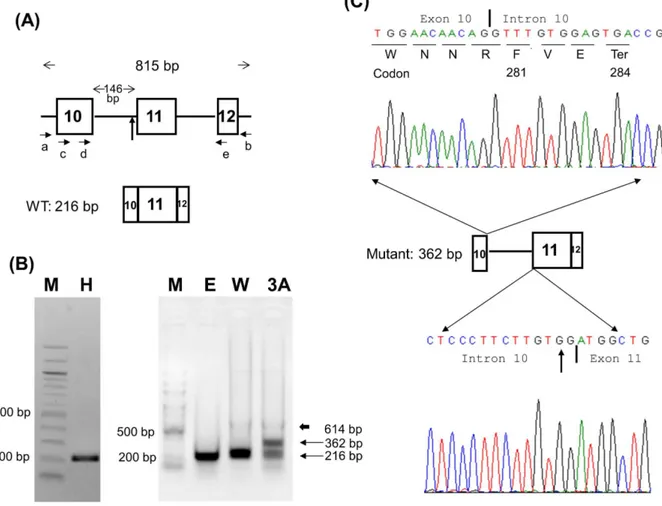
Figure 1. Minigene analysis of c.840-2A.G mutation in patient 3A.(A) The GNAS-IVS10 minigene and primer designs. The construct made by using primers a and b spans from introns 9 to 12 and contains exons 10, 11, and 12 with a total length of 815 bp. Primers c, d, and e were used for semi-nested PCR. (B) COS-7 cells were used for transient transfection study. An extra 362-bp band was found in the mutant (lane 3A) compared with a single 216-bp band in the wild type vectors (lane W). The 362-bp band in lane 3A was subcloned and sequenced. The electropherogram is shown in Panel C. The thick arrow indicates a faint 614-bp band amplified from DNA carried over from COS-7 cells or mini-gene plasmids. Lane H, healthy individual; M, 100 bp DNA ladder; E, empty pcDNA3.1 vector; W, IVS10 wild minigene; 3A, IVS10 mutant minigene. (C) TOPO TA subcloning and sequencing reveal the 362 bp band containing the complete intron 10 of 146 bp. The inclusion of intron 10 changes amino acid Trp to Phe at residue 281 and causes an earlier termination of translation at codon 284 (p.Trp281PhefsTer4 or p.W281Ffs*4). The vertical arrows in Panels A and C denote the site of the c.840-2A.G mutation. The vertical bars in Panel C indicate the exon-intron or intron-exon boundaries.
in patients of other ethnicities. We also assessed the effect of a novel splice acceptor site mutation using a minigene construct and mRNA analysis.
Materials and Methods
Patients
We studied 7 patients from 5 families. These patients included 4 girls and 2 boys with PHP1A and 1 girls with PPHP, diagnosed at 5.8–13.2 years of age. The diagnosis of PHP1A was based on the following criteria: features of AHO, hypocalcemia, hyperphos-phatemia, and resistance to PTH [2]. The diagnosis of PPHP was based on the presence of AHO without biochemical or hormonal abnormalities [2]. Obesity was defined as a BMI value of.95th percentile according to the age- and gender-specific standards for ethnic Chinese children in Taiwan [26]. Patients 1A and 1B are siblings, as are patients 2A and 2B. The institutional review board of Mackay Memorial Hospital approved this study and all subjects including their parents or guardians gave written informed consent.
Detection of Mutations in theGNASGene
Genomic DNA from peripheral blood leukocytes of patients and their relatives was analyzed. All 13 exons and intron–exon boundaries of the GNAS gene were amplified by PCR, using primers and conditions described by Mantovani et al [27]. PCR products were confirmed by electrophoresis on 1.5% agarose gels and were sequenced by using an ABI 3730XL DNA Analyzer (Applied Biosystems, Foster City, CA, USA). The GNAS mRNA and protein reference sequences were NM_000516.4 and NP_000507.1, respectively.
Minigene Constructs, Cell Culture, and Transient Transfection
To analyze the effect of the c.840-2A.G mutation at the splice acceptor site of intron 10 on splicing in patient 3A, a GNAS-IVS10 minigene was constructed. Briefly, 100 ng of genomic DNAs from control individual and patient 3A were used as PCR templates. Primers a, 59-TGTTAGGGATCAGGGTCGCTG-39
(located in intron 9), and b, 59 -AGAGGAGGAACAAGAGAG-GAA-39(located in intron 12), were designed to amplify an 815 bp region (Figure 1A). AccuPrimeTM Taq DNA Polymerase High Fidelity (Invitrogen, Carlsbad, CA, USA) was used according to the manufacturer’s protocol with an initial denaturation at 94uC for 30 seconds, followed by 30 cycles of denaturation (94uC for 15 second), annealing (60uC for 15 seconds), and extension (68uC for
1 minute). The 815 bp PCR products were electrophoresed on 1.5% agarose gels with 100 bp DNA ladder to confirm the correct size, and then purified with QIAquick Gel Extraction Kit (QIAGEN, Valencia, CA, USA). The purified PCR products were subcloned into the pcDNATM3.1/V5-His vector by using pcDNATM3.1/V5-His TOPOH TA Expression Kit (Invitrogen, Carlsbad, CA, USA) according to the manufacturer’s protocol. Both wild-type and mutant minigene constructs were confirmed by Sanger sequencing to ensure correct insert direction and sequences.
COS-7 cells were from ATCC (American Type Culture Collection, CRL-1651) and cultured in DMEM with 10% fetal bovine serum (FBS) plus antibiotics penicillin and streptomycin. Transient transfection was performed at ,80% confluence and
10% FBS/DMEM was replaced with Opti-MEMHreduced serum medium (Life Technologies) before transfection. One mg of plasmid (containing either wild-type or mutated GNAS-IVS10 minigene) in 100ml Opti-MEMH was mixed with 3ml of FuGENE6 (Roche, Indianapolis, IN, USA) before adding to a well of a 6-well plate. Opti-MEMH was removed and replaced with 10% FBS/DMEM 6 hours after transfection.
RT-PCR and Semi-nested PCR
Transfected COS-7 cells were harvested 24 h after transfection and total RNA was extracted with the standard Trizol method (Invitrogen, Carlsbad, CA, USA). Reverse-transcription (RT) of the total RNA was performed by using M-MLV RT kit (Promega, Madison, WI, USA) and the cDNA product was amplified with semi-nested PCR by using primers c, 59 -GGTGGCCAGCAG-CAGCTACA-39, d, 59-CGCCTGCAGGAGGCTCTGAAC-39, and e, 59-CCGGGTCACGCGTGGGTC-39 (Figure 1A). In the semi-nested PCR, primers c and e were used for the first PCR. The PCR product was diluted 100-fold and then 1ml of the diluted PCR product was used for second PCR with primers d and e. The RT-PCR products were gel purified and subcloned into pCR2.1 plasmids with pCR2.1 TOPO TA Cloning Kit (Invitro-gen, Carlsbad, CA, USA) and Sanger sequenced.
RT-PCR of the Peripheral Blood Leukocytes
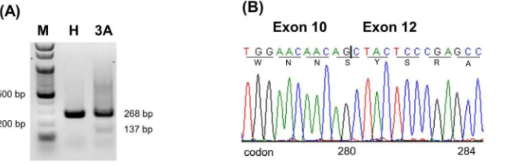
Total RNA from the peripheral blood leukocytes of a healthy control and patient 3A was extracted with the same standard Trizol method and reverse-transcribed with the same M-MLV RT kit (Promega, Madison, WI, USA). Then the cDNA product was amplified with primers c and e (Figure 1A). The PCR product was electrophoresed on a 1.5% agarose gel (Figure 2A).
Figure 2. RT-PCR analysis on the RNA from the peripheral blood leukocytes of a healthy control and patient 3A with primers c and e.(A) The electropherogram shows a band of 268 bp in the healthy control (lane H) but two bands, 268 bp and 137 bp, in patient 3A (lane 3A). The 137-bp band is faint. M indicates 100 bp DNA ladder; H, healthy control; 3A, patient 3A. (B) Sequence analysis of the 137-bp band reveals no exon 11. The deletion of exon 11 results in a frameshift changing Arg to Ser at residue 280 and causing an earlier termination of translation at codon 300 (p.Arg280SerfsTer21 or p.R280Sfs*21). The vertical bar indicates the boundary of exons 10 and 12.
doi:10.1371/journal.pone.0090640.g002
Mutations in Pseudohypoparathyroidism
Family 1 1 2 2 3 4 5 Reference
Member 1A 1B 2A 2B 3A 4A 5A
Diagnosis PHP1A PHP1A PHP1A PHP1A PHP1A PHP1A PPHP
Age at diagnosis (year) 12.2 9.3 8.8 5.8 14.5 13.0 13.2
Sex Female Female Female Male Female Male Female
Round facies + + + + + + +
Short thick neck + + + + + + –
Short 4th and 5th metacarpals + + + + + + +
Short 4th and 5th metatarsals + + + + + + +
Brachydactyly + + + + +
BMI (kg/m2) 24.1a 15.7 25a 21.6a 21.2 36a 21.7
Short stature + + + + + + +
Subcutaneous ossification – – + + + + – –
Intelligence quotient 68 62 59 68 44 57 Averageb 90–109
Ca (mmol/L) 2.1 1.4 1.65 2.4 1.6 1.5 2.25 2.2–2.6
P (mmol/L) 1.73 2.3 2.2 2.0 2.56 3.1 1.55 0.8–1.44
Alkaline phosphatase (IU/L) 180 447 215 410 96 105–420
Intact-PTH (pmol/L) 28.7 97.5 57.5 68.6 26.4 21.69 2.88 1.1–7.2
Free-T4 (pmol/L) 3.9 11.9 15.8 11 12.4 17.1 19 10.2–25.7
TSH (mIU/L) 28.4 15.25 46 4.12 10.71 10.37 2.81 0.54–4.58
LH (IU/L) 7.6 ,3.8 17.3 29.25 5.07 3.8 2–9
FSH (IU/L) 12.94 8.88 57.9 28 2.78 3.11 1.5–12.4
E2 (pmol/L) 60 36 213 73 419 91–355
Prolactin (pmol/L) 247 180 385 ,652
Menarche (yr) 12.25 13 13 11.5 11.33
GNAS mutation
DNA level c.85C.T c.85C.T c.103C.T c.103C.T c.840-2A.G c.1027_1028delGA c.1174G.A
Protein level p.Gln29Ter p.Gln29Ter p.Gln35Ter p.Gln35Ter p.Arg280SerfsTer21 p.Asp343Ter p.Glu392Lys
1-letter symbol p.Q29* p.Q29* p.Q35* p.Q35* p.R280Sfs*21 p.D343* p.E392K
Patients 1A and 1B and patients 2A and 2B are siblings.
aBMI.95th percentile.
bThe performance in school was average.
doi:10.1371/journal.pone.0090640.t001
Mutations
in
Pseudohypop
arathyroidi
sm
ONE
|
www.ploson
e.org
4
March
2014
|
Volume
9
|
Issue
3
|
Results
Clinical Manifestations
All 7 patients had features of AHO (Table 1). Four (1A, 2A, 2B, and 4A; 66.7%) of 6 PHP1A patients were obese. The patients with PHP1A received treatment of calcitriol and calcium carbonate (CaCO3) starting at diagnosis. Laboratory parameters
were monitored every 3–6 months and renal sonography every year. The dosages of calcitriol and CaCO3 were adjusted
according to serum calcium levels and urinary Ca/Cr ratio. Patient 3A took calcitriol (10 ng/kg/day, twice a day) and CaCO3
(elemental Ca 20 mg/kg/day, 4 times a day). Ossification in soft tissue of the sole of her right foot was noted and excised at 17.5 years of age. A renal stone with hyperechogenicity was detected by sonography (Figure 3) when she was 23.4 years old after 8.9 years of therapy. The stone was radio-opaque by radiography (Figure 4). Her serum total Ca levels had been between 2.0 and 2.3 mmol/l (ionized Ca levels between 1.1 and 1.2 mmol/l, reference 1.20– 1.38 mmol/l) with few occasions of hypocalcemia (the lowest ionized Ca level 1.08 mmol/l) due to inadequate compliance. Urinary Ca/Cr ratios had been between 0.013 and 0.125 (reference ,0.213 mg/mg [10]) except it was 0.307 mg/mg 4 months before and 0.481 mg/mg at the detection of the renal stone when she was taking calcitriol 10.2 ng/kg/day and elemental Ca 20.5 mg/kg/day. The dose of CaCO3 was
immediately decreased to 16.8 mg/kg/day of elemental Ca and a follow-up urinary Ca/Cr was 0.071 mg/mg. The stone was disintegrated with extracorporeal shockwave lithotripsy.
Five (83.3%) PHP1A patients also had primary hypothyroidism with elevated thyroid stimulatory hormone (TSH) levels and low
or normal free T4, suggesting TSH resistance at diagnosis. All PHP1A girls had menarche at the normal age (11.5–13 years), but patient 1A had menstrual irregularity and needed progesterone supplement to induce menstruation at age 14 years. All PHP1A patients had mental retardation, with IQs of 44–68. The PPHP patient had not taken an IQ test, but her performance in school was average.
GNASMutations
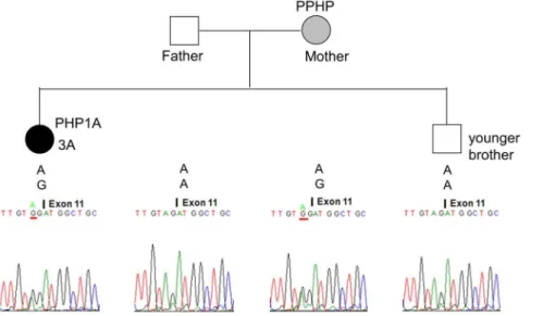
A total of 5 heterozygous mutations were identified in 5 families: c.85C.T (p.Q29*), c.103C.T (p.Q35*), c.840-2A.G (p.R280Sfs*21), c.1027_1028delGA (p.D343*), and c.1174G.A (p.E392K) (Table 1). Mutations c.840-2A.G and c.1027_1028delGA were novel, whereas the others have been reported in patients with PHP1A [2]. The mutant c.840-2A.G allele of patient 3A (Figure 5) and c.1027_1028delGA allele of patient 4A (Figure 6) were passed from their mothers who had PPHP.
Minigene Constructs, RT-PCR, and Semi-nested PCR
Using the GNAS-IVS10 minigene model, we found that the c.840-2A.G mutation destroyed the splicing acceptor and caused the retention of intron 10, resulting in a 362-bp band and a frameshift (Figure 1). This changed the amino acid from Trp to Phe at residue 281, followed by a premature termination of translation at codon 284 (p.W281Ffs*4). Sequencing of the PCR band from lane E (mock transfection with empty vector) in Figure 1B showed the same length of 216 bp similar to human GNASsequence, except 2 variants of c.864C.T and c.885T.C (data not shown).
Figure 3. Sonography of the left kidney.A hyperechoic mass measuring 8 mm in size with acoustic shadow in the center of the left kidney (right panel, arrow).
doi:10.1371/journal.pone.0090640.g003
Mutations in Pseudohypoparathyroidism
RT-PCR of Peripheral Blood Leukocytes
In contrast to the results from the minigene model, RT-PCR of the RNA from the peripheral blood cells of patient 3A revealed only a 268-bp band and a faint but definite 137-bp band (Figure 2A). Sequencing confirmed the shorter PCR fragment containing no exon 11 (Figure 2B). The deletion of exon 11 caused a frameshift changing Arg to Ser at residue 280 and resulting in an earlier termination of translation at codon 300 (p.R280Sfs*21).
Discussion
Pathogenicity of the Detected Mutations
We detected 5 mutations in patients with either PHP1A or PPHP from 5 ethnic Chinese families and all of them co-segregated with disease status in each family. Among them, c.840-2A.G and c.1027_1028delGA were novel. The two mutations were not found in the HGMD, LOVD and NCBI SNP Databases, and 1000 Genomes Projects. They caused frameshifts resulting in premature terminations of translation.
Genetic Epidemiology of PHP
The prevalence of PHP is largely unknown except a reported prevalence of 3.4 (95% CI, 2.6–4.2) per million from Japan [28]. The other estimated prevalence is 0.79 per 100,000 (according to Orphanet Report Series, November 2008) described in a recent publication [9]. A total of 17 different mutations have been identified in 24 PHP1A and 4 PPHP individuals from Asia [23,24,25,29,30,31,32]. Our series added five different GNAS mutations including two novel ones to the list and increased the number to 22. More than half (13 out of 22) of these mutations have not been reported in the other part of the world. And 24% (6 out of 22) are located in exon 1. Mutations c.565_568delGACT, c.308T.C, and c.348_349insC were found in 3, 2, and 2 families. The remaining mutations were reported in only one family each. The trend is similar to those in recently published cohort studies [33,34].
GsaProtein
Parathyroid hormone (PTH) stimulates the formation of intracellular cyclic adenosine monophosphate (cAMP) by adenylyl cyclase via the activation of the Gs protein which is bound to the
Figure 4. Radiography of the kidneys and urinary bladder.A radio-opaque stone measuring 1166 mm in size in the central portion of the left
kidney (arrow).
intracytoplasmic portion of the PTH/PTHrP receptor [35]. The Gs protein is heterotrimeric and composed ofa,b, andcsubunits. A guanosine diphosphate (GDP) is bound to the Gsasubunit in the inactive state. Upon PTH binding to the PTH/PTHrP receptor, a guanosine triphosphate (GTP) replaces the GDP. The Gsa-GTP complex is released from the receptor and thebc subunits and then activates adenylate cyclase which catalyzes the formation of cAMP. The cAMP activates protein kinase A (PKA) which causes phosphaturia. The intrinsic guanosine triphosphatase activity of the Gsasubunit hydrolyzes bound GTP to GDP and terminates the signal transduction. By coupling to the 7-transmembrane-domain receptors, the Gsasubunit is involved in signal transduc-tion of several extracellular messengers and diverse intracellular
effector pathways [3]. The signal-dependent manner of the Ga subunit in binding guanine nucleotide confers specificity to each G protein.
Variability of the Phenotype
The diagnostic age of our small cohort is not bimodally distributed as those in other reports [34,36] because our patients were recruited only from pediatric departments and presented with AHO phenotypes instead of hypocalcemia-related symptoms. Most of our patients had all manifestations of AHO phenotype except patients 3A and 5A who did not have brachydactyly. Families 2, 3 and 4 presented with subcutaneous ossification but no sign of progression to progressive osseous heteroplasia. No
Figure 5. Pseudohypoparathyroidism 1a (PHPIA) caused by the c.840-2A.G mutation in Family 3.The mutant c.840-2A.G allele of patient 3A (solid circle) was inherited from her mother (hatched circle) who had PPHP. Her father and younger brother were normal. The genotype of each member at this site is shown above the electropherogram.
doi:10.1371/journal.pone.0090640.g005
Figure 6. Pseudohypoparathyroidism 1a (PHPIA) caused by the c.1027_1028delGA mutation in Family 4. The mutant c.1027_1028delGA allele of patient 4A (solid square) was inherited from his mother (hatched circle) who had pseudopseudohypoparathyroidism (PPHP). His father was normal. The deletion of two nucleotides in the mutant allele causes a frameshift resulting in a premature termination of translation at codon 343 (p.Asp343Ter or p.D343*). The genotype of each member at this site is shown above the electropherogram.
doi:10.1371/journal.pone.0090640.g006
Mutations in Pseudohypoparathyroidism
apparent delayed puberty were found in female individuals according to their menarche age. Many PHP1A and PPHP patients have a similar heterozygous loss-of-function mutation in theGNASgene, however, the severity of AHO is variable [9]. Two siblings (1A and 1B) of family 1 with Q29* mutation inherited from the mother had the features of AHO and multiple hormone resistance. However, the mother had only mild brachydactyly and relative short stature compared with her sisters. The intrafamilial or interfamilial variability of a phenotype could be due to epigenetic alterations [37], altered transcriptional regulation [38], or effects of other genes [39,40].
Mutations at Splice Sites
Mutations at splice sites cause intron retention, exon skipping, or activation of a cryptic splice site resulting in partial retention of introns or partial loss of exons [3,41]. The analyses of the c.840-2A.G mutation showed inconsistent results from different methods. The mutant allele was expressed with retained intron 10 in the minigene model, however, no mRNA with retention of intron 10 was detected in the patient’s peripheral blood cells. On the contrary, deletion of exon 11 was found in the peripheral blood cells but not in the minigene model. The COS-7 cell transfection experiment was affected by the endogenousGnasfrom Chlorocebus aethiops (African green monkey) because Sanger sequencing showed 2 variants (corresponding to the human GNAS position c.864C.T and c.885T.C) which were not in our design. The best cell model should be of null GNAS, such as the Gnas E22/E22cells from the mouse [42].
Different splicing results caused by the c.840-2A.G mutation can be due to cell-specific GNAS expression in the transfected COS-7 cell and nucleated blood cell using differenttrans-activating factors. Recognition of exon-intron splice site has been shown to be influenced by the upstream introns and splicing signals in the minigene system [43,44]. It is also possible that the mRNA with retention of intron 10 was expressed in the peripheral blood cells but degraded through nonsense-mediated decay [45] to a level which was too low to be detected by our method. We could not know which aberrant GNAS mRNA transcript existed in the renal tubule since the patient did not donate her renal tissue. This mRNA splicing discrepancy between in vitro and in vivo systems have also been observed in other genes [46,47]. Based on the expression of the mutated GNAS gene in the patient’s leukocytes, we concluded that the c.840-2A.G mutation most probably caused deletion of exon 11, resulting in a frameshift changing Arg to Ser at residue 280 and invoking a premature termination of translation at codon 300 (p.Arg280SerfsTer21) (Figure 2). Quan-titative PCR on patient’s EBV-transformed lymphoblasts treated with cycloheximide as a nonsense-mediated decay inhibitor can elucidate the mechanism of low expression level of this splice site mutation [48,49].
Mutations in the Maternal Allele of theGNASGene
All of our PHP1A patients had mental retardation and 80% had primary hypothyroidism with elevated TSH levels. Our findings corroborate previous reports showing that hypothyroidism was present in the majority of patients even at their initial presentation
[12,50,51]. The GNAS gene is biallelically expressed in most tissues, but the paternal allele is variably expressed in the proximal renal tubules, thyroid gland, gonads, and pituitary gland [5]. Therefore mutation in the maternal allele results in hypocalcemia, hyperphosphatemia, hypophosphaturia, resistance to TSH and gonadotropins in PHP1A patients. The variable expression of the paternal allele in the thyroid might be responsible for the wide spectrum of thyroid function alterations in PHP1A patients [52,53] as in our patients.
Hypercalciuria in PHP1A
In the kidney, most filtered calcium is paracellularly reabsorbed in the proximal tubule and the rest is transcellularly reabsorbed [9,54]. PTH stimulates the production of 25(OH)D 1a -hydroxy-lase and inhibits phosphate reabsorption in the proximal tubule as well as promotes reabsorption of calcium in the distal tubule [54]. In patients with PHP1A, the proximal tubule does not respond to PTH whereas the distal tubule does [55]. Phosphaturic effect of PTH is defective but anticalciuric action remains functional, therefore renal stones are rare in PHP1A patients [9]. However, a renal stone developed in patient 3A after 8.9 years of calcitriol and CaCO3 therapy when she had hypercalciuria on a dose of
calcitriol lower than the recommended range of 15–30 ng/kg/day [54] and intake of elemental Ca estimated to be at the upper limit of the recommended range of 20–32.5 mg/kg/day [56] (20.5 mg/ kg/day of supplementary Ca and estimated 11 mg/kg/day from diet [57]). This suggested that renal stones could occur in a PHP1A patient during a period of hypercalciuria. Thus the dosage of calcitriol and elemental Ca should be individualized to maintain normalized PTH and serum calcium levels without hypercalciuria [5].
Conclusions
We report 5GNAS mutations in ethnic Chinese patients with PHP1A or PPHP from 5 families and expanded the spectrum of mutations with 2 novel ones (c.840-2A.G and c.1027_1028delGA). Clinically diagnosis of PHP is straightforward and molecular diagnosis is powerful to elucidate the genetic causes for counseling in affected families. Regular monitoring and adjustment in therapy are mandatory to achieve optimal therapeutic effects and avoid nephrolithiasis in patients with PHP1A.
Acknowledgments
The authors acknowledge the High-throughput Genome Analysis Core Facility of National Core Facility Program for Biotechnology, Taiwan (NSC-100-2319-B-010-001), for sequencing.
Author Contributions
Conceived and designed the experiments: YLW DYH YJL. Performed the experiments: DYH HCC CLL. Analyzed the data: DYH YLW YJL. Contributed reagents/materials/analysis tools: YLW HPH WHT CYH WYT MCC FSL JDT SY SLS SPL. Wrote the paper: YLW DYH YJL. Reseached clinical data: YLW HPH WHT CYH WYT MCC FSL JDT SY SLS SPL.
References
1. Albright F, Burnett CH, Smith PH, Parson W (1942) Pseudohypoparathyroid-ism: an example of Seabright-Bantam syndrome. Endocrinology 6: 922–932. 2. Germain-Lee EL, Groman J, Crane JL, Jan de Beur SM, Levine MA (2003)
Growth hormone deficiency in pseudohypoparathyroidism type 1a: another manifestation of multihormone resistance. J Clin Endocrinol Metab 88: 4059– 4069.
3. De Sanctis L, Romagnolo D, Olivero M, Buzi F, Maghnie M, et al. (2003) Molecular analysis of the GNAS1 gene for the correct diagnosis of Albright hereditary osteodystrophy and pseudohypoparathyroidism. Pediatr Res 53: 749– 755.
pseudohypopara-thyroidism type Ic defines a new subgroup of pseudohypoparapseudohypopara-thyroidism affecting selectively Gsalpha-receptor interaction. Hum Mutat 32: 653–660. 5. Bringhurst FR, Demay MB, Kronenberg HM (2011) Hormones and disorders of
mineral metabolism. In: Melmed S, Polonsky KS, Larsen PR, Kronenberg HM, editors. Williams Textbook of Endocrinology. 12th ed. Philadelphia: Saunders Elsevier. 1237–1304.
6. Kozasa T, Itoh H, Tsukamoto T, Kazino Y (1988) Isolation and characteriza-tion of the human Gs alpha gene. Proc Natl Acad Sci U S A 85: 2081–2085. 7. Levine MA, Ahn TG, Klupt SF, Kaufman KD, Smallwood PM, et al. (1988)
Genetic deficiency of the alpha subunit of the guanine nucleotide-binding protein Gs as the molecular basis for Albright hereditary osteodystrophy. Proc Natl Acad Sci U S A 85: 617–621.
8. Levine MA, Downs RW Jr, Moses AM, Breslau NA, Marx SJ, et al. (1983) Resistance to multiple hormones in patients with pseudohypoparathyroidism. Association with deficient activity of guanine nucleotide regulatory protein. Am J Med 74: 545–556.
9. Mantovani G (2011) Clinical review: Pseudohypoparathyroidism: diagnosis and treatment. J Clin Endocrinol Metab 96: 3020–3030.
10. Linglart A, Maupetit-Mehouas S, Silve C (2013) GNAS -Related Loss-of-Function Disorders and the Role of Imprinting. Horm Res Paediatr: 119–129. 11. Bastepe M (2013) Genetics and epigenetics of parathyroid hormone resistance.
Endocr Dev 24: 11–24.
12. Linglart A, Carel JC, Garabedian M, Le T, Mallet E, et al. (2002) GNAS1 lesions in pseudohypoparathyroidism Ia and Ic: genotype phenotype relationship and evidence of the maternal transmission of the hormonal resistance. J Clin Endocrinol Metab 87: 189–197.
13. Linglart A, Gensure RC, Olney RC, Juppner H, Bastepe M (2005) A novel STX16 deletion in autosomal dominant pseudohypoparathyroidism type Ib redefines the boundaries of a cis-acting imprinting control element of GNAS. Am J Hum Genet 76: 804–814.
14. Bastepe M, Frohlich LF, Linglart A, Abu-Zahra HS, Tojo K, et al. (2005) Deletion of the NESP55 differentially methylated region causes loss of maternal GNAS imprints and pseudohypoparathyroidism type Ib. Nat Genet 37: 25–27. 15. Chillambhi S, Turan S, Hwang DY, Chen HC, Juppner H, et al. (2010) Deletion of the noncoding GNAS antisense transcript causes pseudohypopara-thyroidism type Ib and biparental defects of GNAS methylation in cis. J Clin Endocrinol Metab 95: 3993–4002.
16. Richard N, Abeguile G, Coudray N, Mittre H, Gruchy N, et al. (2012) A new deletion ablating NESP55 causes loss of maternal imprint of A/B GNAS and autosomal dominant pseudohypoparathyroidism type Ib. J Clin Endocrinol Metab 97: E863–867.
17. Bastepe M, Lane AH, Juppner H (2001) Paternal uniparental isodisomy of chromosome 20q–and the resulting changes in GNAS1 methylation–as a plausible cause of pseudohypoparathyroidism. Am J Hum Genet 68: 1283–1289. 18. Fernandez-Rebollo E, Perez de Nanclares G, Lecumberri B, Turan S, Anda E, et al. (2011) Exclusion of the GNAS locus in PHP-Ib patients with broad GNAS methylation changes: evidence for an autosomal recessive form of PHP-Ib? J Bone Miner Res 26: 1854–1863.
19. Linglart A, Menguy C, Couvineau A, Auzan C, Gunes Y, et al. (2011) Recurrent PRKAR1A mutation in acrodysostosis with hormone resistance. N Engl J Med 364: 2218–2226.
20. Michot C, Le Goff C, Goldenberg A, Abhyankar A, Klein C, et al. (2012) Exome sequencing identifies PDE4D mutations as another cause of acrodysos-tosis. Am J Hum Genet 90: 740–745.
21. Lee H, Graham JM Jr, Rimoin DL, Lachman RS, Krejci P, et al. (2012) Exome sequencing identifies PDE4D mutations in acrodysostosis. Am J Hum Genet 90: 746–751.
22. Fokkema IF, Taschner PE, Schaafsma GC, Celli J, Laros JF, et al. (2011) LOVD v.2.0: the next generation in gene variant databases. Hum Mutat 32: 557–563. 23. Ishikawa Y, Tajima T, Nakae J, Nagashima T, Satoh K, et al. (2001) Two mutations of the Gsalpha gene in two Japanese patients with sporadic pseudohypoparathyroidism type Ia. J Hum Genet 46: 426–430.
24. Lim SH, Poh LK, Cowell CT, Tey BH, Loke KY (2002) Mutational analysis of the GNAS1 exons encoding the stimulatory G protein in five patients with pseudohypoparathyroidism type 1a. J Pediatr Endocrinol Metab 15: 259–268. 25. Lam ACF, Chan DHC, Lai KKS, Tong TMF, Lo IFM, et al. (2006) Phenotypic
Spectrum of 3 Pseudohypoparathyroidism type 1a, and 2 Pseudopseudohypo-parathyroidism Chinese Patients with Novel GNAS Mutations. HK J Paediatr 11: 284–289.
26. Chen W, Chang MH (2010) New growth charts for Taiwanese children and adolescents based on World Health Organization standards and health-related physical fitness. Pediatr Neonatol 51: 69–79.
27. Mantovani G, Romoli R, Weber G, Brunelli V, De Menis E, et al. (2000) Mutational analysis of GNAS1 in patients with pseudohypoparathyroidism: identification of two novel mutations. J Clin Endocrinol Metab 85: 4243–4248. 28. Nakamura Y, Matsumoto T, Tamakoshi A, Kawamura T, Seino Y, et al. (2000) Prevalence of idiopathic hypoparathyroidism and pseudohypoparathyroidism in Japan. J Epidemiol 10: 29–33.
29. Sun LH, Cui B, Zhao HY, Tao B, Wang WQ, et al. (2009) Identification of a novel GNAS mutation for pseudohypoparathyroidism in a Chinese family. Endocrine 36: 25–29.
30. Park CH, Park HD, Lee SY, Kim JW, Sohn YB, et al. (2010) Clinical, biochemical, and genetic analysis of korean patients with pseudohypoparathy-roidism type Ia. Ann Clin Lab Sci 40: 261–266.
31. Jin HY, Lee BH, Choi JH, Kim GH, Kim JK, et al. (2011) Clinical characterization and identification of two novel mutations of the GNAS gene in patients with pseudohypoparathyroidism and pseudopseudohypoparathyroid-ism. Clin Endocrinol (Oxf) 75: 207–213.
32. Miao ZM, Wang C, Wang BB, Meng DM, Su DM, et al. (2011) Identification of a novel mutation in a pseudohypoparathyroidism family. Int J Endocrinol 2011: 509549.
33. Elli FM, deSanctis L, Ceoloni B, Barbieri AM, Bordogna P, et al. (2013) Pseudohypoparathyroidism type Ia and pseudo-pseudohypoparathyroidism: the growing spectrum of GNAS inactivating mutations. Hum Mutat 34: 411–416. 34. Fernandez-Rebollo E, Lecumberri B, Gaztambide S, Martinez-Indart L, Perez
de Nanclares G, et al. (2013) Endocrine profile and phenotype-(epi)genotype correlation in Spanish patients with pseudohypoparathyroidism. J Clin En-docrinol Metab 98: E996–1006.
35. Farfel Z, Bourne HR, Iiri T (1999) The expanding spectrum of G protein diseases. N Engl J Med 340: 1012–1020.
36. Gelfand IM, Eugster EA, DiMeglio LA (2006) Presentation and clinical progression of pseudohypoparathyroidism with multi-hormone resistance and Albright hereditary osteodystrophy: a case series. J Pediatr 149: 877–880. 37. Adegbite NS, Xu M, Kaplan FS, Shore EM, Pignolo RJ (2008) Diagnostic and
mutational spectrum of progressive osseous heteroplasia (POH) and other forms of GNAS-based heterotopic ossification. Am J Med Genet A 146A: 1788–1796. 38. Bertaux K, Broux O, Chauveau C, Hardouin P, Jeanfils J, et al. (2006) Runx2
regulates the expression of GNAS on SaOs-2 cells. Bone 38: 943–950. 39. Craigen WJ, Lindsay EA, Bricker JT, Hawkins EP, Baldini A (1997) Deletion of
chromosome 22q11 and pseudohypoparathyroidism. Am J Med Genet 72: 63– 65.
40. Phelan MC, Rogers RC, Clarkson KB, Bowyer FP, Levine MA, et al. (1995) Albright hereditary osteodystrophy and del(2) (q37.3) in four unrelated individuals. Am J Med Genet 58: 1–7.
41. Strachan T, Read AP (2011) Human genetic variability and its consequences. In: Strachan T, Read AP, editors. Human Molecular Genetics. 4th ed. New York: Garland Science. 405–440.
42. Bastepe M, Gunes Y, Perez-Villamil B, Hunzelman J, Weinstein LS, et al. (2002) Receptor-mediated adenylyl cyclase activation through XLalpha(s), the extra-large variant of the stimulatory G protein alpha-subunit. Mol Endocrinol 16: 1912–1919.
43. Romano M, Marcucci R, Baralle FE (2001) Splicing of constitutive upstream introns is essential for the recognition of intra-exonic suboptimal splice sites in the thrombopoietin gene. Nucleic Acids Res 29: 886–894.
44. Cooper TA (2005) Use of minigene systems to dissect alternative splicing elements. Methods 37: 331–340.
45. Culbertson MR (1999) RNA surveillance. Unforeseen consequences for gene expression, inherited genetic disorders and cancer. Trends Genet 15: 74–80. 46. D’Souza-Li L, Canaff L, Janicic N, Cole DE, Hendy GN (2001) An acceptor
splice site mutation in the calcium-sensing receptor (CASR) gene in familial hypocalciuric hypercalcemia and neonatal severe hyperparathyroidism. Hum Mutat 18: 411–421.
47. Hwang DY, Hung CC, Riepe FG, Auchus RJ, Kulle AE, et al. (2011) CYP17A1 intron mutation causing cryptic splicing in 17alpha-hydroxylase deficiency. PLoS One 6: e25492.
48. Ishigaki Y, Li X, Serin G, Maquat LE (2001) Evidence for a pioneer round of mRNA translation: mRNAs subject to nonsense-mediated decay in mammalian cells are bound by CBP80 and CBP20. Cell 106: 607–617.
49. Harries LW, Hattersley AT, Ellard S (2004) Messenger RNA transcripts of the hepatocyte nuclear factor-1alpha gene containing premature termination codons are subject to nonsense-mediated decay. Diabetes 53: 500–504.
50. de Sanctis L, Vai S, Andreo MR, Romagnolo D, Silvestro L, et al. (2004) Brachydactyly in 14 genetically characterized pseudohypoparathyroidism type Ia patients. J Clin Endocrinol Metab 89: 1650–1655.
51. Bastepe M, Juppner H (2003) Editorial: Pseudohypoparathyroidism and mechanisms of resistance toward multiple hormones: molecular evidence to clinical presentation. J Clin Endocrinol Metab 88: 4055–4058.
52. Mantovani G, Ballare E, Giammona E, Beck-Peccoz P, Spada A (2002) The gsalpha gene: predominant maternal origin of transcription in human thyroid gland and gonads. J Clin Endocrinol Metab 87: 4736–4740.
53. Germain-Lee EL, Ding CL, Deng Z, Crane JL, Saji M, et al. (2002) Paternal imprinting of Galpha(s) in the human thyroid as the basis of TSH resistance in pseudohypoparathyroidism type 1a. Biochem Biophys Res Commun 296: 67– 72.
54. Allgrove J (2009) The parathyroid and disorders of calcium and bone metabolism. In: Brook CGD, Clayton PE, Brown RS, editors. Clinical Pediatric Endocrinology. 6th ed. Massachusetts: Wiley-Blackwell. 374–427.
55. Stone MD, Hosking DJ, Garcia-Himmelstine C, White DA, Rosenblum D, et al. (1993) The renal response to exogenous parathyroid hormone in treated pseudohypoparathyroidism. Bone 14: 727–735.
56. Greer FR, Krebs NF (2006) Optimizing bone health and calcium intakes of infants, children, and adolescents. Pediatrics 117: 578–585.
57. Wu SJ, Pan WH, Yeh NH, Chang HY (2007) Dietary nutrient intake and major food sources: the Nutrition and Health Survey of Taiwan Elementary School Children 2001–2002. Asia Pac J Clin Nutr 16 Suppl 2: 518–533.
Mutations in Pseudohypoparathyroidism