in Solid Tumors: Systematic Review and Meta-Analysis
Alberto Ocana1*, Francisco Vera-Badillo2, Mustafa Al-Mubarak2, Arnoud J. Templeton2, Vero´nica Corrales-Sanchez1, Laura Diez-Gonzalez1, Marı´a D. Cuenca-Lopez1, Bostjan Seruga3, Atanasio Pandiella4, Eitan Amir2
1Translational Research Unit, Albacete University Hospital, Albacete, Spain,2Division of Medical Oncology and Hematology, Princess Margaret Cancer Centre and University of Toronto, Toronto, Canada,3Sector of medical Oncology, Institute of Oncology Ljubljana, Ljubljana, Slovenia,4Centro de Investigacio´n del Ca´ncer, CSIC-University of Salamanca, Salamanca, Spain
Abstract
Background:Aberrations in the phosphatidylinositol 3-kinase (PI3K)/mammalian target of rapamycin (mTOR)/AKT pathway are common in solid tumors. Numerous drugs have been developed to target different components of this pathway. However the prognostic value of these aberrations is unclear.
Methods:PubMed was searched for studies evaluating the association between activation of the PI3K/mTOR/AKT pathway (defined as PI3K mutation [PIK3CA], lack of phosphatase and tensin homolog [PTEN] expression by immunohistochemistry
or western-blot or increased expression/activation of downstream components of the pathway by immunohistochemistry) with overall survival (OS) in solid tumors. Published data were extracted and computed into odds ratios (OR) for death at 5 years. Data were pooled using the Mantel-Haenszel random-effect model.
Results:Analysis included 17 studies. Activation of the PI3K/mTOR/AKT pathway was associated with significantly worse 5-year survival (OR:2.12, 95% confidence intervals 1.42–3.16, p,0.001). Loss of PTEN expression and increased expression/ activation of downstream components were associated with worse survival. No association betweenPIK3CAmutations and
survival was observed. Differences between methods for assessing activation of the PI3K/mTOR/AKT pathway were statistically significant (p = 0.04). There was no difference in the effect of up-regulation of the pathway on survival between different cancer sites (p = 0.13).
Conclusion:Activation of the PI3K/AKT/mTOR pathway, especially if measured by loss of PTEN expression or increased expression/activation of downstream components is associated with poor survival. PIK3CA mutational status is not
associated with adverse outcome, challenging its value as a biomarker of patient outcome or as a stratification factor for patients treated with agents acting on the PI3K/AKT/mTOR pathway.
Citation:Ocana A, Vera-Badillo F, Al-Mubarak M, Templeton AJ, Corrales-Sanchez V, et al. (2014) Activation of the PI3K/mTOR/AKT Pathway and Survival in Solid Tumors: Systematic Review and Meta-Analysis. PLoS ONE 9(4): e95219. doi:10.1371/journal.pone.0095219
Editor:Kostas Pantopoulos, Lady Davis Institute for Medical Research/McGill University, Canada
ReceivedFebruary 3, 2014;AcceptedMarch 24, 2014;PublishedApril 28, 2014
Copyright:ß2014 Ocana et al. This is an open-access article distributed under the terms of the Creative Commons Attribution License, which permits unrestricted use, distribution, and reproduction in any medium, provided the original author and source are credited.
Funding:The funders had no role in study design, data collection and analysis, decision to publish, or preparation of the manuscript. Work in A.O. lab is supported by grant PI13/01444 from ISCIII and Diputacio´n de Albacete. Work in A.P. lab is supported by the Fundacio´n Cientı´fica de la AECC and the Ministry of Economy and Competitiveness (BFU2012-39151) and Red Tema´tica de Investigacio´n Cooperativa en Ca´ncer. A.O. and A.P. receive support from CRIS Cancer foundation.
Competing Interests:The authors have declared that no competing interests exist. * E-mail: [email protected]
Introduction
Historically, the development of anti-neoplastic drugs has not focused on the targeting of specific molecular aberrations [1]. However, more recently, some targeted drugs have been developed against known oncogenes in selected patient popula-tions. Examples of this are trastuzumab for HER2 over-expressing or amplified breast and gastric cancer [2,3], imatinib for chronic myeloid leukemia (CML) [4], vemurafenib for metastatic melano-ma with (V600E) B-RAFmutations and crizotinib for non-small cell lung cancer patients with anaplastic lymphoma kinase (ALK)
rearrangements [5,6]. In these examples, development of the drug was carried out in parallel with the identification of a biomarker
that permitted the selection of patients with a higher chance of response.
more aggressive phenotype potentially guiding the selection of more intensive treatment [8].
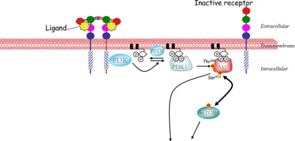
The phosphatidylinositol 3-kinase (PI3K)/mammalian target of rapamycin (mTOR)/AKT pathway has been linked to the pathophysiology of several neoplastic diseases [9,10]. Activation of this pathway can be a result of mutations in thePI3KorAKT
genes, loss of phosphatase and tensin homolog (PTEN), or constitutive activation of upstream regulatory pathways such as receptor tyrosine kinases (Figure 1) [9,10]. Given the pro-oncogenic role of the PI3K/AKT/mTOR pathway in cancer, it has become a target of interest for drug development. Inhibition of mTOR with rapalogs has shown clinical efficacy against some solid tumors, including everolimus for angiomyolipoma associated with tuberous sclerosis, metastatic renal cell carcinoma, breast cancer, or pancreatic neuroendocrine carcinomas and temsiroli-mus for renal cell carcinoma [11–14]. Many other agents in clinical development are designed to inhibit the PI3K/AKT/ mTOR pathway at different levels and include pure PI3K inhibitors, dual PI3K-mTOR inhibitors, AKT inhibitors or mTOR inhibitors [15,16]. Despite the approval of some drugs and the clinical development of other agents targeting the PI3K/ AKT/mTOR pathway, little is known about which patients are more likely to benefit from targeting this pathway. Similarly, the relationship between alterations of this pathway and a more aggressive phenotype is unclear.
Here we report a systematic review and meta-analysis of studies assessing the association of activation of the PI3K/AKT/mTOR pathway and clinical outcome in solid tumors.
Methods
Identification and selection of studies
This analysis was conducted in line with the Preferred Reporting Items for Systematic Reviews and Meta-Analyses guidelines [17]. Medline (Host: PubMed) was searched for studies published between January 2002 and December 2012, which evaluated the expression of components of the PI3K/AKT/ mTOR pathway and survival in solid tumors. We used the MeSH terms ‘‘PIK3CA and cancer’’ and ‘‘PIK3CA-mTOR and cancer’’ and ‘‘PTEN loss and cancer’’ adding the limitation of human
studies. In addition we used the entry ‘‘PIK3CA or mTOR’’ and the name of each specific solid tumor (e.g. PIK3CA or mTOR and breast cancer) to recognize additional studies. The search was restricted to publications in English. Additional studies were identified through reviews of citation lists (Figure S1 and figure S2). Eligibility criteria were the availability of survival data for at least 5 years in relation to three types of pathway aberrations; mutations in the PI3K gene in any domain as measured by polymerase chain reaction (PCR) or other genomic techniques, the lack of PTEN expression by immunohistochemistry (IHC) or western-blot, or the evaluation of downstream components of the pathway like phospho-S6, mTOR, phospho-mTOR, AKT or phospho-4EBP1 by IHC. Studies reporting outcome of patients who had received a specific targeted agent against the PI3K/ AKT/mTOR pathway or related pathways were excluded as were studies reporting only disease free survival or cancer-specific survival.
Data Extraction
Two authors (VSC and AO) extracted information indepen-dently using pre-prepared data abstraction forms. The following details were extracted: tumor type, number of patients, duration of follow-up, mechanism for activation of PI3K/AKT/mTOR pathway (PI3K mutation, activation of mTOR/AKT or PTEN loss), methods used for the evaluation of pathway activation, and cut-off used for defining pathway activation. The outcome of interest was five-year overall survival (OS). In all cases, survival data were estimated from Kaplan-Meier curves independently by two authors (FV and MA).
Data Synthesis
The effect of any aberration in the pathway on overall survival was analyzed initially. Subsequently subgroup analyses were conducted to explore the relationship between survival and different components of the PI3K/AKT/mTOR pathway that were evaluated in each study. Group one was termed ‘‘mTOR or AKT activation’’ and included studies that evaluated downstream components of the pathway including phosphorylated proteins such as mTOR, AKT, S6, and others (see table 1). Group two was termed ‘‘PIK3CA mutations’’ and included those studies that
evaluated mutations in thePI3Kgene. The third included studies that evaluated loss of PTEN as measured by IHC. A second subgroup analysis included assessment based on the primary cancer site.
Statistical analysis
The proportion of patients surviving 5 years was estimated from the Kaplan-Meier curves for both normal (control group) and the presence of the molecular alteration (experimental group). The relative frequency of survival at 5 years between the control and experimental groups was expressed as an odds ratio (OR) and its 95% confidence interval (CI). Data were combined into a meta-analysis using RevMan 5.1 meta-analysis software (Cochrane Collabo-ration, Copenhagen, Denmark). Estimates of ORs were weighted and pooled using the Mantel-Haenszel method. Cochran’s Q (p,
0.10) and the I2 index (.50%) were used to define inter-study heterogeneity. Due to significant heterogeneity, random effects modeling was used for all analyses. Analyses were conducted for all studies and differences between the subgroups were assessed using methods described by Deeks et al. [18]. All statistical tests were two sided, and statistical significance was defined as p,0.05. No corrections were made for multiple comparisons.
Results
Description of studies
We identified 17 studies that evaluated activation of the PI3K/ mTOR/AKT pathway and survival in solid tumors. These studies comprised a total of 4746 patients with a median sample size of 279 patients. The characteristics of included studies are shown in table 1. Six studies evaluated the expression of the PI3K/AKT/ mTOR pathway in breast cancer, five in gastrointestinal tumors, three in gynecological cancers, and one each in prostate, non-small cell lung cancer (NSCLC) and oropharyngeal cancers. Six studies were included in the group called ‘‘mTOR or AKT activation’’, six in the ‘‘PIK3CAmutation’’ group and five in the ‘‘PTEN loss’’
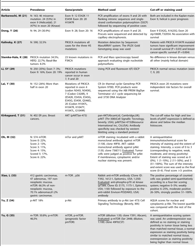
group. The estimated median follow-up was 4.3 years (range = 2.6 to 16 years). The prevalence of pathway activation and the methods used for the analyses of molecular alterations of this pathway are shown in Table 2.
Association of activation of PI3K/AKT/mTOR pathway and survival
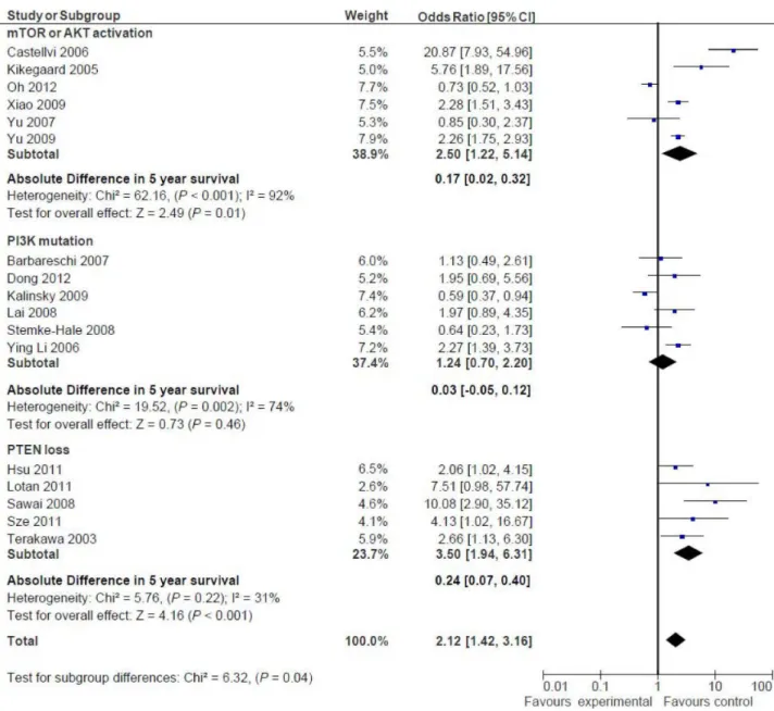
Overall, there was an association between the presence of molecular alterations in the PI3K/AKT/mTOR pathway and worse 5-year survival (OR 2.12; 95% CI 1.42–3.16, p,0.001) (Figure 2). There was significant inter-study heterogeneity (Cochran’s Q p,0.001, I2= 84%).
Association of pathway activation and survival by tumor type
Studies in gastrointestinal tumors (n = 5) and gynecologic cancers (n = 3) showed a numerically higher association with worse survival (OR 2.51; 95% CI 1.83–3.43 and OR 4.78; 95% CI 1.14–20.1, respectively) compared with studies in breast cancer (n = 6) and other solid tumors (NSCLC, prostate and oropharyn-geal, [n = 3]) which showed no association (OR 1.43; 95% CI 0.74–2.79 and OR 1.08; 95% CI 0.44–2.67, respectively). Of interest, in the one study in prostate cancer, there was a large magnitude of effect on survival (OR 7.51, 95% CI 0.98–57.74), but there was no obvious effect seen in studies of NSCLC and orophayngeal cancer (OR 0.73, 95% 0.52–1.03 and 0.85, 95% CI 0.30–2.37, respectively). However, these differences did not meet statistical significance (subgroup difference p = 0.13).
Association of pathway activation and survival by type of activation
The results of the subgroup analysis based on the specific part of the PI3K/AKT/mTOR pathway are shown in figure 3. For studies assessing PIK3CA gene mutations (n = 6), there was no association with worse 5-years survival (OR: 1.24; 95% CI 0.70– 2.20, p = 0.46). In contrast, among studies evaluating activated
Table 1.Characteristics of included studies.
Article Group Subtype Tumor Subtype Follow-up time
Barbareschi, M[21] PIK3CA mutations Breast Cancer Not reported
Dong, Y[26] PIK3CA mutations Gynecological cancer Not reported
Kalinsky, K[27] PIK3CA mutations Breast Cancer Median 12.8 years (range not reported)
Stemke-Hale, K[28] PIK3CA mutations Breast Cancer Not reported
Li, SY[29] PIK3CA mutations Breast Cancer Median 4.2 years (range 0.2–6.5 years)
Lai, Y[30] PIK3CA mutations Breast Cancer Median, 6.4 years (range 0.1–9.3 years)
Kirkegaard, T[31] mTOR or AKT activation Breast Cancer Median 6.5 years (range 0.6–18.4 years)
Oh, M[32] mTOR or AKT activation Non-small Cell Lung Cancer Median 2.9 years (range 0.1–12.7 years)
Xiao, L[33] mTOR or AKT activation Gastrointestinal tumors Median 5.6 years (range 0.02–12.2 years)
Yu, Z[34] mTOR or AKT activation Head and Neck Cancer Mean 3.0 years (range not reported)
Yu, G[35] mTOR or AKT activation Gastrointestinal tumors Mean 3.1 years (1.8–6.1 years)
Castellvi, J[24] mTOR or AKT activation Gynecological cancer Mean 2.6 years (range 2.0–6.7 years)
Hsu, CP[36] PTEN loss Gastrointestinal tumors Median 4.3 years (range 0.3–6.6 years)
Lotan, LT[37] PTEN loss Prostate Cancer Median 16.0 years (range not reported)
Sawai, H[38] PTEN loss Gastrointestinal tumors Median 3.0 years (range not reported)
Sze, KM[39] PTEN loss Gastrointestinal tumors Not reported
Terakawa, N[40] PTEN loss Gynecological cancer Not reported
Table 2.Prevalence and analyses of the molecular alterations of the PI3K/mTOR/AKT pathway.
Article Prevalence Gene/protein Method used Cut-off or staining used
Barbareschi, M (21) N: 163; 46 missense mutation. 24 (53%) in exon 9 (Helicoidal); 21 (47%) in exon 20 (Kinase)
Exon 9: 12 E542K 11 E545K Exon 20: 20 H1047R
PCR amplification of exons 9 and 20 with flanking intronic sequences and single-strand conformation polymorphism (SSCP) followed by sequencing of positive cases
Both are included in the Kaplan-maier. Exon 9, helical is poor prognosis
Dong, Y (26) N: 94; 29 (30.9%) Exon 9: 28; Exon 20: 16 PCR amplifications of exon 9 and 20. Procuts were sequenced and detected by capillary electrophoresis.
Exon 9 E542G, A1625G; Exon 20 stp1060R, T3205A. No association with survival
Kalinsky, K (27) N: 590; 32.5% PIK3CA mutations all cases for the three HS mutations
Mutation detection by Sequenom MassARRAY system. The iPLEX Gold Genotyping assay was used
Patients with PIK3CA H1047R mutated tumors have significant improvement in overall survival (P = 0.03) and breast cancer-specific survival (P = 0.004)
Stemke-Hale, K (28) PIK3CA mutation 34.5%; HER2: 22.7%; Basal-like tumors: 8.3%
23 known mutations in P3KCA
PCR and Mass spectroscopy based approach evaluating single nucleotide polymorfisms
No difference in kinase domain versus all other (mainly helical domain)
Li, SY (29) N: 250 (35%); Exon 7: 3%; Exon 9: 16%; Exon 20: 19%
PIK3CA mutations reported in human cancer occur in exon 7, 9 and 20
Using PCR and fluorescen t(F)-SSCP PIK3CA normal versus mutation (exon 7, 9, 20)
Lai, Y (30) N: 152 (26%) More than half in exon 20
Mutations of PIK3CA reported in exon 4 (codon N345I, N345K), 7 (Codon C420R), 9 (E542K, E545A, E545G, E545G, E545K, Q546E), 20 (Codon H1047L, H1047R, H1047Y, H1047L, G1049R)
CR (in thermal cycler GeneAmp PCR System 9700). PCR products were sequenced using the ABI PRISM BigDye Terminator v3.1 cycle sequencing kit and 3730 DNA Analyzer
PIK3CA exon 20 mutations were independent risk factors for overall survival.
Kirkegaard, T (31) N: 402 ER pos. Breast cancers.
AKT (pAKTSer-473) pan-AKT(AbcamLid, Cambridge,UK); pAKT (Thr-308)(Cell Signallic Tecnology, beverly, USA); pAKT(Ser-473)(Biosource International Inc, CA,USA); Antibody specificity was checked by western blotting using a standard protocol
The cut-off value for high and low levels of pAKT expression is defined as above and below the median histoscore.
Oh, M (32) N: 574 mTOR: Score 0: 22%, Score 2: 13%, Score 3: 11%, Score 4: 15%, Scored 5: 17%, Score 6: 22%.
mTor and pAKT mTOR staining: incubated with a rabbit monoclonal antibody against mTOR (1:100, clone 49F9). AKT: rabbit monoclonal antibody against pAkt (1:50, clone 736E11) Evaluated: Tumor cells were judged as positive for pAkt if membranous, cytoplasmic and/or nuclear staining was present.
A semiquantitative
immunohistochemical score for intensity of staining and the extent of staining. Intensity, a score of 0 to 3 (corresponding to negative, weak, moderate, and strong positivity) Extent of staining was scored as 0 (0%), 1 (1–10%), 2 (11–50%), and 3 (51–100%), The sum of the intensity and extent score was used as the final score (0–6). Final score$3: positive.
Xiao, L (33) 412 gastric carcinomas, 47 adenomas, 197 non-neoplastic mucosa. mTOR: 66.3% of non-neoplastic mucosa, 70.1% adenomas,61.2% gastric carcinomas.
m-TOR , pS6 Rabbit anti-mTOR antibody (Clone ID: Y392, 1612-1, Epitomics, USA; 1:250) Evaluated: anti-phospho-p70 s6 kinase (pT389, Clone ID; E175, 1175-1, Epitomics, USA; 1:50) followed by exposure to the anti-rabbit Envison-PO(DAKO, USA) antibody
The positive percentage of counted cells was graded semi-quantitatively according to a four-tier scoring system; negative 0–5%; weakly positive 6–25%, moderate positive 26–50%, strongly positive 51–100%.
Yu, Z (34) p-AKT 18% p-Akt Primary antibody to p-Akt (ser 473) Cell Signaling Technology (Beverly, MA)
AQUA scores for nuclear and cytoplasmic p-Akt. The lowest quartile was compared with the rest of the cohort.
Yu, G (35) m-TOR: 50.8%; p-mTOR: 46,5%
mTOR, p-mTOR. (prognostic factor pmTOR)
mTOR (dilution 1:50; clone Y391; Abcam), Evaluated: p-mTOR (Ser 2448; dilution, 1:100; clone 49F9;CST)
components of mTOR or AKT (n = 6) a significant association with worse outcome was observed (OR: 2.50; 95% CI 1.22–5.14, p = 0.01). Similarly, studies assessing PTEN loss by IHC (n = 5) showed a significant association with worse survival (OR 3.50; 95% CI 1.94–6.31, p,0.001). This difference between subgroups was significant (p = 0.04).
Discussion
The identification of biomarkers that can inform the clinical behavior of a given tumor is important for patient education and treatment planning. In this study we explored the prognostic role of different components of the PI3K/AKT/mTOR pathway with the intention to identify tumors that rely on this molecular alteration. Such information may help to guide the clinical development of therapeutic strategies against this pathway.
Overall, there was evidence of an association between alterations in the PI3K/AKT/mTOR pathway and poor survival. When analyzing different components of the pathway we observed that those studies evaluating loss of PTEN and activated components of downstream proteins were linked with the poorest 5-years survival. Conversely, PIK3CA mutations were not linked with worse outcome in our analysis. The fact that PTEN is a major regulator of the activation of the PI3K pathway could explain its association with worse outcome [19,20] as those tumors with loss
of PTEN expression could have activation of the different components of this pathway. In contrast, although PIK3CA
mutations are considered driver mutations because they are linked to cell survival and increased proliferation, in our study there was no association with poor outcome [9,10]. PIK3CA mutations compromise numerous molecular lesions including those affecting both the catalytic and helical domains. Mutations in the helical domain may favor the oncogenic capability of PIK3CA by facilitating its interaction with certain signaling intermediates linked to the transmission of pro-oncogenic signals [21]. In the individual studies included in our analysis, mutations at different domains were pooled and this could decrease the statistical power needed to detect a worse outcome in this group for specific mutations. In addition, mutations can have different functional roles and different clinical behavior depending on the tumor type. For example, recent studies have shown thatPIK3CA mutations are associated with different outcomes in breast cancer depending on whether the tumor is estrogen receptor positive or negative, and whether HER2 is over-expressed or amplified compared to HER2-normal [22,23]. Regardless of this, the data presented challenges the clinical relevance ofPIK3CA mutations as unique measures of PI3K/AKT/mTOR pathway activation. This is relevant, as some ongoing clinical trials with agents that target this pathway are using such mutational analysis as an indication of pathway activity, with mutations being used as biomarkers for
Table 2.Cont.
Article Prevalence Gene/protein Method used Cut-off or staining used
Castellvi, J (24) p-4EBP1 (47.1%) p-4EBP1 4EBP1 Cell signaling Tech Scored the percentage of positive cells and intensity of the staining, which was assessed semiquantitatively. Samples that showed any positivity were grouped together for statistical purposes
Hsu, CP (36) N: 133 CRC group: 89.2%R53.4%
PTEN Primary anti-PTEN anti- body (1/200) at room temperature for 2 h
Positive: more than 10%
Lotan, LT (37) N: 397 146 PTEN loss (36.8%).
PTEN Rabbit monoclonal anti-PTEN
antibody(clone D4.3,-9188, cell Signaling Technologies
Using this system, each spot of tumor tissue was scored as negative positive for PTEN protein by comparing staining in malignant gland with that of adjacent benign gland and/or stroma which provided an internal positive control within each tissue core. Staining was classified as negative if the intensity was markedly decreased or entirely negative
Sawai, H (38) PTEN strongly expressed in 62,9% colorectal cancer
PTEN Anti-PTEN antibodies (clone 28116; Santa Cruz Biotechnology, Santa Cruz, CA,USA
The intensity of tissue staining was graded semi quantitatively on a 4 point scale (2,+,++,+++). Likewise, the proportion of cells stained was assessed on a 4 point scale (1: 0–15%; 2: 25–50%; 3: 50–85% and 4: 85–100% cell stained).Tissues were classified into strongly staining and weakly staining
Sze, KM (39) 47,5% PTEN underexpression
PTEN Cell signalling biotechnology, Denver; MA
Western-blot
Terakawa, N (40) 103 endometrial cancers, 36% negative PTEN
PTEN A mouse monoclonal anti PTEN antibody, PTEN A2B1(Santa Cruz Biotechnology, Santa Cruz CA,USA)
A positive case was defined as one in which all of the tumor cells were stained, a heterogeneous case was defined as one with both staining and non-staining tumor cells and a negative case was defined as one with no staining of any tumor cells
selection of patients undergoing experimental treatments with PI3K inhibitors.
The analysis of studies evaluating downstream components by IHC showed a significant association with worse survival. These findings have substantial clinicopathological relevance, as evalu-ation of the activity of a protein appears more biologically relevant than the estimation of gene expression. Therefore, assessment of phosphorylated forms of signaling surrogates such as pS6 or pAKT may be more precise than evaluation of their total levels. However, results from these studies were heterogeneous as the markers evaluated belong to different components of the pathway (mTORC1 and mTORC2) [24].
These data may have relevance beyond prognostic value. Among solid tumors where targeted therapy has been developed against a known oncogene, presence of the oncogene has generally been associated with worse outcome. This is the case in HER2/ neu over-expressing or amplified breast or gastric cancers or BRAF-mutated melanoma. Consequently, it is possible that the effect of drugs targeting the PI3K/AKT/mTOR pathway will only be seen in patients where biomarkers consistently show a detrimental clinical outcome. Based on this hypothesis, it would be expected that PIK3CA mutations may not be associated with improvement in outcome from drugs targeting the PI3K/AKT/ mTOR pathway. This hypothesis is supported by data in breast cancer, which show little predictive value of PIK3CA with the mTOR inhibitor everolimus [15,16,25]. On the other hand, it is known that not all druggable molecular alterations in cancer are linked with worse outcome like the expression of estrogen receptors in breast cancer.
When analyzing the results by tumor type, alterations of the PI3K/AKT/mTOR pathway in breast cancer were not linked with worse outcome. However, most of these studies evaluated
PIK3CA mutations. Conversely, studies in gynecological tumors and gastrointestinal cancers were more enriched in studies evaluating PTEN and protein markers of ‘‘mTOR or AKT activation’’; and these were linked with worse outcome. Only single studies in NSCLC, oropharyngeal and prostate cancers were available and these showed variable results. These studies were generally small and consequently reported wide confidence intervals which crossed the null boundary. Consequently, the relevance of these results in isolation remains unclear. The inconsistent measurement of pathway activation means that the independent effect of PI3K/mTOR/AKT activation in different tumor types cannot be evaluated with certainty
Our study has limitations. This is a meta-analysis of the literature and is therefore more likely to be compromised by selection bias with enrichment for studies reporting positive results. In addition, there is also substantial intra- and inter-study heterogeneity including differences in biomarkers of interest and variability in PIK3CA domain mutations. Despite the use of statistical methods to reduce the effects of such heterogeneity, there remains uncertainty regarding the accuracy of the pooled estimates. Furthermore, hazard ratios were not reported by most studies and therefore we estimated the odds of death at 5 years instead. This is a less robust measure for survival, but was the only feasible method using the available data.
Finally, despite these limitations, results of this study do have some implications for both clinical and translational research. It is shown that the activation of the PI3K/AKT/mTOR pathway is related to poor outcome, and it is particularly relevant in gastrointestinal and gynecological cancers. In addition, the evaluation of PTEN levels ideally complemented with concomi-tant evaluation of the activation status of proteins such as pS6 and
Figure 2. Odds ratio (OR) for 5-year overall survival (OS) in all studies.Forest plots of odds ratios for overall survival at 5 years based on activation of the PI3K/mTOR/AKT pathway. Odds ratios for each trial are represented by the squares, the size of the square represents the weight of the trial in the meta-analysis, and the horizontal line crossing the square represents the 95% confidence interval. The diamonds represent the estimated pooled effect based for each cohort individually (labeled subtotal) and for all cohorts together (labeled total).
AKT is linked with worse outcome probably identifying tumors that rely most on the PI3K/AKT/mTOR pathway.
Supporting Information
Figure S1 Flow diagram of literature search.
(TIFF)
Figure S2 PRISMA flowchart using MeSH terms.
(PPT)
Checklist S1
(DOC)
Author Contributions
Conceived and designed the experiments: AO FV MA AT VC BS AP EA. Performed the experiments: AO FV MA AT VC BS AP EA LD MC. Analyzed the data: AO FV MA AT VC BS AP EA LD MC. Contributed reagents/materials/analysis tools: AO FV MA AT VC BS AP EA LD MC. Wrote the paper: AO FV MA AT VC BS AP EA.
References
1. Amir E, Seruga B, Martinez-Lopez J, Kwong R, Pandiella A, et al.(2011) Oncogenic targets, magnitude of benefit, and market pricing of antineoplastic drugs. J Clin Oncol 29: 2543–2549.
2. Slamon DJ, Leyland-Jones B, Shak S, Fuchs H, Paton V, et al.(2001) Use of chemotherapy plus a monoclonal antibody against HER2 for metastatic breast cancer that overexpresses HER2. N Engl J Med 344:783–92.
Figure 3. Odds ratio (OR) for 5-year overall survival (OS) according to the expression of different components of the PI3K/mTOR pathway (group subtype).Forest plots of odds ratios for overall survival at 5 years split by subgroups defined by type of activation of the PI3K/ mTOR/AKT pathway. Odds ratios for each trial are represented by the squares, the size of the square represents the weight of the trial in the meta-analysis, and the horizontal line crossing the square represents the 95% confidence interval. The diamonds represent the estimated pooled effect based for each cohort individually (labeled subtotal) and for all cohorts together (labeled total).
3. Bang YJ, Van Cutsem E, Feyereislova A, Chung HC, Shen L, et al.(2010) Trastuzumab in combination with chemotherapy versus chemotherapy alone for treatment of HER2-positive advanced gastric or gastro-oesophageal junction cancer (ToGA): a phase 3, open-label, randomised controlled trial. Lancet 376: 687–97.
4. O’Brien SG, Guilhot F, Larson RA, Gathmann I, Baccarani M, et al.(2003) Imatinib compared with interferon and low-dose cytarabine for newly diagnosed chronic-phase chronic myeloid leukemia. N Engl J Med 348: 994–1004. 5. Chapman PB, Hauschild A, Robert C, Haanen JB, Ascierto P, et al.(2011)
Improved survival with vemurafenib in melanoma with BRAF V600E mutation. N Engl J Med 364: 2507–16.
6. Kwak EL, Bang YJ, Camidge DR, Shaw AT, Solomon B, et al. (2010) Anaplastic lymphoma kinase inhibition in non-small-cell lung cancer. N Engl J Med 363: 1693–703.
7. Sawyers CL (2008) The cancer biomarker problem. Nature 452: 548–52. 8. Paik S, Shak S, Tang G, Kim C, Baker J, et al.(2004).A multigene assay to
predict recurrence of tamoxifen-treated, node-negative breast cancer. N Engl J Med 351: 2817–26.
9. Engelman JA (2009) Targeting PI3K signalling in cancer: opportunities, challenges and limitations. Nat Rev Cancer 9: 550–62.
10. Courtney KD, Corcoran RB, Engelman JA.(2010). The PI3K pathway as drug target in human cancer. J Clin Oncol 28: 1075–83.
11. Bissler JJ, Kingswood JC, Radzikowska E, Zonnenberg BA, Frost M, et al. (2013) Everolimus for angiomyolipoma associated with tuberous sclerosis complex or sporadic lymphangioleiomyomatosis (EXIST-2): a multicentre, randomised, double-blind, placebo-controlled trial. Lancet 381: 817–24.
12. Motzer RJ, Escudier B, Oudard S, Hutson TE, Porta C, et al. (2008) Efficacy of everolimus in advanced renal cell carcinoma: a double-blind, randomised, placebo-controlled phase III trial. Lancet 372: 449–56.
13. Baselga J, Campone M, Piccart M, Burris HA 3rd, Rugo HS, et al. (2012) Everolimus in Postmenopausal Hormone-Receptor-Positive Advanced Breast Cancer. N Engl J Med 366: 520–9.
14. Hudes G, Carducci M, Tomczak P, Dutcher J, Figlin R, et al. (2007) Temsirolimus, interferon alfa, or both for advanced renal-cell carcinoma. N Engl J Med 356 :2271–81.
15. Ocana A, Amir E, Seruga B, Martin M, Pandiella A (2013) The evolving landscape of protein kinases in breast cancer: Clinical implications. Cancer Treat Rev 39: 68–76.
16. Garcia-Echeverria C, Sellers WR (2008) Drug discovery approaches targeting the PI3K/Akt pathway in cancer. Oncogene 27: 5511–26.
17. Liberati A, Altman D, Tetzlaff J (2009) The PRISMA statement for reporting systematic reviews and meta-analyses of studies that evaluate health care interventions: explanation and elaboration. PLoS Med 21; 6(7):e1000100. 18. Deeks JJ, Higgins JPT, Altman D.G, editors Ltd (2006) Analysing and presenting
results. Cochrane Handbook for Systematic Reviews of Interventions 4 2 5 In: The Cochrane Library, Chichester, UK, John Wiley & Sons.
19. Li J, Yen C, Liaw D, Podsypanina K, Bose S, et al. (1997) PTEN, a putative protein tyrosine phosphatase gene mutated in human brain, breast, and prostate cancer. Science 275: 1943–7.
20. Alonso A, Sasin J, Bottini N, Friedberg I, Friedberg I, et al.(2004) Protein tyrosine phosphatases in the human genome. Cell 117: 699–711.
21. Barbareschi M, Buttitta F, Felicioni L,Cotrupi S, Barassi F, et al. (2007) Different prognostic roles of mutations in the helical and kinase domains of the PIK3CA gene in breast carcinomas. Clin Cancer Res 13: 6064–9.
22. Baselga J, Corte´s J, Im S-A, Clark E, Kiermaier A, et al. (2012) Biomarker Analyses in CLEOPATRA: A Phase III, Placebo-Controlled Study of Pertuzumab in HER2-Positive, First-Line Metastatic Breast Cancer (MBC). Cancer Research: December 15, 2012; Volume 72, Issue 24, Supplement 3.
23. Ramirez-Ardila D, Helmijr JC, Look MP, Lurkin I, Ruigrok-Ritstier K, et al. (2013) Hotspot mutations in PIK3CA predict treatment outcome on Aromatase Inhibitors but are not predictive for Tamoxifen. Breast Cancer Res Treat 139: 39–49
24. Castellvi J, Garcia A, Rojo F, Ruiz-Marcellan C, Gil A, Baselga J, et al. (2006) Phosphorylated 4E binding protein 1: a hallmark of cell signaling that correlates with survival in ovarian cancer. Cancer 107: 1801–11.
25. Hortobagyi GN, Piccart-Gebhart MJ, Rugo S . Hope, Burris A . Howard, Campone Mario, et al. (2013) Correlation of molecular alterations with efficacy of everolimus in hormone receptor–positive, HER2-negative advanced breast cancer: Results from BOLERO-2. J Clin Oncol 31, (suppl; abstr LBA509). 26. Dong Y, Yang X, Wong O, Zhang X, Liang Y, et al. (2011) PIK3CA mutations
in endometrial carcinomas in Chinese women: phosphatidylinositol 39-kinase pathway alterations might be associated with favorable prognosis. Hum Pathol 43: 1197–205.
27. Kalinsky K, Jacks LM, Heguy A, Patil S, Drobnjak M, et al. (2009) PIK3CA mutation associates with improved outcome in breast cancer. Clin Cancer Res 15: 5049–59.
28. Stemke-Hale K, Gonzalez-Angulo AM, Lluch A, Neve RM, Kuo WL, et al.(2008) An integrative genomic and proteomic analysis of PIK3CA, PTEN, and AKT mutations in breast cancer. Cancer Res 68: 6084–91.
29. Li SY, Rong M, Grieu F, Iacopetta B (2006) PIK3CA mutations in breast cancer are associated with poor outcome. Breast Cancer Res Treat 96: 91–5. 30. Lai YL, Mau BL, Cheng WH, Chen HM, Chiu HH, et al. (2008) PIK3CA exon
20 mutation is independently associated with a poor prognosis in breast cancer patients. Ann Surg Oncol 15: 1064–9.
31. Kirkegaard T, Witton CJ, McGlynn LM, Tovey SM, Dunne B, et al. (2005) AKT activation predicts outcome in breast cancer patients treated with tamoxifen. J Pathol 207: 139–46.
32. Oh MH, Lee HJ, Yoo SB, Xu X, Choi JS, et al. (2012) Clinicopathological correlations of mTOR and pAkt expression in non-small cell lung cancer. Virchows Arch 460: 601–9.
33. Xiao L, Wang YC, Li WS, Du Y (2009) The role of mTOR and phospho-p70S6K in pathogenesis and progression of gastric carcinomas: an immunohis-tochemical study on tissue microarray. J Exp Clin Cancer Res 28:152. 34. Yu Z, Weinberger PM, Sasaki C, Egleston BL, Speier WF 4th, et al. (2007)
Phosphorylation of Akt (Ser473) predicts poor clinical outcome in oropharyngeal squamous cell cancer. Cancer Epidemiol Biomarkers Prev 16: 553–8. 35. Yu G, Wang J, Chen Y, Wang X, Pan J, et al. (2009) Overexpression of
phosphorylated mammalian target of rapamycin predicts lymph node metastasis and prognosis of chinese patients with gastric cancer. Clin Cancer Res 15: 1821– 9.
36. Hsu CP, Kao TY, Chang WL, Nieh S, Wang HL, Chung YC (2011) Clinical significance of tumor suppressor PTEN in colorectal carcinoma. Eur J Surg Oncol 37: 140–7.
37. Lotan TL, Gurel B, Sutcliffe S, Esopi D, Liu W, et al. (2011) PTEN protein loss by immunostaining: analytic validation and prognostic indicator for a high risk surgical cohort of prostate cancer patients. Clin Cancer Res 17: 6563–73. 38. Sawai H, Yasuda A, Ochi N, Ma J, Matsuo Y, et al. (2008). Loss of PTEN
expression is associated with colorectal cancer liver metastasis and poor patient survival. BMC Gastroenterol 26; 8:56.
39. Sze KM, Wong KL, Chu GK, Lee JM, Yau TO, et al. (2011) Loss of phosphatase and tensin homolog enhances cell invasion and migration through AKT/Sp-1 transcription factor/matrix metalloproteinase 2 activation in hepatocellular carcinoma and has clinicopathologic significance. Hepatology 53: 1558–69.