XAV939-Mediated ARTD Activity Inhibition in
Human MB Cell Lines
Cristiano Renna, Roberta Salaroli, Claudia Cocchi, Giovanna Cenacchi*
Department of Biomedical and Neuromotor Sciences, Alma Mater Studiorum University of Bologna, Bologna, Italy
Abstract
Diphtheria toxin-like ribosyltransferases 1 and 5 (ARTD-1, ARTD-5) are poly ADP-ribose enzymes (PARP) involved in non-homologous end-joining (NHEJ), which is the major pathway of double-strand break (DSB) repair. In addition, ARTD-5, or Tankyrase (TNKS), is a positive regulator of the WNT signaling implicated in the development and bio-logical behavior of many neoplasms, such as Medulloblastoma (MB), in which radiotherapy is an essential part of the treatment. The use of radiosensitizing agents may improve the therapeutic index in MB patients by increasing the efficacy of radiotherapy, while reducing toxicity to the neuroaxis. ARTD-5 seems to be a good molecular target for improving the cur-rent treatment of MB. In this study, we used the small molecule XAV939, a potent ARTD-5 inhibitor with a slight affinity for ARTD-1, in different human MB cell lines. XAV939 inhibited the WNT pathway and DNA-PKcs in our MB cells, with many biological consequences. The co-administration of XAV939 and ionizing radiations (IR) inhibited MB cells proliferation and clonogenic capacity, decreased their efficacy in repairing DNA damage, and increased IR-induced cell mortality. In conclusion, ourin vitrodata show that XAV939 could be a very promising small molecule in MB treatment, and these results lay the basis for furtherin vivo
studies with the aim of improving the current therapy available for MB patients.
Introduction
ARTDs, a superfamily of 17 proteins, play a crucial role in different cellular functions such as DNA damage detection and repair, chromatin modification, mitotic apparatus formation, and cell death by transferring ADP-ribose unit or units onto specific molecular targets (a post-translational modification process referred to as“PARsylation”). Given this essential role in DNA mechanism repair, several studies have been carried out to explore the therapeutic poten-tial of ARTDs specific inhibitors. Thus, bothin vitroand mice studies indicate the rationale to combine ARTDs inhibitors with DNA damaging agents in many different tumor types. On Oc-tober 24, 2014, AstraZeneca announced that the Committee for Medicinal Products for Human Use (CHMP) of the European Medicines Agency (EMA) has adopted a positive opin-ion recommending the marketing authorizatopin-ion of Lynparza (olaparib, an ARTD-1 and ARTD-2 inhibitor) as monotherapy for the maintenance treatment of patients with relapsed
OPEN ACCESS
Citation:Renna C, Salaroli R, Cocchi C, Cenacchi G (2015) XAV939-Mediated ARTD Activity Inhibition in Human MB Cell Lines. PLoS ONE 10(4): e0124149. doi:10.1371/journal.pone.0124149
Academic Editor:Pier Giorgio Petronini, University of Parma, ITALY
Received:April 17, 2014
Accepted:March 12, 2015
Published:April 2, 2015
Copyright:© 2015 Renna et al. This is an open access article distributed under the terms of the Creative Commons Attribution License, which permits unrestricted use, distribution, and reproduction in any medium, provided the original author and source are credited.
Data Availability Statement:All relevant data are within the paper.
Funding:The work was supported by Bologna University RFO funds and Fondazione del Monte di Bologna e Ravenna (www.fondazionedelmonte.it), Italy. The funders had no role in study design, data collection and analysis, decision to publish, or preparation of the manuscript.
BRCA-mutated high grade serous epithelial ovarian, fallopian tube, or primary peritoneal cancer. [1–6].
The ARTD family member, ARTD-5, otherwise known as tankyrase (TNKS) has been shown to be involved in a multitude of critical cellular processes; it consists of two isoforms (TNKS1 and TNKS2), which share 85% amino acid sequence identity and have overlapping functions [7–10].
TNKS1 regulates DNA repair via PARsylation mediated stabilization of DNA-dependent Protein Kinase catalytic subunit (DNA-PKcs). The depletion of TNKS by siRNA-mediated knockdown or its inhibition resulted in proteasome-mediated DNA-PKcs degradation. The failure of the non-homologous end-joining (NHEJ) function on DNA damage mechanism, the major pathway of DSB repair, is also evident. Correct DNA-PKcs activity is critical for the NHEJ mechanism; hence, TNKS inhibition results in an increased sensitivity to DNA damage agents [11–13].
Furthermore, it was shown that TNKS is a positive regulator of WNT signaling. TNKS-mediated PARsylation of AXIN induces the degradation of AXIN, the concentration-limiting component of theβ-catenin destruction complex, and therefore, WNT pathway activation. Thus, TNKS inhibition antagonizes the WNT pathway by promoting Axin stabilization [14].
Alterations of the WNT pathway often occur in Medulloblastoma (MB), a highly invasive embryonal neuroepithelial tumor of the cerebellum (WHO, grade IV) [15–18]. By analyzing gene-expression profiles, a recent study has proposed four subtypes of MB, each of which is characterized by a distinct genetic profile, oncogenic pathway activation, and clinical outcomes. Specifically, MB subgroup A is characterized by the WNT pathway, subgroup B is character-ized by SHH signaling, and C and D are charactercharacter-ized by the expression of neuronal differenti-ation genes [19,20].
Radiotherapy is particularly effective in MB treatment. Ionizing radiations (IR) induce dif-ferent DNA damage typologies; the most critical lesions are DSBs [13]. Unfortunately, radio-therapy is notorious for causing late-onset side effects, not only regarding the developing cortex and deep brain structures, but also the posterior fossa; the risk is higher in younger pa-tients [21–26].
The use of radiosensitizing agents, which target specific tumor cell characteristics, such as their replication dependency and DNA repair defects, may improve the therapeutic index by increasing the efficacy of radiotherapy, while reducing the toxicity and damage to the developing brain.
This therapeutic strategy could be particularly useful in highly proliferative high-grade childhood brain tumors such as MB, which arise in largely non-replicative normal tissues with proficient DNA repair [27].
In this regard, TNKS seems to be an optimal molecular target to improve the currently available therapy for MB, given its crucial role in the NHEJ pathway and, consequently, in DSB repair. Moreover, as mentioned previously, TNKS depletion results in a WNT pathway inhibi-tion that is strongly involved in MB development and biological behavior.
Materials and Methods
Cell culture
The study of the therapeutic potential of ARTD-1 and TNKS inhibition was conducted on two different human MB cell lines: ONS-76 (derived from classic MB) and DAOY (desmoplastic MB) [30–31]. (ONS-76 were kindly provided by Dr. Charles G. Eberhart, John Hopkins Uni-versity, Baltimore, MD, with the agreement of Dr. Mike Bobola, University of Washington, Seattle, WA). DAOY were purchased from American Type Culture Collection (ATCC; Manassas, VA).
ONS-76 and DAOY cell lines were maintained, respectively, in RPMI 1640 (Roswell Park Memorial Institute medium; Euroclone, Milan, IT), supplemented with 10% heat-inactivated fetal bovine serum (FBS; Euroclone, Milan, IT), MEM (Minimum Essential Medium with ear-le’s salts; Euroclone, Milan, IT), supplemented with 10% heat-inactivated fetal bovine serum (FBS; Euroclone, Milan, IT), and 1% sodium pyruvate (Eurobio, courtaboeuf Cedex, FR). Both media were supplemented with 1% penicillin-streptomycin (Euroclone, Milan, IT), 1% 2 mM L-glutamine (Euroclone, Milan, IT), and 1% non-essential amino acids (Euroclone, Milan, IT). Cell cultures were maintained at 37°C in 5% CO2.
Protein extraction and Western Blots
ONS-76 and DAOY nuclear and total proteins were extracted and Western Blotting (WB) was performed, as previously described [32]. The following primary antibodies were used at the re-spective dilutions in blocking solution: rabbit polyclonal anti-β-catenin (H-102; Santa Cruz Bio-technology, Santa Cruz, CA, USA) 1:1000; mouse monoclonal anti-DNA-PKcs 1:1000 (1C12; Abcam, Cambridge, UK); goat polyclonal anti-Axin (R20; Santa Cruz Biotechnology, Santa Cruz, CA, USA) 1:500; polyclonal anti-β-actin (I-19; Santa Cruz Biotechnology, Santa Cruz, CA, USA) 1:1000; and mouse monoclonal anti-lamin B1 (ZL-b, ABcam, Cambridge, UK) 1:400. Primary antibodies were detected with horseradish peroxidase (HRP)-labelled secondary anti-bodies at the respective dilutions in blocking solution: anti-rabbit (GE Healthcare Europe GmbH, Freiburg, DE) 1:1000; anti-mouse (GE Healthcare Europe GmbH; Freiburg, DE) 1:10000; and anti-goat (Santa Cruz Biotechnology, Santa Cruz, CA) 1:1000. Total DNA-PKcs, β-actin, and Axin were detected on total protein extracts, whileβ-catenin and lamin B detection was performed on nuclear extracts.β-actin and lamin B were used to check for total and nuclear loading, respectively. The experiments were performed at least three times.
blotting tank (BioRad, Hercules, CA, USA) contained plate electrodes and a supercooling coil. The equipment was attached to a thermocirculator set at -13.5°C. Nitrocellulose membranes were incubated with anti-DNA-PKcs and anti-β-actin antibody.
XAV939 and IR treatment
DAOY and ONS-76 cell lines were plated 24 h prior to treatment with 5μM of XAV939 or with equal DMSO volume, with radiations or with a co-administration of drug and radiations (cells were treated with the small molecule and irradiated 8 h later).
XAV939 (Sigma-Aldrich Corporation, St. Louis, MO, USA) was solubilized in DMSO to a stock concentration of 50 mM, which was diluted to a working concentration of 5μM. Cultures were maintained under these conditions for the duration of the designated time course. Con-trols were exposed to DMSO alone.
Cells were irradiated with an Irradiateur Biologique 437C (CIS-BIO; Cedex, France)γ-ray machine at a dose of 2 or 10 Gy and a dose rate of approximately 2 Gy/min.
Post treatments, the cells were analyzed at different time points. Cell growth, colony forma-tion, mortality rate, and DNA repair efficacy were evaluated.
Cell growth analysis
ONS-76 and DAOY MB cell lines were plated at a density of 200,000 and 500,000 viable cells per T25 flask, respectively, and treated as described above.
Cell number and viability were assessed using the Trypan blue reagent (Sigma-Aldrich Cor-poration, St. Louis, MO, USA). The cells were collected by trypsin-EDTA treatment, pelleted by gentle centrifugation, and resuspended in Trypan blue reagent to count viable cells using a light microscope every day for 4 days after treatments. Multiple replicate flasks were assayed at each time point in each experiment, and each sample was examined in three independent ex-periments. At each time point, we report the mean cell number from all the scored flasks; error bars display SE.
Cell mortality assay
In order to evaluate cell mortality, 200,000 cells were seeded in T25 culture flasks and collected 48, 72, and 96 h after treatments. Irradiated and control samples from each line were harvested at corresponding time-points, trypsinized, and total cells were suspended 1:1 in Trypan Blue solution. Dead and total cells from every sample were counted using a Neubauer chamber. Multiple replicate flasks were assayed at every time point in each experiment, and each sample was examined in three independent experiments. At each time point, we report the mean cell number from all the scored flasks; error bars display SE.
In a second set of experiments, cells were treated with XAV939 at the indicated concentra-tion and irradiated with a double 2 Gy dose. Mortality rate was then calculated.
In the histograms, cell mortality of the treated cells was normalized to DMSO control cells.
Clonogenic assay
The survival fraction at 2 Gy (SF2), determined as the ratio of the number of colonies formed by irradiated cells to the number of colonies formed by non-irradiated cells, was also calculated.
Neutral Comet Assay
In order to avoid high lyses temperatures, which could alter the assay reliability, we used the protocol described by Olive and Banàth, with some modifications [33,34]. MB cell lines treated and untreated with XAV939 (5μM) were exposed to 10 Gy. Neutral Comet Assay was per-formed 0, 16, and 24 h after radiation. Non-irradiated cells were used as controls. In short, after treatments cells were resuspended in low melting agarose and immediately placed on mi-croscopic slides (pre-coated with 0.5% normal melting agarose) under coverslips and, after aga-rose solidification, they were lysed with specific basic buffer (2% sarkosyl, 0.5M Na2EDTA, pH 8.0, stored at 4°C) for at least 4 h at 37°C. After lysis, the slides were washed in electrophoresis buffer (90 mM Tris buffer, 90 mM boric acid, 2 mM Na2EDTA, pH 8.5) and placed in a hori-zontal gel electrophoresis chamber filled with fresh electrophoresis buffer. Electrophoresis was conducted at 7 mA (0.6 V/cm) for 25 min and at a temperature not higher than 15°C. The slides were then washed in distillated water, stained with a 20μg/mL ethidium bromide solu-tion for 20 min, and covered with cover glasses. The image analysis was performed on pictures of 50 randomly selected comets per slide for each sample. Experiments were repeated at least three times. Data show mean comet TM and SD.
Pictures were captured using a fluorescence microscope at 50X magnification. Free software, CASP, was used to measure the Tail Moment (TM, % Tail DNA x length Tail) and quantify DNA damage. Cells treated with hydrogen peroxide (at 10μM for 15 min) were used as inner controls to verify the specificity of the method used to recognize mainly DSBs [34].
Statistical analysis
Data are expressed as means ± SD. Statistical analyses were performed using Dunnett’s test (WB densitometry analyses), one-way ANOVA, and Bonferroni’s post-test (clonogenic form-ing assay, cell mortality) or two-way ANOVA test and Bonferroni’s post-test (growth curves assay), and employed Prism 5.0 software (GraphPad Software, La Jolla, CA). Statistical signifi-cance was set atp.05.
Results
XAV939 inhibits WNT pathway and DNA-PKcs in MB cell lines
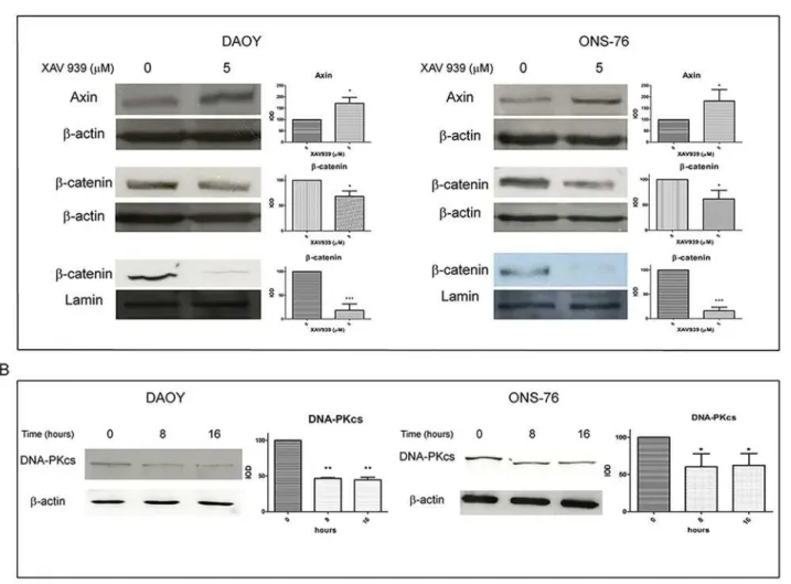
XAV939 is known to induce WNT signaling inhibition by increasing Axin protein levels, there-by promotingβ-catenin degradation [14]. We found an effective increase in total Axin levels, 8 h after treatment with XAV939 (5μM) and a decrease inβ-catenin protein levels, both in total and in nuclear extracts, at 16 h. Densitometry analyses revealed a 70% increase of total Axin protein levels in DAOY cells and an 80% increase in ONS-76 cells, treated with XAV939 (5μM) compared to DMSO treated control cells. Totalβ-catenin protein levels decreased by about 30% in both cell lines compared to controls; whereas, in nuclear fraction, we found an 80% reduction ofβ-catenin levels (Fig 1A).
The increase in Axin protein levels, followed byβ-catenin reduction, and DNA-PKcs levels inhibition confirmed the efficacy of XAV939 in affecting TNKS PARP activity on these MB cell lines.
The co-administration of XAV939 and IR inhibits MB cell proliferation
and clonogenic capacity
XAV939 and IR effects on the viability of MB cell lines were assessed by comparing growth curves. Cells were treated with 5μM of XAV939 or with an equal DMSO volume, with radiation (γ-ray, dose 2 Gy, dose rate ~ 2 Gy/min) or with a co-administration of drug and radiation.
Fig 2Ashows that the co-administration of XAV939 and IR strongly suppresses MB cell proliferation in both cell lines. In DAOY cells, the greatest proliferation inhibition was
Fig 1. XAV939 inhibits TNKS PARP-activity in MB cell lines.WB analysis and densitometry of total and nuclear MB cells extracts after XAV939 treatment: MB cell lines (DAOY, ONS-76) were treated with 5μM XAV939 or with an equal volume of DMSO.A.Both cell lines showed an increase in total Axin protein levels at 8 h after treatment (80% and 70% respectively in ONS-76 and DAOY compared to DMSO treated control,p<. 05), followed by aβ-catenin decrease in total (30%,p<. 05) and, in particular, in nuclear extracts, at 16 h after drug administration (80% reduction compared with control,p<. 001).B.XAV939 induced a DNA-PKcs protein level reduction of about 40% at 8 h and 16 h after treatment compared to non-treated control cells (p<. 05 in A,p<. 01 in B).
Densitometry data (mean±s.e.) were normalized withβ-actin (for total extracts) and lamin-b (for nuclear extracts) and are representative of the results derived from three independent experiments.
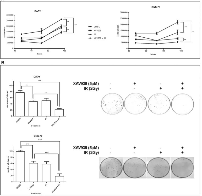
Fig 2. XAV939 impairs the clonogenic and proliferative capacity and enhances MB cells radio-sensitivity. A.Growth curves assay. XAV939 (5μM) treated cells and IR (2 Gy) treated cells showed a similar growth rate with about 40% (DAOY) and 30% (ONS-76) reductions compared to DMSO control cells (p<. 01). In both cell lines, the co-administration of XAV939 and IR induces a massive cell proliferation inhibition with a decrease of about 70% (p<. 001).
Each point represents the mean±s.e. of three independent assays.B.Clonogenic forming assay. XAV939 alone (5μM) inhibits a clone-forming ability in both MB cell lines as well as IR treatment (2 Gy), with a clonogenic capability reduction of about 38% (p<. 01). The IR and drug co-administration induces a
drastic inhibition of clone-forming in both cell lines (70% reduction in DAOY cells and 81% in ONS-76 compared with control cells,p<. 001).
observed 72 h post IR in XAV939 pre-treated cells, with a 69.2% reduction in cell proliferation compared to DMSO control cells (p<. 001, two-way ANOVA) and a 42.65% decrease in cell
number relative to DMSO irradiated cells (p<. 01, two-way ANOVA). In ONS-76 cells, we
found a massive inhibition in XAV939 and irradiated cells at 96 h after IR. At this time point, we detected a 75% cell number reduction compared to DMSO control cells (p<. 001, two-way
ANOVA) and a 34% reduction compared with DMSO irradiated cells (p<. 001, two-way
ANOVA). Furthermore, our data showed that XAV939 alone is able to inhibit MB cell growth: the greater proliferation decrease observed was at 72 h after drug administration (38.9% reduc-tion in DAOY cells and 48.1% in ONS-76 cells,p<. 01, two-way ANOVA).
Analogous results were found for clonogenic capability. Both MB cell lines showed a signifi-cant reduction (38%) of the colony formation after exposure to 5μM XAV939 (p<. 01,
one-way ANOVA). Nevertheless, XAV939 and IR treated cells showed the greatest inhibition of clonogenic formation compared to the other samples. In DAOY cells, we observed a 70.6% clone number reduction in XAV939 and irradiated cells compared to control cells (p<. 001,
one-way ANOVA) and 55.6% relative to DMSO irradiated cells (p<. 01, one-way ANOVA);
whereas, in ONS-76 cells, reductions of 81% (p<. 001, one-way ANOVA) and 67.9% (p<. 05,
one-way ANOVA) were observed, respectively (Fig 2B).
By using clone formation data, we calculated SF2. We found that XAV939 reduced the SF2 from 0.66 to 0.29 in DAOY cells and from 0.58 to 0.19 in ONS-76 cells, indicating that PARP activity inhibition halved the percentage of cells that survive radiation treatments. This con-firms a concrete enhanced radiosensitivity in these MB cells.
XAV939 affects efficacy of DNA repair after IR
The critical cellular lesions in relation to cytotoxic effects generated by IR are DNA-DSBs [12,13]. The DNA-PK pathway is one of the two most important pathways involved in DNA-DSBs repair [1]. As TNKS is a positive regulator of this pathway by inducing DNA-PKcs stability [10], we investigated whether XAV939 affects DNA repair efficacy after IR treatment. On the other hand, ARTD-1 inhibition by XAV939 could also have a critical role in NHEJ mechanisms [1].
The neutral comet assay was performed for this purpose, because of its ability to highlight predominantly DSBs [33,34] (Fig 3). As a measure of DNA damage, we used TM, the most common parameter, for analyzing the comet assay results. We utilized hydrogen peroxide (at 10μM for 15 min) as a negative control, since it induces principally SSBs; whereas, DSBs are considered very rare events [34].
First, we assessed the repair kinetics of IR-induced DSBs in DAOY and ONS-76 cells analyz-ing TM at various time points after IR (0, 16, and 24 h, 10 Gy dose). Fig3Aand3Bshow that the highest TM value was observed, as expected, immediately after IR in both cell lines. At 16 h after irradiation, we observed a decrease in TM, whereas 24 h after IR, we detected a TM value similar to that of non-irradiated cells, suggesting that DNA damage was repaired. Moreover, hydrogen peroxide did not induce a significant increase in TM compared to non-irradiated cells, confirming the effectiveness of the method in highlighting mainly DSBs.
TM in XAV939 pre-treated cells in relation to DMSO control cells; whereas, it was higher than 30% at 16 h. These results suggest a poor efficacy of DNA damage repair in XAV939 and IR treated cells compared to controls.
XAV939 increases IR-induced cell mortality
We evaluated, by trypan blue assay, whether cell proliferation inhibition and clonogenic forma-tion reducforma-tion correlated with an increase in IR-induced cell mortality.
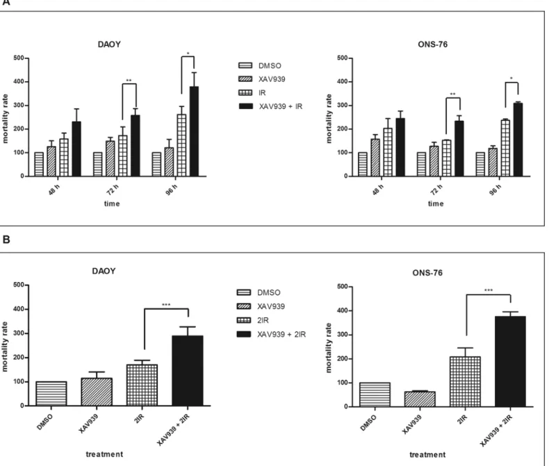
DAOY cells irradiated with a 2 Gy dose showed a 16.81%, 10.9%, and 9.3% mortality rate at 48, 72, and 96 h after radiation, respectively. Cells treated with 5μM of XAV939 before IR showed 23.5%, 17%, and 13% mortality rate with a 6.77%, 6.1%, and 3.7% increase (data not shown). Normalizing data to controls, we observed, at 72 h and 96 h after radiation, about a 40% and a 45% increase in cell mortality in cells pre-treated with XAV939 compared to only irradiated ones (Fig 4A): this difference was considered statistically significant (p<. 01 and
p<. 05, respectively, one-way ANOVA). ONS-76 cells, at the same time points, showed
Fig 3. XAV939 affects the DNA repair efficacy of MB cell lines.Neutral Comet Assay was performed on IR treated cells (10 Gy), with or without XAV939 administration, at different time points post radiations (0, 16, and 24 h). Histograms represent mean±s.e. of TM measured in at least three independent experiments for each treatment and time point. The greatest TM was observed immediately after IR treatment in both cell lines (A: DAOY, B: ONS-76). XAV939 induces enhanced TM values at 16 h and 24 h after IR compared to DMSO irradiated cells indicating a minor DNA repair capacity. On the right of the figure, a representative example of comets for each treatment obtained in DAOY (upper) and ONS-76 (down) cells.
a 10.61%, 9.11%, and 10.43% mortality rate after IR. In this cell line, XAV939 induced a slight increase in IR-mediated cell mortality: 12.96%, 13.49%, and 13.23% with a difference of 2.35%, 4.38%, and 2.8%, respectively (data not shown). When we normalized data to controls, a statis-tically significant increase in cell mortality between IR treated cells, XAV939, and irradiated cells was observed at 72 h (52.8%,p<. 01 one-way ANOVA) and at 96 h (30%,p<. 05,
one-way ANOVA).
Fig 4. XAV939 improves cell mortality IR-induced.Histograms represent means±s.e. of three independent experiments. Mortality rates of XAV939 treated cells, IR treated cells, XAV939, and IR treated cells are reported as relative percentages compared to DMSO control cells.A.Cell mortality after a 2 Gy dose administration: XAV939 alone does not induce a significant cell mortality increase in each time point considered. The greatest cell death was observed in XAV939 and IR treated cells in both cell lines, with a statistically significant difference at 72 h and 96 h compared with IR treated cells (p<. 01
andp<. 05, respectively).B.Mortality rate in MB cell lines after two treatments with 2 Gy dose of IR. In both cell lines, cells treated with XAV939 and irradiated, as described, showed a double mortality rate compared to DMSO irradiated cells (p<. 001).
Despite these results, we did not observe as strong an increase in cell mortality as expected. Testing the hypothesis that a single 2 Gy dose was enough to affect proliferation and clono-genic capability, but not enough to induce a massive increase in cell mortality, we irradiated with a second 2 Gy dose. DAOY cells treated with XAV939 andγ-rays, with two doses of 2 Gy, showed a 30.95% mortality rate compared to 16.17% of cells not treated with the small mole-cule but only irradiated two times (p<. 001, one-way ANOVA). Similarly, in ONS-76 cells,
XAV939 administration induced an almost double mortality rate in cells irradiated, as de-scribed, compared to cells treated with IR alone (17.75% and 9.8%, respectively,p<. 001,
one-way ANOVA).Fig 4Bshows data normalized to non-irradiated controls: we report, in both cell lines, about a 100% increase in mortality rate of XAV939 and irradiated cells compared to cells treated with IR alone, as described. These data confirm an enhanced MB cell radiosensitivity mediated by XAV939 administration. Finally, a statistically significant difference in mortality rate was not detected between XAV939 treated cells and DMSO control cells, in both cell lines and at all time points considered.
Discussion
In recent years, increasing interest has been focused on ARTD proteins because of their strong involvement in the DNA repair mechanism. ARTD inhibitors were first developed as sensitiz-ers to DNA-damaging chemotherapy or IR, and several highly potent and specific ones have been used in clinical trials in cancer patients [2–4]. Olaparib (AZD2281) is an oral PARP-1 and PARP-2 inhibitor that has been commercialized by Astra Zeneca [6]. Nonetheless, Dre-galla and co-workers focused on the therapeutic potential of ARTD-5 inhibition due to its cru-cial role in the NHEJ pathway by using XAV939 small molecule [11]. On the other hand, TNKS, but not PARP-1/PARP-2, is implicated in the WNT pathway by regulating Axin protein levels [14]. Recentin vitroandin vivostudies showed that WNT pathway inhibition suppresses MB growth, promoting cell cycle arrest followed by apoptosis [35,36].
In ourin vitrostudy, XAV939-mediated ARTD-1 and ARTD-5 inhibition impairs the growth and clonogenic capability of MB cell lines, but does not significantly increase cell mor-tality, indicating the low toxicity of the drug. The most notable results observed on cell prolifer-ation occurred following the co-administrprolifer-ation of XAV939 and IR. In these experiments, we treated cells with a 2 Gy dose that corresponded to the daily fraction of the total radiation dose (36 Gy) usually used in standard MB therapy. At this low dose, we found a massive cell prolif-eration inhibition in XAV939 pre-treated cells compared to DMSO irradiated cells. The assess-ment of cell proliferation after irradiation is considered a valid assay for defining cell radiation sensitivity because it provides information on the proliferative capacities of surviving cells after radiation [37,38]. Our data suggest that XAV939 treatment induces an increase in radiosensi-tivity of MB cells.
Comparable results were found when analyzing the clonogenic capacity of MB cell lines after drug and IR treatments. Indeed, the same trend, obtained by growth curves analyses, was observed using the clonogenic assay, the gold standard for the determination of radiation sensi-tivity [37,38]. MB cells treated with a combination of XAV939 andγ-rays showed greater clone-forming capability inhibition compared with cells treated with drug or IR alone. The re-duction of SF2in both MB cell lines confirmed greater radiosensitivity in XAV939 treated cells.
needs adapting for individual cell types in order to achieve optimal sensitivity [34]. In our ex-periments, we used the radiation dose of 10 Gy, which is considered an adequate dose for studying DSB repair by the neutral comet assay [34]. In this case, our specific interest was to confirm and quantify whether and how much XAV939 affects DNA mechanism repair in MB cell lines.
The observed kinetic repair trend in control tests was in accordance with results of previous studies [37–48]. Moreover, it has been shown that radiation-induced DSBs are rejoined no later than 20 h in mammalian cell lines [46,48]. In our experiments, both MB cell lines showed, 24 h after IR, a TM comparable to non-irradiated cells, indicating a well-functioning DNA repair mechanism.
Conversely, XAV939 and IR treated cells showed an evident delay in their capability to re-store DNA. We focused on 16 h and 24 h time points because it was suggested that damage measured after 4 h or more by treatments is an excellent index of the cell DNA injury restora-tion ability [48].
Thus, the higher TM value and, therefore, conspicuous DNA damage in XAV939 and IR treated cells at the time points considered, compared to the irradiated control cells, strongly supports the hypothesis that MB cells after XAV939 administration, at the given dose, have limited efficacy to repair DSBs. These data are in accordance with the cell growth and colony-forming ability inhibition previously shown.
In conclusion, XAV939 affected the DNA injury restoration ability, ultimately implying an improvement in the radiosensitivity of MB cells. XAV939-mediated PARP activity inhibition impairs growth and clonogenic capability and increases the mortality rate of MB cell lines. These results are most likely a consequence of TNKS PARP activity inhibition that leads to the WNT pathway inhibition and to the DNA-PKcs instability, causing cell growth reduction and radiosensibility. Since XAV939 also binds to ARTD-1 (also involved in DSB mechanism), but with approximately 10-fold lower affinity than to ARTD-5A/ARTD-5b [14–16], we cannot ex-clude that part of the shown radiosensitivity is due to this binding; further studies will shed light on this.
Taken together, ourin vitrodata suggest XAV939 as a potential molecule for improving the current therapy for MB. Our results lay the basis for furtherin vivostudies with the aim of de-signing clinical trials combining XAV939 (or other drugs with similar or better affinity for TNKS) with conventional radiotherapy. Potentially, this will lead to a more effective therapy, with the possibility of reducing radiation doses and, at last, long term side effects.
To date, onlyin vitrostudies on XAV939 have been performed; therefore, a study focusing on systemic drug administration is needed to investigate the concrete clinical relevance of this small molecule [49].
Author Contributions
Conceived and designed the experiments: CR RS. Performed the experiments: CR CC. Ana-lyzed the data: CR RS GC. Contributed reagents/materials/analysis tools: CR CC RS. Wrote the paper: CR RS GC.
References
1. Chanet F, Bollet MA, Hall J. (2010) Targeting poly(ADP-ribose) polymerase activity for cancer therapy.
2. Ratnam K, Low JA. (2007) Current development of clinical inhibitors of poly(ADP-ribose) polymerase in oncology. Clin Cancer Res 13, 1383–1388. PMID:17332279
3. Annunziata CM, O'Shaughnessy J. (2010) Poly (ADP-Ribose) Polymerase as a Novel Therapeutic Tar-get in cancer. Clin Cancer Res 16, 4517–4526. doi:10.1158/1078-0432.CCR-10-0526PMID:
4. Marcie KW, Alice Chen, (2011) PARP Inhibitor Treatment in Ovarian and Breast Cancer. Curr Probl Cancer 35(1), 7–50 doi:10.1016/j.currproblcancer.2010.12.002PMID:21300207
5. O’Shaughnessy J, Osborne C, Pippen JE, Yoffe M, Patt D, Rocha C, et al. (2011) Iniparib plus chemo-therapy in metastatic triple-negative breast cancer. N Engl J Med 364:205–214. doi:10.1056/ NEJMoa1011418PMID:21208101
6. Available:http://www.astrazeneca.com/.../20141024—lynparza-olaparib-receives-positive-chmp-opinion
7. Chiang YJ, Hsiao SJ, Yver D, Cushman SW, Tessarollo L, Smith S, et al. (2008) Tankyrase 1 and tan-kyrase 2 are essential but redundant for mouse embryonic development. PLoS One 9, 2639.
8. Kuimov AN, Kuprash DV, Petrov VN, Vdovichenko KK, Scanlan MJ, Jongeneel CV, et al. (2001) Cloning and characterization of TNKL, a member of tankyrase gene family. Genes Immun 2: 52–55. PMID:11294570
9. Smith S, Giriat I, Schmitt A, de Lange T. (1998) Tankyrase, a poly(ADP-ribose) polymerase at human telomeres. Science 282: 1484–1487. PMID:9822378
10. Cook BD, Dynek JN, Chang W, Shostak G, Smith S. (2002) Role for the related poly(ADP-Ribose) poly-merases tankyrase 1 and 2 at human telomeres. Mol Cell Biol 22: 332–34. PMID:11739745
11. Dregalla RC, Zhou J, Idate RR, Battaglia CL, Liber HL, Bailey SM. (2010) Regulatory roles of tankyrase 1 at telomeres and in DNA repair: suppression of T-SCE and stabilization of DNA-PKcs. AGING 2, 691–708. PMID:21037379
12. Rich T, Allen RL, Wyllie AH. (2000) Defying death after DNA damage. Nature 407:777–783. PMID:
11048728
13. Obe G, Johannes C, Schulte-Frohlinde D. (1992) DNA double-strand breaks induced by sparsely ioniz-ing radiation and endonucleases as critical lesions for cell death, chromosomal aberrations, mutations and oncogenic transformation. Mutagenesis 7(1), 3–12. PMID:1321942
14. Huang S, Mishina Y, Liu S, Cheung A, Stegmeier F, Michaud GA, et al. (2009) Tankyrase inhibition stabilizes Axin and antagonizes Wnt signaling. Nature 461, 614–620. doi:10.1038/nature08356PMID:19759537
15. Wang Q, Li H, Liu N, Chen XY, Wu ML, Zhang KL, et al. (2008) Correlative analyses of notch signaling with resveratrol-induced differentiation and apoptosis of human medulloblastoma cells. Neurosci Lett 438(2):168–173. doi:10.1016/j.neulet.2008.04.012PMID:18456406
16. Yang ZJ, Ellis T, Markant SL, Read TA, Kessler JD, Bourboulas M, et al. (2008) Medulloblastoma can be initiated by deletion of patched in lineage-restricted progenitors or stem cells. Cancer Cell 14(2):135–145. doi:10.1016/j.ccr.2008.07.003PMID:18691548
17. Gilbertson RJ, Ellison DW. (2008) The origins of medulloblastoma subtypes. Annu Rev Pathol 3:341–365. PMID:18039127
18. Guessous F, Li Y, Abounader R. (2008) Signaling pathways in medulloblastoma. J Cell Physiol 217(3):577–583. doi:10.1002/jcp.21542PMID:18651559
19. Northcott PA, Korshunov A, Witt H, Hielscher T, Eberhart CG, Mack S, et al. (2011) Medulloblastoma Comprises Four Distinct Molecular Variants. J Clin Oncol 19, 1408–1414.
20. Kool M, Koster J, Bunt J, Hasselt NE, Lakeman A, van Sluis P, et al. (2008) Integrated genomics identi-fies five medulloblastoma subtypes with distinct genetic profiles, pathway signatures and clinicopatho-logical features. PLoS One 3(8):e3088. doi:10.1371/journal.pone.0003088PMID:18769486
21. Holthouse DJ, Dallas PB, Ford J, Fabian V, Murch AR, Watson M, et al. (2009) Classic and desmoplas-tic medulloblastoma: complete case reports and characterizations of two new cell lines. Neuropatholo-gy 29(4):398–409. doi:10.1111/j.1440-1789.2008.00989.xPMID:19077040
22. Gerber NU, Zehnder D, Zuzak TJ, Poretti A, Boltshauser E, Grotzer MA. (2008) Outcome in children with brain tumours diagnosed in the first year of life: long-term complications and quality of life. Arch Dis Child 93:582–589. PMID:17634182
23. Mulhern RK, Merchant TE, Gajjar A, Reddick WE, Kun LE. (2004) Late neurocognitive sequelae in sur-vivors of brain tumours in childhood. Lancet Oncol 5:399–408. PMID:15231246
24. Vinchon M, Baroncini M, Leblond P, Delestret I. (2011) Morbidity and tumor-related mortality among adult survivors of pediatric brain tumors: a review. Childs Nerv Syst 27:697–704. doi: 10.1007/s00381-010-1385-6PMID:21409425
25. Merchant TE, Pollack IF, Loeffler JS. (2010) Brain tumors across the age spectrum: biology, therapy, and late effects. Semin Radiat Oncol 20:58–66. doi:10.1016/j.semradonc.2009.09.005PMID:19959032
26. Grill J, Renaux VK, Bulteau C, Viguier D, Levy-Piebois C, Sainte-Rose C, et al. (1999) Long-term intel-lectual outcome in children with posterior fossa tumors according to radiation doses and volumes. Int J Radiat Oncol Biol Phys 45:137–145. PMID:10477017
28. Wahlberg E, Karlberg T, Kouznetsova E, Markova N, Macchiarulo A, Thorsell AG, et al. (2012) Family-wide chemical profiling and structural analysis of PARP and tankyrase inhibitors. Nat Biotechnol; 30:283–8. doi:10.1038/nbt.2121PMID:22343925
29. Krukenberg KA, Jiang R, Steen JA, Mitchison TJ2. (2014) Basal activity of a PARP1-NuA4 complex varies dramatically across cancer cell lines. Cell Rep 8:1808–18. doi:10.1016/j.celrep.2014.08.009
PMID:25199834
30. Yamada M, Shimizu K, Tamura K, Okamoto Y, Matsui Y, Moriuchi S, et al. (1989) Establishment and bi-ological characterization of human medulloblastoma cell lines. Brain Nerve 41, 695–702. PMID:
2818910
31. Jacobsen PF, Jenkyn DJ, Papadimitriou JM. (1985) Papadimitriou JM. Establishment of a human me-dulloblastoma cell line and its heterotransplantation into nude mice. J Neuropathol Exp Neurol 44, 472–485. PMID:2993532
32. Salaroli R, Di Tomaso T, Ronchi A, Ceccarelli C, Cammelli S, Cappellini A, et al. (2008) Radiobiologic response of medulloblastoma cell lines: involvement of beta-catenin? J Neurooncol 90:243–251. doi:
10.1007/s11060-008-9659-5PMID:18688572
33. Olive PL, Banáth JP. (2006) The comet assay: a method to measure DNA damage in individual cells. British Columbia Cancer Research Center, 675.
34. Wojewódzka M, Buraczewska I, Kruszewski M. (2002). A modified neutral comet assay: elimination of lysis at high temperature and validation of the assay with anti-single-stranded DNA antibody. Mutat Res 518, 9–20. PMID:12063063
35. Cimmino F, Scoppettuolo M, Carotenuto M, De Antonellis P, Dato VD, De Vita G, et al. (2011) Nor-cantharidin impairs medulloblastoma growth by inhibition of Wnt/b-catenin signaling. J Neurooncol 106, 59–70. doi:10.1007/s11060-011-0645-yPMID:21735115
36. Baryawno N, Sveinbjörnsson B, Eksborg S, Chen CS, Kogner P, Johnsen JI. (2010) Small molecule in-hibitor of Phosfatidylinositol 3-Kinase/Akt Signaling Inhibit Wnt/β-Catenin Pathway Cross-Talk and Suppress Medulloblastoma Growth. Cancer Res 70; 2138–2149.
37. Luttjeboer M, Lafleur MV, Kwidama ZJ, Van Rijn J, Van Den Berg J, Slotman BJ, et al. (2010) Strategies for the analysis of in vitro radiation sensitivity and prediction of interaction with potential radiation modi-fying agents. Int J Radiat Biol 86, 458–466. doi:10.3109/09553000903568019PMID:20470196
38. Franken NA, Rodermond HM, Stap J, Haveman J, van Bree C. (2006). Clonogenic assay of cells in vitro. Nat Protoc 1, 2315–2319. PMID:17406473
39. Afanasieva K, Zazhytska M, Sivolob A. (2010) Kinetics of comet formation in single-cell gel electropho-resis: Loops and fragments. Electrophoresis 31, 512–519. doi:10.1002/elps.200900421PMID:
20119958
40. Wojewódzka M, Gràdzka I, Buraczewska I. (2002) Modified neutral comet assay for human lympho-cytes. Nukleonika 47(1), 1–5.
41. Olive PL, Banath JP. (1993) Detection of DNA double-strand breaks through the cell cycle after expo-sure to X-rays, bleomycin, etoposide and 125IdUrd. Int J Radiat Biol 64, 349–358. PMID:7693837
42. Olive PL, Banath JP. (1995) Radiation-induced DNA doublestrand breaks produced in histone-depleted tumor cell nuclei measured using the neutral comet assay. Radiat Res 142, 144–152. PMID:
7724728
43. Olive PL, Wlodek D, Banath JP. (1991) DNA double-strand breaks measured in individual cells sub-jected to gel electrophoresis. Cancer Res 51, 4671–4676. PMID:1873812
44. Rydberg B. (2000) Radiation-induced heat-labile sites that convert into DNA double-strand breaks. Radiat Res 153, 805–812. PMID:10825756
45. Wada S, Kurahayashi H, Kobayashi Y, Funayama T, Yamamoto K, Natsuhori M, et al. (2003) The rela-tionship between Cellular Radiosensitivity and Radiation-Induced DNA Damage Measured by Comet Assay. J Vet Med Sci 65, 471–477. PMID:12736429
46. Banath JP, Fushiki M, Olive PL. (1998) Rejoining of DNA single- and double-strand breaks in human white blood cells exposed to ionizing radiation. Int J Radiat Biol 73, 649–660. PMID:9690683
47. Zaffaroni N, Orlandi L, Villa R, Bearzatto A, Rofstad EK, Silvestrini R. (1994) DNA double-strand break repair and radiation response in human tumour primary cultures. Int J Radiat Biol 66, 279–285. PMID:
7930830
48. Eastham AM, Marples B, Kiltie AE, Orton CJ, West CM. (1999) Fibroblast radiosensitivity measured using the comet DNA-damage assay correlates with clonogenic survival parameters. Br J Cancer 79, 1366–1371. PMID:10188877
49. Fancy SP, Harrington EP, Yuen TJ, Silbereis JC, Zhao C, Baranzini SE, et al. (2011) Axin2 as regulato-ry and therapeutic target in newborn brain injuregulato-ry and remyelination. Nat Neurosci 14:1009–1016. doi: