ameloblastomas and tooth germs
Karuza Maria Alves Pereira, DDS, MSc,aBruna Aguiar do Amaral, DDS, MSc,a Bruna Rafaela Martins dos Santos, DDS, MSc,aHébel Cavalcanti Galvão, DDS, PhD,b
Roseana de Almeida Freitas, DDS, PhD,band Lélia Batista de Souza, DDS, PhD,bNatal, Brazil POST-GRADUATE PROGRAM, ORAL PATHOLOGY, FEDERAL UNIVERSITY OF RIO GRANDE DO NORTE
Objective. The aim was to analyze the expression of E-cadherin and-catenin in ameloblastomas and tooth germs to determine their roles in cell differentiation processes and invasiveness compared with odontogenesis.
Study design. Twenty-one ameloblastoma cases (16 solid and 5 unicystic tumors) and 5 tooth germs were submitted to the immunohistochemical detection of E-cadherin and-catenin. Immunoreactivity was evaluated using descriptive and semiquantitative analysis, investigating the location and intensity of staining. The Fisher exact test was performed, andPvalues of⬍.05 were considered to indicate statistical significance.
Results. There was no statistically significant difference in the expression of E-cadherin and-catenin between solid and unicystic ameloblastomas (P⫽.59;P⫽.63; respectively). The same was found when comparing solid and unicystic ameloblastomas with the tooth germs for both E-cadherin (P⫽.53;P⫽.44; respectively) and-catenin (P⫽ .12;P⫽.16; respectively). Nuclear staining of-catenin was observed in only 4 cases (3 solid and 1 unicystic tumor). Conclusion. The results showed no differences in the expression of E-cadherin or-catenin between tooth germs and solid and unicystic ameloblastomas. The expression of these molecules seems mainly to be related to the process of cell differentiation.(Oral Surg Oral Med Oral Pathol Oral Radiol Endod 2010;109:425-431)
Odontogenic tumors arise from epithelial and/or ecto-mesenchymal elements involved in odontogenesis and are classified according to the tissue of origin. Tumors originating from odontogenic apparatus or its deriva-tives or remnants exhibit marked histologic variation and are classified as benign or malignant.1 Ameloblas-tomas are the most frequent benign tumors derived from odontogenic epithelium and are characterized by slow-growth but local invasion and a high rate of re-currence. These tumors morphologically resemble tis-sue structures of the odontogenic epithelium, such as the enamel organ and/or dental lamina,1 and can be divided into multicystic (solid), unicystic, and periph-eral tumors based on clinical and radiographic findings. Regarding clinical behavior, unicystic ameloblastomas have been reported to present a less aggressive biologic behavior and a better prognosis than solid ameloblas-tomas.1,2
During embryogenesis, numerous morphogenetic al-terations are induced by the binding of ligands to their receptors.4 Several known growth factors affect the expression of genes that regulate various processes,
including cell migration. Recent studies have focused on regulators of cell proliferation and differentiation in view of the fact that their signaling pathways involve proteins that directly participate in gene transcription and cell adhesion.4,5
Cell adhesion is essential for the regulation of cell behavior. Numerous molecules mediating this process have been identified and investigated. One of these molecules is -catenin, a transcription cofactor in the Wnt pathway and a structural protein that connects cadherins to the actin cytoskeleton during cell-cell ad-hesion.6 Cell adhesion mediated by E-cadherin re-quires, in addition to the availability of calcium ions on the cell surface, the adequate presence of other proteins, such as the so-called␣-,-, and␥-catenins present on the cytoplasmic side of the plasma membrane, which anchor E-cadherin to the actin cytoskeleton. These as-sociations of classical cadherins with their catenins form structures identified by microscopy as adherent junctions.7These molecules play an important role in the maintenance of epithelial tissue architecture, as well as in organogenesis and morphogenesis. E-Cadherin and␣-catenin, together with-catenin, form a complex which is necessary for cell adhesion. Studies on epithe-lial odontogenic tumors have demonstrated that these molecules might be associated with cell differentiation or tumor invasiveness.8Furthermore, mutations and the nuclear expression of-catenin have been observed in some epithelial odontogenic tumors.
aDoctoral Student. bProfessor.
Received for publication Dec 21, 2008; returned for revision Oct 15, 2009; accepted for publication Oct 20, 2009.
1079-2104/$ - see front matter © 2010 Published by Mosby, Inc. doi:10.1016/j.tripleo.2009.10.032
In view of the role of E-cadherin and -catenin in cell adhesion, the objective of the present study was to analyze the immunohistochemical expression of these molecules in ameloblastomas and tooth germs, thus contributing to a better understanding of the role of these proteins in the processes of cell differentiation and local invasiveness of these tumors compared with odontogenesis.
MATERIAL AND METHODS
Paraffin-embedded specimens of 21 ameloblastomas (16 solid and 5 unicystic tumors) and 5 tooth germs were used for this study. The ameloblastoma cases were obtained from the Pathologic Anatomy Service, Discipline of Oral Pathology, Dentistry School, Federal University of Rio Grande do Norte, Brazil, between 2000 and 2007. The tooth germs were kindly provided by the Laboratory of Surgical Pathology, Dentistry School, University of Pernambuco. For morphologic analysis, the paraffin-embedded material was cut into 5-m-thick sections, mounted on slides, stained with hematoxylin-eosin, and examined by light microscopy. The histologic features observed in ameloblastomas and tooth germs were submitted to descriptive analysis. For immunohistochemistry, the paraffin-embedded ma-terial was cut into 3-m-thick sections and mounted on glass slides previously prepared with organosilane adhe-sive. Next, the sections were submitted to the streptavidin-biotin method using the following primary antibodies: E-cadherin monoclonal antibody diluted 1:50 (clone H108; Santa Cruz Biotechnology, Santa Cruz, CA; 120 minutes, antigen retrieval with citrate, pH 6.0, Pascal) and-catenin polyclonal antibody diluted 1:125 (clone 14; BD Transduction Laboratories, San Jose, CA; over-night, antigen retrieval with citrate, pH 6.0, for 40 minutes in a steamer). Fragments of normal prostate tissue were used as positive control. Sections in which the primary antibodies were replaced with 1%
buff-ered bovine serum albumin were then submitted to the same steps as negative control.
Immunohistochemical staining of E-cadherin and
-catenin was evaluated using descriptive and semiquan-titative methods based on an adaptation of the criteria of Freitas et al. (2009).9In the latter analysis, scores of 1 or
2 were used for the stained cells for percentage of immu-nostained cells⬍10% (score 1) and ⬎10% (score 2). Parameters were independently analyzed through light microscopy by 3 of the authors. Then they analyzed the cases as a group to reach a consensus. Statistical analysis was performed using SPSS for Windows version 17.0 (SPSS, Chicago, IL). The Fisher exact test was performed forPvalues of⬍.05 to be considered to indicate statistical significance.
Immunostaining of E-cadherin and -catenin was also evaluated according to the type of reactive cell and the cellular location of immunostaining. Regarding the type of reactive cell, the immunoexpression was eval-uated in peripheral columnar and/or cuboidal cells, cells resembling the stellate reticulum, and cells of the enamel organ. The cellular location of immunostaining for E-cadherin was determined in membrane and cyto-plasm. For-catenin, the cellular immunostaining was determined in membrane, cytoplasm, and nucleus.
RESULTS
Microscopic analysis of the morphologic features of the specimens revealed that all tooth germs were in the bell stage. Solid ameloblastomas exhibited islands, nests, and anastomosed cords of odontogenic epithelium, consisting of hyperchromatic peripheral columnar cells in a pali-saded fashion and more loosely arranged central cells resembling the stellate reticulum of the enamel organ. In the case of unicystic ameloblastomas, a pathologic cavity lined with ameloblastomatous epithelium was observed.
The immunostaining results of -catenin and E-cadherin in tooth germs and ameloblastomas are sum-Table I. Immunoexpression of -catenin and E-cadherin in solid and unicystic ameloblastomas and tooth germs
Molecule
Intensity of immunostaining
Histologic type, n (%)
Solid ameloblastoma Unicystic ameloblastoma Tooth germ Total
-Catenin Score 1 9 (53.6%) 2 (40%) 5 (100%) 16 (61.5%)
Score 2 7 (46.8%) 3 (60%) 0 (0%) 10 (38.5%)
Total 16 (100%)a,b 5 (100%)a,c 5 (100%)b,c 26 (100%)
E-Cadherin Score 1 4 (25%) 2 (40%) 0 (0%) 6 (23.1%)
Score 2 12 (75%) 3 (60%) 5 (100%) 20 (76.9%)
Total 16 (100%)d,e 5 (100%)d,f 5 (100%)e,f 26 (100%)
a,b,cFisher exact testPvalues for
-catenin: (a) between solid and unicystic ameloblastomas (P⫽.63); (b) between solid ameloblastoma and tooth germs (P⫽.12); (c) between unicystic ameloblastoma and tooth germs (P⫽.16).
d,e,fFisher exact test:pvalues for E-cadherin: (d) between solid and unicystic ameloblastomas (P
marized in Tables I-III. Regarding the intensity of -catenin staining, there was a predominance of weak staining (score 1) in solid ameloblastomas (9 cases) and moderate/strong staining (score 2) in 3 cases of unicys-tic ameloblastomas. Statisunicys-tical analysis revealed no sig-nificant difference when comparing-catenin expres-sion between solid and unicystic ameloblastomas (P⫽ .63;Table I).
Expression of -catenin was detected in the cell membrane, cytoplasm, and nucleus of epithelial cells in both ameloblastomas (Fig. 1). In cases of unicystic ameloblastomas, 80% of the specimens presented cy-Fig. 1. Immunohistochemical reactivity for-catenin in
mul-ticystic ameloblastoma, showing membranous and cytoplas-mic reactivity in neoplastic cells (⫻400).
Fig. 2. Ameloblastoma showing membranous and cytoplas-mic reactivity in neoplastic cells. Diffuse nuclear expression for-catenin is also found in neoplastic cells (⫻400). Table II. Cellular type location of-catenin and E-cadherin immunostaining in solid and unicystic ameloblastomas
Molecule Cellular type
Histologic type, n (%)
Solid ameloblastoma Unicystic ameloblastoma Total
-Catenin Peripheral columnar cells* 1 (6.25%) 1 (20%) 2 (9.52%)
Cells resembling the stellate reticulum† 0 (0%) 0 (0%) 0 (0%)
Both‡ 14 (87.5%) 4 (80%) 18 (85.71%)
Total 15 (93.75%) 5 (100%) 20 (95.23%)
E-Cadherin Peripheral columnar cells* 2 (12.5%) 0 (0%) 2 (9.52%)
Cells resembling the stellate reticulum† 6 (37.5%) 1 (20%) 7 (33.33%)
Both‡ 8 (50%) 4 (80%) 12 (57.14%)
Total 16 (100%) 5 (100%) 21 (100%)
*Exclusive staining in peripheral cells.
†Exclusive staining in cells resembling the stellate reticulum.
‡Simultaneous staining in both peripheral cells and cells of the stellate reticulum.
Table III. Immunostaining for -catenin and E-cadherin in solid and unicystic ameloblastomas according to the location of cells present in the tissue specimens
Molecule Cellular location
Histologic type, n (%)
Solid ameloblastoma Unicystic ameloblastoma Tooth germ Total
-catenin Membrane/cytoplasmic 13 (81.25%) 4 (80%) 5 (100%) 22 (84.61%)
Membrane/cytoplasmic/nuclear 3 (18.75%) 1 (20%) 0 (0%) 4 (15.38%)
Total 16 (100%) 5 (100%) 5 (100%) 26 (100%)
E-cadherin Cytoplasmic 6 (37.5%) 2 (40%) 4 (80%) 12 (46.15%)
Membrane/cytoplasmic 10 (62.5%) 3 (60%) 1 (20%) 14 (53.85%)
toplasmic and membrane staining. Nuclear reactivity was observed only in neoplastic cells of 4 ameloblas-tomas (3 solids and 1 unicystic tumor;Fig. 2;Table III). In cases of solid and unicystic ameloblastomas, immu-noreactivity to-catenin was detected both in loosely organized cells resembling the stellate reticulum of the enamel organ and in peripheral cells resembling amelo-blasts (Table II).
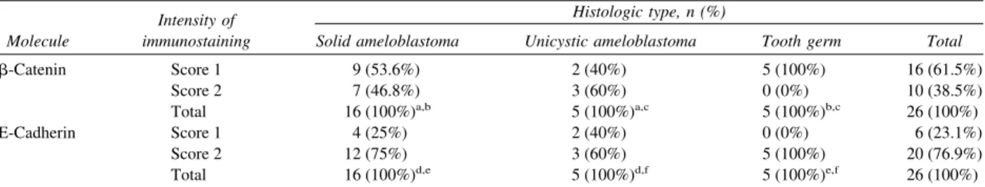
Regarding E-cadherin, all cases of ameloblastomas (Fig. 3) and tooth germs were immunopositive for this protein. Moderate/strong staining (score 2) was ob-served in 12 cases (75%) of solid ameloblastomas (Fig. 4) and in 5 tooth germ specimens (100%), whereas no staining intensity predominated in unicystic ameloblas-tomas (Fig. 4; Table I). In solid and unicystic
amelo-blastomas, this protein was expressed in the cell mem-brane and cytoplasm of peripheral columnar neoplastic cells and loosely organized cells (Tables IIandIII). The Fisher exact test also showed no significant difference to E-cadherin in the solid ameloblastomas compared with the unicystic ameloblastomas (P ⫽.59;Table I). All tooth germ specimens were positive for the 2 adhesion molecules analyzed. The intensity of immu-noexpression was weak (score 1) for -catenin and moderate/strong (score 2) for E-cadherin (Fig. 5;Table I). In the cell, staining was mainly detected in the membrane and cytoplasm (Table III), with these pro-teins being more strongly expressed in the cell mem-brane of the stellate reticulum and weakly in cells of the inner and outer epithelium and intermediate layer of the enamel organ.
The Fisher exact test also showed no significant difference between solid ameloblastomas and tooth germs for both -catenin (P ⫽ .12) and E-cadherin (P ⫽ .53). The same was observed when comparing unicystic ameloblastomas and tooth germs for both -catenin (P⫽.16) and E-cadherin (P⫽.44;Table I).
DISCUSSION
Cell junctions link epithelial cells to one another and are formed by different proteins, generically called adhesion molecules, which play a fundamental role in the regulation of cell growth and differentiation. These adhesion molecules include cadherins and catenins. Cadherins are a family of calcium-dependent ho-mophilic transmembrane adhesion molecules. As a member of this family, E-cadherin is responsible for epithelial cell polarity and the establishment of tissue Fig. 3. Immunohistochemical reactivity for E-cadherin in
multicystic ameloblastoma, showing membranous and cyto-plasmic reactivity in neoplastic cells (⫻100).
Fig. 4. Immunohistochemical reactivity for E-cadherin in unicystic ameloblastoma, showing membranous and cyto-plasmic reactivity in neoplastic cells (⫻400).
morphology.11 Catenins are adhesion molecules that are directly or indirectly associated with E-cadherin, forming a cadherin-catenin complex.12
Among catenins,-catenin is a multifunctional mol-ecule that plays a role in cell-cell adhesion mediated by classic cadherins as well as in Wnt signaling.13 This protein directly interacts with the cytoplasmic domain of cadherin as well as with␣-catenin, thus connecting cadherins to the actin filament. Free-catenin in cyto-plasm is phosphorylated by association with a complex of multiproteins, resulting in the maintenance of low cytoplasmic levels. Alternatively, glycogen synthase kinase 3(GSK-3), one of the proteins of this com-plex, can be inactivated in the presence of a Wnt signal, with the consequent absence of phosphorylation of -catenin and an increase in the cytoplasmic pool of this molecule. In this condition, -catenin enters the nucleus, where it interacts with the T-cell factor/lym-phoid-enhancing factor family.14
Thus, -catenin can be present at 3 different sites in the cell: bound to the cytoplasmic membrane, in the cytoplasm, and in the nucleus.
During the development of the tooth germ, 3 classic cadherins, E-, P-, and N-cadherin, are expressed in the epithelial compartment of the enamel organ,15together
with-catenin, which possibly plays multiple roles in tooth development. The different functions of these molecules, particularly in cells and tissues, are related to their location, in the case of -catenin, and to the signaling pathways involved. Normal expression and function of E-cadherin are essential for the induction and maintenance of the polarized and differentiated epithelium during embryo development.16
Studies investigating these 2 adhesion molecules have reported contradictory results regarding the rela-tionship between alterations in the expression pattern of the E-cadherin–-catenin complex and the biologic be-havior of tumors. Most studies have shown that a reduction in the expression of this complex is associ-ated with a more aggressive tumor behavior and a poorer prognosis for the patients.17
Ultrastructural analysis of tooth germs of experimen-tal animals has shown a distinct pattern of expression of -catenin during the development of the animal, a fact reflecting specific roles during morphogenesis and cy-todifferentiation.15
Obara et al. (1998)18 analyzed the distribution of
E-cadherin by indirect immunofluorescence in tooth germs and demonstrated moderate E-cadherin staining in all epithelial cells during the bell stage, especially in the stellate reticulum of the enamel organ. We obtained similar results, with all tooth germs showing moderate expression of this protein. On the basis of these results, we agree with those authors that E-cadherin plays a role
in the regulation of tooth morphogenesis, with this molecule mediating the organization and maintenance of the architecture of the enamel organ.
In another study, Obara et al. (2004)13investigated the
intracellular distribution of-catenin and E-cadherin in tooth germs in the cap stage and showed variations in -catenin staining among different regions of the enamel organ, with strong staining being detected in the cytoplasm and nucleus of cells located in the enamel knot and in the inner epithelium of the enamel organ. Those authors suggested that nuclear staining for -catenin is due to specific activation of the Wnt sig-naling pathway during this stage of development. In the present study, no nuclear expression of-catenin was observed in any type of cell of the tooth germs ana-lyzed, with most tooth germs presenting only weak membrane and cytoplasmic staining, which was more expressive in cells of the stellate reticulum. This dif-ference may reflect different stages of odontogenesis in the samples studied.
Adhesion molecules have been studied in ameloblas-tomas in an attempt to understand whether these pro-teins play a role in the oncogenesis and cytodifferenti-ation of these tumors.3,8,10,19 Kumamoto and Ooya
(1999),8 analyzing the expression of E-cadherin and
␣-catenin in odontogenic tumors and tooth germs, de-tected E-cadherin expression in tooth germs, amelo-blastomas, and other odontogenic tumors, with the ob-servation of strong membrane staining in the stellate reticulum of tooth germs as well as in the membrane of polyhedral ameloblastoma cells. A closely similar staining pattern was found in the present study in both tooth germs and ameloblastomas, with the observation of predominantly moderate membrane and cytoplasmic staining. These findings suggest that E-cadherin func-tions as a modulator during tooth development and participates in the morphodifferentiation of the odon-togenic epithelium of ameloblastomas, because a sim-ilar distribution in the enamel organ was observed irrespective of the clinical variant of this tumor, thus conserving the morphodifferentiation of the odonto-genic epithelium. In both tooth germs and ameloblas-tomas, the staining intensity was more marked in cells resembling the stellate reticulum, suggesting a possible higher concentration of this protein at specific sites where it promotes the adhesion between distant cells in this arrangement.
ameloblastomas and tooth germs. The intensity of -cate-nin immunoexpression in tooth germs was weak and was observed in 4 specimens (80%), with stronger expression in the membrane of stellate reticulum cells and weaker expression in cells of the inner and outer epithelium and intermediate layer of the enamel organ. These character-istics suggest that-catenin may play a double role, par-ticipating in cell-cell adhesion and in the Wnt signaling pathway during odontogenesis or in ameloblastomas.
Cases of ameloblastomas presenting accumulation of
-catenin have been reported in the literature, although mutations in its gene (CTNNB1) are rare. Kumamoto and Ooya (2005)10 detected nuclear expression of -catenin in 9 out of 40 ameloblastoma cases analyzed, but no nuclear immunoreactivity was observed in the tooth germ specimens studied. Similar findings were obtained in the present study, in which nuclear immu-nostaining was observed in only 4 of the 21 ameloblas-toma cases analyzed (3 solid tumors and 1 unicystic tumor), and in none of the tooth germ specimens.
Sekine et al. (2003)19found nuclear and cytoplasmic staining for-catenin in 50% of ameloblastomas, but a mutation in theCTNNB1gene was observed in only 1 of the 20 ameloblastoma cases studied. Because a mu-tation in the-catenin gene was present in only 1 case, those authors concluded that there are other mecha-nisms responsible for the nuclear expression of this protein in ameloblastomas. The nuclear accumulation of -catenin might be related to physiologic mecha-nisms that regulate the expression of this molecule during odontogenesis or to the presence of genetic alterations in other molecules affecting the expression of-catenin, such as Wnt and APC.
In agreement with these findings, Miyake et al. (2006),3 studying odontogenic tumors, observed focal to moderate-catenin immunoreactivity in ameloblas-tomas. Mutations in the-catenin gene were observed in only 1 of the ameloblastoma cases analyzed, a find-ing confirmfind-ing that, despite the nuclear stainfind-ing of this adhesion molecule, mutations in its gene are rare in ameloblastomas, although abnormalities in the Wnt sig-naling pathway might be associated with some tumors. Thus, the nuclear accumulation of-catenin may result not only from a mutation in theCTNNB1gene, but also from mutations in theAPC and AXIN1gene or even from the inactivation of theGSK-3gene, whose prod-uct is responsible for the proteolytic degradation of -catenin.3
Tanahashi et al. (2008)20studied-catenin and other molecules of the Wnt pathway in ameloblastomas and observed nuclear immunoreaction to -catenin in 11 out of 18 cases (61%), with that staining mainly being detected in peripheral columnar cells. In the present study, nuclear-catenin staining was also mainly
de-tected in peripheral cells, but the number of immuno-reactive cases was smaller (15.3%) than that reported by those authors. Our results agree with literature data showing nuclear staining for-catenin in some cases of ameloblastoma; however, this molecule does not seem to be involved in the pathogenesis of this odontogenic tumor.
In general, immunostaining for E-cadherin and catenins is more intense in well differentiated tumors which main-tain their cell adhesiveness and are less invasive, but is lower in poorly differentiated tumors which lose cell-cell adhesion and present a strongly invasive behavior.17Most of the specialized literature show that a reduction in E-cadherin is mainly associated with parameters of higher biologic aggressiveness, increased invasiveness, metasta-ses, tumor recurrence, and a poorer prognosis for the patients.17
CONCLUSION
The present results showed no differences in the expression of E-cadherin or -catenin between tooth germs and solid and unicystic ameloblastomas. The expression of these molecules seems mainly to be re-lated to the process of cell differentiation. Therefore, other molecular mechanisms more decisively influence the biologic behavior of ameloblastomas.
REFERENCES
1. Barnes L, Eveson JW, Reichart P, Sidransky D, editors. World Health Organization Classification of Tumours. Pathology and Genetics Head and Neck Tumours. Lyon: IARC: 2005; p. 296-300.
2. Ledesma-Montes A, Mosqueta-Taylor A, Carlos-Bregni R, de León ER, Palma-Guzmán JM, Páez-Valencia C, et al. Amelo-blastoma: a regional Latin-American multicentric study. Oral Dis 2007;13:303-7.
3. Miyake T, Tanaka Y, Kato K, Tanaka M, Sato Y, Ijiri R, et al. Gene mutation analysis and immunohistochemical study of
-Catenin in odontogenic tumors. Pathol Int 2006;56:732-7. 4. Nelson WJ, Nusse R. Convergence of Wnt,-catenin, and
cad-herin pathways. Science 2004;303:1483-7.
5. Andrade ESS, da Costa Miguel MC, Pinto LP, de Souza LB. Ameloblastoma and adenomatoid odontogenic tumor: the role of
␣21,␣31, and␣51 integrins in local invasiveness and ar-chitectural characteristics. Ann Diagn Pathol 2007;11:199-205. 6. Obara N, Suzuki Y, Takeda M. Gene expression of-catenin is up-regulated in inner dental epithelium and enamel knots during molar tooth morphogenesis in the mouse. Cell Tissue Res 2006; 325:197-201.
7. Knudsen KA, Wheelock MJ. Cadherins and the mammary gland. J Cell Biochem 2005;95:488-96.
8. Kumamoto H, Ooya K. Expression of E-cadherin and alpha-catenin in epithelial odontogenic tumors: an immunohistochem-ical study. J Oral Pathol Med 1999;28:152-7.
10. Kumamoto H, Ooya K. Immunohistochemical detection of -catenin and adenomatous polyposis coli in ameloblastomas. J Oral Pathol Med 2005;34:401-6.
11. Wheelock MJ, Johnson KR. Cadherins as modulators of cellular phenotype. Ann Rev Cell Dev Biol 2003;19:207-35.
12. Ben-ze’ev A, Shtutman M, Zhurinsky J. The integration of cell adhesion with gene expression: the role of-catenin. Exp Cell Res 2000;261:75-82.
13. Obara N, Lesot H. Subcellular localization of-catenin and cadherin expression in the cap-stage enamel organ of the mouse molar. Histochem Cell Biol 2004;121:351-8.
14. Young CS, Kitamura M, Hardy S, Kitajewski. Wnt-1 induces growth, cytosolic beta-catenin, and Tcf/Lef transcriptional acti-vation in Rat-1 fibroblasts. Mol Cell Biol 1998;18:2474-85. 15. Palacios J, Benito N, Berraquero R, Pizarro A, Cano A, Gamallo
C. Diferential spatiotemporal expression of E- and P-cadherin during mouse tooth development. Int J Dev Biol 1995;39:663-6. 16. Ramburan A, Govendar D. Cadherins and catenins in pathology.
Curr Diagnostic Pathol 2002;8:305-17.
17. Bánkfalvi A, Krassort M, Végh A, Felszeghy E, Piffkó J. De-ranged expression of the E-cadherin/beta-catenin complex and the epidermal growth factor receptor in the clinical evolution and
progression of oral squamous cell carcinomas. J Oral Pathol Med 2002;31:450-7.
18. Obara N, Suzuki Y, Nagai Y, Takeda M. Expression of E- and P-cadherin during tooth morphogenesis and cytodifferentiation of ameloblasts. Anat Embryol 1998;197:469-75.
19. Sekine S, Sato S, Takata T, Fukuda Y, Ishida T, Kishino M, et al.-Catenin mutations are frequent in calcifying odontogenic cysts, but rare in ameloblastomas. Am J Pathol 2003;163: 1707-12.
20. Tanahashi J, Daa T, Yada N, Kashima K, Kondoh Y, Yokoyama S, et al. Mutational analysis of Wnt signaling molecules in ameloblastoma with aberrant nuclear expression of beta-catenin. J Oral Pathol Med 2008;37:565-70.
Reprint requests:
Profa. Dra. Lélia Batista de Souza Departamento de Odontologia
Universidade Federal do Rio Grande do Norte Av. Senador Salgado Filho, 1787
Lagoa Nova—Natal—RN Brazil 59056-000