Tolerance to Gamma Radiation in the
Tardigrade
Hypsibius dujardini
from Embryo
to Adult Correlate Inversely with Cellular
Proliferation
Eliana Beltrán-Pardo1
*, K. Ingemar Jönsson1,2, Mats Harms-Ringdahl1, Siamak Haghdoost1, Andrzej Wojcik1
1Department of Molecular Biosciences, The Wenner-Gren Institute, Stockholm University, Stockholm, Sweden,2School of Education and Environment, Kristianstad University, Kristianstad, Sweden
*elianabeltran@yahoo.com
Abstract
Tardigrades are highly tolerant to desiccation and ionizing radiation but the mechanisms of this tolerance are not well understood. In this paper, we report studies on dose responses of adults and eggs of the tardigradeHypsibius dujardiniexposed to gamma radiation. In adults the LD50/48hfor survival was estimated at ~ 4200 Gy, and doses higher than 100 Gy reduced
both fertility and hatchability of laid eggs drastically. We also evaluated the effect of radia-tion (doses 50 Gy, 200 Gy, 500 Gy) on eggs in the early and late embryonic stage of devel-opment, and observed a reduced hatchability in the early stage, while no effect was found in the late stage of development. Survival of juveniles from irradiated eggs was highly affected by a 500 Gy dose, both in the early and the late stage. Juveniles hatched from eggs irradi-ated at 50 Gy and 200 Gy developed into adults and produced offspring, but their fertility was reduced compared to the controls. Finally we measured the effect of low temperature during irradiation at 4000 Gy and 4500 Gy on survival in adult tardigrades, and observed a slight delay in the expressed mortality when tardigrades were irradiated on ice. SinceH.
dujardiniis a freshwater tardigrade with lower tolerance to desiccation compared to limno-terrestrial tardigrades, the high radiation tolerance in adults, similar to limno-limno-terrestrial tardi-grades, is unexpected and seems to challenge the idea that desiccation and radiation toler-ance rely on the same molecular mechanisms. We suggest that the higher radiation tolerance in adults and late stage embryos ofH.dujardini(and in other studied tardigrades) compared to early stage embryos may partly be due to limited mitotic activity, since tardi-grades have a low degree of somatic cell division (eutely), and dividing cells are known to be more sensitive to radiation.
OPEN ACCESS
Citation:Beltrán-Pardo E, Jönsson KI, Harms-Ringdahl M, Haghdoost S, Wojcik A (2015) Tolerance to Gamma Radiation in the TardigradeHypsibius dujardinifrom Embryo to Adult Correlate Inversely with Cellular Proliferation. PLoS ONE 10(7): e0133658. doi:10.1371/journal.pone.0133658
Editor:Shawn Ahmed, University of North Carolina, UNITED STATES
Received:April 9, 2015
Accepted:June 29, 2015
Published:July 24, 2015
Copyright:© 2015 Beltrán-Pardo et al. This is an open access article distributed under the terms of the Creative Commons Attribution License, which permits unrestricted use, distribution, and reproduction in any medium, provided the original author and source are credited.
Data Availability Statement:A file with data for this study is available from the Dryad database (accession number doi:10.5061/dryad.50r1b).
Funding:This study was funded by the Swedish National Space Board (http://www.rymdstyrelsen.se/ en/Home/Home/), from grant 87/11 to AW.
Introduction
Tardigrades are small aquatic invertebrates known to tolerate a number of severe stressors, including very high and low temperatures, complete desiccation under vacuum and high doses of ionizing radiation [1]. Beginning with the seminal paper by May et al. [2] several studies on radiation tolerance of both adult tardigrades and their eggs have been published. These studies include both low-LET radiation (X-rays, [2]; gamma rays, [3,4]), high-LET radiation (alpha particles, [4]; protons, [5]), UV radiation [6,7], and combinations of cosmic and UV radiation under space conditions [8]. The results from these studies position tardigrades among the most radiation-tolerant animals, with LD50 values of adults within the first 2 days after irradiation in the range of 3 to 10 kGy for ionizing radiation. Several studies have also shown that tolerance to ionizing radiation is independent of whether the animals are irradiated in a hydrated or des-iccated state [2,3,4], but Horikawa et al. [7] found that tolerance to UVC was higher when tar-digrades were irradiated in a desiccated state. A few studies of radiation tolerance in tardigrade eggs have also been reported, and as expected eggs have a much lower tolerance compared to adults [9–11]. Previous studies have also documented differences in tolerance to radiation among different tardigrade species [7,8]. Most species studied belong to the ecological category of“limno-terrestrial”species inhabiting microenvironments that regularly dry out, and the tol-erance to extreme unnatural stressors such as ionizing radiation is supposed to be a by-product of adaptations to survive in dry environmental conditions [12].
The only non-limno-terrestrial tardigrade species that have been used in radiation studies so far is the freshwater speciesHypsibius dujardini. Horikawa et al. [7] used this species together with the limno-terrestrial speciesRamazzottius varieornatusin a study on tolerance to UVC radiation and DNA damage/repair, and documented a much higher radiation tolerance inR. varieornatus. They also provided evidence of a lower tolerance to desiccation inH.dujardini(no survival under desiccation at 33.8% relative humidity, compared to 99% survival inR. varieorna-tus). Also Wright [13] found lower desiccation tolerance inH.dujardinicompared to several other tardigrades that inhabit limno-terrestrial microhabitats. In view of the indicated lower tol-erance to desiccation and UV radiation inH.dujardiniwe found it valuable to investigate if also tolerance to gamma radiation is lower in this species.H.dujardinirepresents an interesting tar-digrade species for radiation studies also because it is one of the few tartar-digrades where the embryonic development has been studied in details [14]. Cellular activities during embryonic development (e.g., mitosis, regulation of genes/proteins) may provide important information for understanding the mechanisms of radiation tolerance, and in two previous studies we have shown that the tolerance to radiation in tardigrade embryos increases as they develop [10,11].
In the present paper, we report a study on radiation tolerance in adults and embryos at dif-ferent developmental stages inH.dujardini. We also evaluate the effect of low temperature during irradiation on post-irradiation survival and reproduction.
Methods
Animals used in the study
Radiation source and measurement of radiation effects
Tardigrade samples were irradiated with gamma rays in a Gammacell 1000137Cs source (Iso-medix, Inc., Kanata, Ontario, Canada) at a dose rate of 6.04 Gy/min. Effects of radiation on ani-mal survival has previously been estimated by Jönsson et al. [3] from the viability criteria: (i) dead with extended body and no movement; (ii) alive but without coordinated leg movements; and (iii) alive with coordinated leg movements. In this study we did not distinguish between conditions (ii) and (iii) but included both within the category of viable animals. Animal fertility was estimated as the number of eggs laid during a certain period of time, while viability of eggs represent the proportion of eggs that hatched. All irradiations were made on active hydrated animals or eggs.
Dose-response experiment with adult tardigrades
In order to evaluate the effect of gamma radiation on survival of adults, groups of 20 adults were selected and placed in 19mm diam. petri dishes with 1ml of distilled water and agar as a base for walking. Irradiation of the samples was performed at room temperature at the follow-ing doses: 0, 100, 500, 700, 900, 1000, 2000, 3000, 4000, 4500 and 5000 Gy. After irradiation the animals were transferred to 30 mm petri dishes with 2 ml of distilled water and 100μl of algae culture (Chlorococcumsp.). Survival of animals along with their capacity to propagate (fertility, egg hatchability) was monitored daily until 12–20 days post-irradiation. To measure egg viabil-ity the eggs laid were transferred to new petri dishes with the same conditions (the algae was added only after the first hatch) in order to record hatching rate. The eggs were observed until hatched, ranging from 4 to 10 days post-laying.
Dose-response experiment with eggs at different stages of development
The effect of gamma radiation on hatchability ofH.dujardinieggs was evaluated according to a previously established protocol forMilnesium tardigradum[11], modified for time when the eggs were irradiated since total development time is different in the two species. As a reference for defining the two developmental stages used in our study, we used the study of Gabriel et al. [14]: Initial stage of development, 0 to 6 hours post-laying; Late stage of development, 72 to 77 hours post-laying. Pictures of embryos were taken to verify the reached developmental stage at these times of development. For dose-response analysis we used 3 doses plus control per devel-opmental stage (0 Gy, 50 Gy, 200 Gy and 500 Gy) and 3 repeats per dose, with 15 eggs in each repeat. The eggs were monitored until 18 days post-laying. Within this period the eggs started to hatch and the juveniles were separated and fed with 30μl of algae culture, allowing them to develop into young adults. The survival rate of these animals was measured over the interval of day 11–20 or 10–20 post-laying (for early and late dev. group, respectively), and was calculated as the number of young adults divided by the number of hatched eggs. The days 10 or 11 were selected as initial points of survival monitoring since these represented the last days when via-ble irradiated eggs hatched. The fertility, based on number of eggs laid, was measured, and rela-tive fertility calculated as mean number of eggs laid (in each replicate sample) divided by the highest number of young adults available per repeat. Eggs laid within each replicate sample were removed continuously.four cell (*3 hpl); stages 4–6, 8 to 32 cells (*4–6.5 hpl); stages 7–10, 60 to*500 cells (*
7.5–16.5 hpl); stage 11, elongation and comma shape of the whole embryo (*17 hpl); stages
12–13, appearance of segmental units (*21 hpl); stage 14, limb bud formation (*26 hpl), with
no detection of cell division in this region at this time; stage 15, pharynx and buccal apparatus can be seen (*28.5–38.5 hpl); stage 16, ganglia and midgut visible (*38–50 hpl); stage 17,
muscle twitching, presence of stylets, claws, and eyespots. The embryo rotates inside the egg shell (*40–50 hpl); stage 18, pharynx and buccal apparatus morphologically distinct (*52–65
hpl); stage 19, hatching. The embryos hatched 4 to 4.5 days after being laid.
Effect of low temperature during irradiation on adult survival and fertility
In this experiment we evaluated if there was an effect on survival of adults when irradiation was performed at different temperatures. Animals with no visible eggs in the body were selected. Two treatment groups were used,irradiation on iceandirradiation without ice(= room temperature), and the experiment included 4 doses (100 Gy, 200 Gy, 4000 Gy and 4500 Gy) plus control. These doses were chosen based on the results from the dose response experi-ment with adult tardigrades, which showed that only doses above 3000 Gy affected survival and that effects on fertility and hatchability were observed in the interval between 100 and 500 Gy (seeResultssection). For each dose we used 3 repeats with 15 animals in each. Before the irradiation, animals selected for irradiation on ice were kept at 4°C for 15 hours to avoid stress. A metal container with a water mantle was frozen to -80°C, filled with ice and placed in the irradiation chamber. Thirty mm petri dishes with animals were placed on ice and also covered with ice (0–0.2°C). The“without ice”treatment group was irradiated at ambient room temper-ature. The control sample (0 Gy) was left next to the irradiation source in a similar metal con-tainer. During irradiation no food or water was added but the ice was replaced after 7 hours of irradiation to maintain the low temperature. Immediately after irradiation the animals were counted, 500μl of water and 50μl of algae was added, and then all animals were kept at room temperature. Survival data of animals was collected after 12 hours and then once per day for 12 days, and were thereafter monitored for 22 days and 28 days in order to measure their fertility and the viability of eggs, respectively.Statistical analyses
We used nonparametric variance analyses (Kruskal-Wallis Analysis of Variance, H used for notation of test statistics; Mann-Whitney U-test, U used for notation of test statistics) for statis-tical comparisons among treatment groups. Complete statisstatis-tical results are reported in the Sup-porting Information File (Data inS1 Text).
Results
Effects of irradiation on adult survival and reproduction
Doses of gamma ray up to 3000 Gy did not affect the survival ofH.dujardini(No statistical dif-ference between doses from 0 to 3000 Gy at any of the 1–12 time estimates; Kruskal-Wallace analysis, p>0.05 for all estimates;Fig 1andS1 Text). Survival for all doses above 3000 Gy
dif-fered significantly from the control (p<0.05 at all estimates), and this was true at most time
4500 Gy and 10% after 5000 Gy 24 hours post-irradiation. The reduction in survival over time after the initial 24h decline was much slower, particularly for the 5000 Gy group. The LD50/48h
was estimated to be 4180 Gy (Table Curve 2D 5.01 with Transition functions; Best fit: Log nor-mal cumulative function, r2= 0.9995).
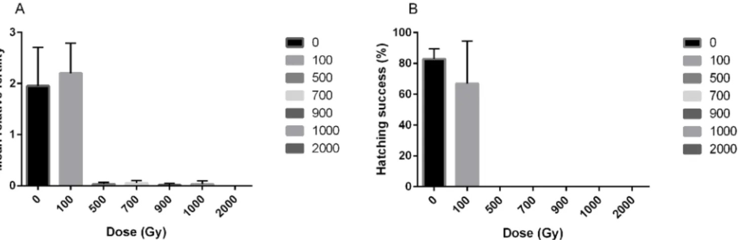
The effect of irradiation on fertility and egg viability inH.dujardiniwas also evaluated (Fig 2andS1 Text). A dose of 100 Gy gave a similar level of fertility (U = 3.0, P = 0.51,Fig 2A) and viability of eggs (U = 6.0, P = 0.51,Fig 2B) as in the controls. Irradiation with 500 Gy or 1000 Gy reduced animal fertility significantly (U = 9.0, p = 0.046 for both cases) to<5% and no laid
eggs hatched. Finally, doses above 1000 Gy induced complete infertility.
Effects of irradiation on eggs at different stages of development
The effects of irradiation on hatchability of eggs in the early and late developmental stages are shown inFig 3A and 3B, respectively. There was a clear dose response for hatchability of eggs irradiated at the early developmental stage, with no effect of 50 Gy (100%, SD = 0) compared to controls (100%, SD = 0), but a significant effect of 200 Gy (69%, SD = 13.7; U = 9.0, p = 0.037) Fig 1. Survival of adultH.dujardiniafter exposure to different doses of gamma radiation.Error bars represent standard deviations from 3 repeats, each with 20 animals.
doi:10.1371/journal.pone.0133658.g001
Fig 2. Radiation effects on fertility of the tardigradeH.dujardini.A) Fertility levels expressed as the average number of eggs laid during 12 days after irradiation. B) Egg viability expressed as the proportion of laid eggs that hatched. Error bars represent standard deviations from 3 repeats, each with 20 animals.
and 500 Gy (9%, SD = 3.1; U = 9.0, p = 0.034), and also a tendency of a dose-dependent delay in hatching time (Fig 3A). In contrast, late-stage embryos showed high resistance to radiation, and no significant reduction of hatchability could be observed for doses up to 500 Gy (0 Gy = 100% (SD = 0), 50 Gy = 93% (SD = 5.4), 200 Gy = 98% (SD = 3.1), 500 Gy = 100% (SD = 0); Kruskal-Wallis Analysis of differences between dose groups at day 13: H = 4.7, p = 0.20;Fig 3B).
We also analyzed the post-hatching effects in eggs that survived irradiation during the early or late stage of development. Survival of juveniles hatched from eggs irradiated at 50 Gy and 200 Gy in the early developmental stage (Fig 4A) did not differ significantly from controls (p>0.05; seeS1 Text) at any measured times (survival estimate at day 20: 91.1% (SD = 3.1) for
controls, 93.3% (SD = 5.4) for the 50 Gy group and 89.1% (SD = 10.4) for 200 Gy). Eggs irradi-ated at 500 Gy had significantly reduced survival (p<0.05) from day 14 onwards (survival
esti-mate at day 20: 16.6% (SD = 23.5) for the 500 Gy group. Note however that only 1–2 eggs hatched in the 500 Gy group). The survival of animals from eggs irradiated at 50 Gy and 200 Gy in the late developmental stage did not differ significantly from controls (p>0.10) at any
time (survival estimate at day 20: 93.3% (SD = 5.4) for controls, 85.6% (SD = 10.2) for the 50 Gy group, 75.4% (SD = 12.3) for the 200 Gy group). Also the 500 Gy group did not differ from the controls up to day 13, but survival then declined significantly from day 17 onwards (p0.05), and reached 43.2% (SD = 2.7) on day 20.
The relative fertility (eggs/individual) of the young adults is shown inFig 4C. For both the early and late stage irradiation category there was a significant overall difference in relative Fig 3. Hatching sequence and success ofH.dujardinieggs irradiated at different doses and at two different stages of egg development.(A) Irradiation at the early developmental stage. (B) Irradiation at the late developmental stage. Time in days after laying are given at the x-axis. Error bars represent standard deviations from 3 repeats, each with 15 eggs.
doi:10.1371/journal.pone.0133658.g003
Fig 4. Survival ofH.dujardinijuveniles hatched from eggs irradiated during early or late stage of embryonic development, and their relative fertility.(A) Young adult survival after irradiation in the early stage of egg development. (B) Young adult survival after irradiation in the late stage of development. (C) Relative fertility of young adults irradiated in the early and late stages, calculated as mean number of eggs laid (in each replicate sample) divided by the highest number of young adults available per repeat.
fertility among dose groups (Early: H = 9.3, p = 0.025; Late: H = 10.1, p = 0.018). In the early stage irradiation category mean relative fertility of controls was considerably higher (3.28 eggs/ ind., SD = 1.52) than in the irradiated groups (50 Gy: 0.91 eggs/ind., SD = 0.16; 200 Gy: 0.25 eggs/ind., SD = 0.36), but due to high variation within the control group only the 200 and 500 Gy groups reached statistical significance (control vs. 50 Gy: p = 0.077; control vs. 200 Gy: U = 9.0, p = 0.046; control vs. 500 Gy: U = 9.0, p = 0.037). For the late stage irradiation group there was no significant difference in relative fertility between controls (1.3 eggs/ind., SD = 0.75) and the 50 Gy group (0.7 eggs/ind., SD = 0.19, U = 8.0, p = 0.13), but the higher dose groups had marginally significantly (200 Gy: 0.2 eggs/ind., SD = 0.07, U = 9.0, p = 0.05) or significantly lower relative fertility (500 Gy: no eggs, U = 9.0, p = 0.037). The 500 Gy group dif-fered significantly also from the 50 Gy and 200 Gy groups (U = 9.0, p = 0.037 for both cases).
The additional eggs irradiated with 50 Gy in the early stage of development and its respec-tive controls were photographed and analyzed with respect to the description of egg develop-mental stages provided by Gabriel et al. [14].S1 Figshows the effect of gamma irradiation on cells in the initial stage of development as an apparent loss of cells 0.5h after the treatment. In the control eggs cell proliferation is evident and the developmental stages are similar in timing to the ones described by Gabriel et al. [14]. During the following days, the irradiated eggs con-tinued their development through almost all the stages but with some time delay compared to the controls.
Effects of low temperature during irradiation on survival and fertility of
adults
We analyzed if the temperature at which adult tardigrades were irradiated influenced survival for doses of 4000 and 4500 Gy. At low temperature survival rate was high (100%) when mea-sured immediately after irradiation, while irradiation at room temperature (no ice) reduced survival significantly, to 71% in the 4000 Gy group and to 64% in the 4500 Gy group (p<0.05
in both cases;Fig 5A and 5B;S1 Text). After this initial difference in survival there were only marginal or no differences in survival between the temperature regimes for 4000 Gy (p0.05;
Fig 5A), while for the 4500 Gy group the room temperature group had lower survival up to 3 days after irradiation (p<0.05;Fig 5B), but did not differ thereafter.
Fig 5. Effects of low temperature at irradiation on adultH.dujardinisurvival.The figure shows estimates of mean survival over time after irradiation up to 12 days post-irradiation, for samples irradiated on ice (low temperature) and without ice (20°C), and for controls kept at 20°C. Fig (A) shows data for irradiation with 4500 Gy, and Fig (B) for irradiation with 4000 Gy. BR represents survival before radiation and AR represents survival immediately after irradiation. Each data point represents the mean of three repeats, each with 15 animals.
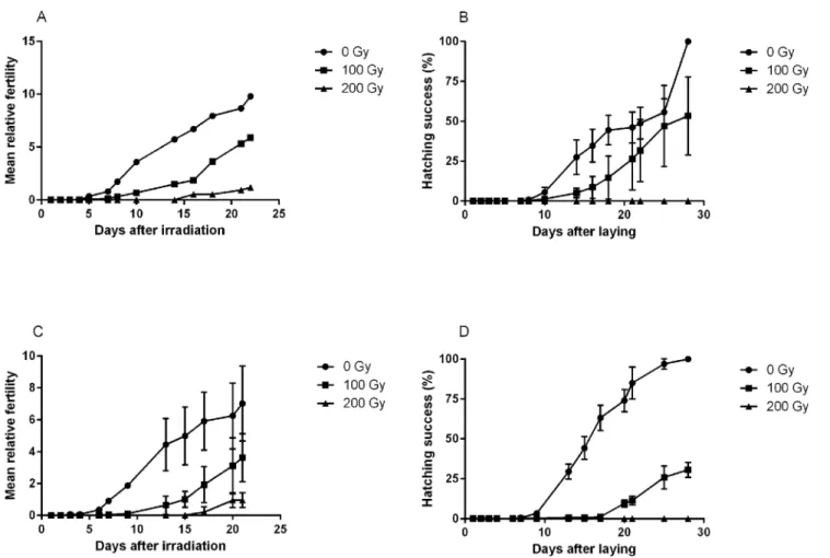
We also evaluated if irradiation temperature had an effect on fertility and viability on eggs for dose levels of 0, 100 and 200 Gy (Fig 6). As shown above, irradiation>1000 Gy resulted in
complete infertility. The general dose-response in relative fertility (eggs/individuals) was very similar for samples irradiated on ice and at room temperature (Fig 6A and 6C), and there was no statistical difference in the production of eggs (pair-wise comparisons between ice and room temperature for each dose-group, p>0.05 in all analyses). There was a clear and
signifi-cant dose-response both in total relative fertility (lower fertility at higher doses, H = 13.7, p = 0.001) and in the temporal patterns of egg laying (more delay at higher doses). Tardigrades irradiated at 200 Gy started to produce eggs only after 15 days, but the eggs laid were not viable (Fig 6B and 6D). Also in hatching success there was a significant dose-dependent difference among the three dose groups (H = 16.3, p<0.001) and an apparent dose-dependent delay in
hatching (Fig 6B and 6D). For eggs irradiated at 100 Gy, those kept on ice tended to hatch slightly faster than those kept at room temperature (Fig 6B and 6D).
Discussion
The present study adds a freshwater species,H.dujardini, to the list of tardigrades showing a very high tolerance to gamma radiation in the active hydrated state, with an LD5048hof
~4500 Gy. This is in the same range of tolerance as previously reported for the limno-Fig 6. Mean relative fertility of adultH.dujardiniand hatching success of laid eggs after irradiation on ice (A, B) or at room temperature (C, D).
Fertility data points represent the mean number of eggs laid per individual within each repeat sample. Three repeats were used for each dose. Hatching success represents the proportion of laid eggs that hatched within each repeat sample.
terrestrial tardigradesMilnesium tardigradum(5000 Gy [4]) andRichtersius coronifer(3500 Gy/22 hours; [3]). Since radiation tolerance is hypothesized to be a by-product of evolved mechanisms for desiccation tolerance [12], the high tolerance ofH.dujardiniis unexpected although our knowledge on desiccation tolerance inH.dujardiniis rather limited. However, Wright [13] reported that this species was less tolerant to desiccation at low relative humidity (RH) compared to several other species (includingM.tardigradum), and that successful anhydrobiosis in this species required pre-desiccation under high humidity conditions (85% RH). Thus, our results provide an interesting input to the question of the origin of tolerance to radiation in tardigrades, and the extent to which tolerances to radiation and desiccation are based on the same mechanisms.
The high tolerance gamma radiation inH.dujardinialso contrasts with previous findings [7] that this species has much lower tolerance to UV radiation than the limno-terrestrial spe-ciesH.varieornatus. Also other studies have documented differences among tardigrade species in tolerance to UV radiation. DehydratedMilnesium tardigradumsurvived exposure to UV radiation in space much better thanR.coronifer[8], despite similar tolerance to gamma radia-tion. Thus, there seems to be more differences among tardigrade species in the sensitivity to UV radiation than to ionizing radiation. The molecular mechanisms behind this pattern is unclear, but can relate to, e.g., mechanisms of DNA repair, antioxidant protection or shielding by pigmentation. The impacts of gamma radiation and UV light on DNA are generally differ-ent (double strand breaks and tymidine dimers, respectively) and also the repair pathways. However, both types of radiation induce reactive oxigen species (ROS) [15,16]. In the study by Horikawa et al. [7] the induction of thymidine dimers by UV-C was similar inH.dujardini andH.varieornatus, confirming similar effects of radiation on DNA in these species despite their different tolerance to UV. In the latter species, the photolyase gene phrA (connected with DNA repair) was strongly upregulated in UV-C irradiated animals, while inH.dujardinino homologous phrA gene was found based on the available EST library for this species. There-fore, possibly the sensitivity ofH.dujardinito UV-C could be associated with an evolutionary loss or mutation of the phrA gene/protein involved, as it seems has happened in humans [16], but the high tolerance to ionizing radiation remains to be explained.
The irradiation of embryonic cells (eggs) in our study also revealed two additional effects. First, there was a delay in time of hatching. Second, some dividing cells were apparently lost after irradiation exposure (seeS1 Fig). Most metazoan embryos show fast cell division cycles at the beginning of their development [21], and DNA damage during cell divisions usually trig-gers cell cycle arrest providing time for repair or apoptosis induction [22,23]. However, in the early developmental stage ofC.elegans,D.melanogasterandX.laevisembryos, checkpoint activation to stop the cell cycle after DNA damage is highly or slightly silenced during the fast cell cycle divisions, and activated at the end of midblastula transition [24]. In a cell ablation experiment with tardigrade embryos using a laser beam, Hejnol [25] removed half of the 4-cell embryo inThulinia stephaniae. Remarkably, the development proceeded normally and the eggs eventually hatched, but with a time delay compared to the controls. Such developmental delays were also observed in the present study, and in previous radiation studies inM. tardigra-dum[11], indicating that the early embryogenesis is highly regulated in tardigrades [25]. After the apparent cell loss caused by irradiation in our study, the rest of the cells in theH.dujardini embryo were able to continue development but were delayed compared to the control.
In many animals genome integrity is subject to stringent DNA damage detection, repair, or induction of apoptosis in cells with low genomic fidelity [26]. Indications of apoptosis avoid-ance have been shown in mice oocytes (early stage, meiotic) lacking specific apoptosis regulator genes (PumaandNoxa). The absence of these genes protected them from γ-irradiation-induced apoptosis and generated healthy offspring [26]. In the desiccation and radiation toler-ant chironomidPolypedilum vanderplanki, radioresistance was associated with repair of dam-aged DNA, and repair was also indicated after desiccation [27]. However, inP.vanderplanki the process of repair is apparently rather slow (24h after 70 Gy gamma rays, and 168h after heavy ions4He), and during this repair period apoptosis is prevented. The apparent recovery in fertility observed in our study 15 days after adult irradiation with 200 Gy might also be an indication of a similar slow repair process in tardigrades.
Even though the irradiated but surviving embryonic cells were able to produce an embryo capable of hatching and developing into young adults, there was an apparent reduction in their fertility as the dose increased. This indicates that surviving cells were still affected in later stages of the life cycle. Moreover, the strong decline in survival of young adults originating from eggs irradiated with 500 Gy during the late stage of egg development is also an indication of residual damage, affecting life span and fertility. Studies on rotifers andC.eleganshave shown that radi-ation does not affect fertility in terms of egg number, but rather induces sterility [28]. Accord-ing to these studies, the cells in the eggs were not directly killed by radiation, but received damage that was expressed later in life.
The role of an efficient DNA repair system has previously been suggested as a possible explanation for the high tolerance to radiation in tardigrades [11,12,29,30,31], but has yet to be documented. However, two important proteins for DNA damage/response are induced in tar-digrades after radiation exposure (RAD51, [31]; HSP70, [32]). Also, in response to desiccation, the polyubiquitin gene (UBB) related to the regulation of DNA repair mechanisms has been reported to be up-regulated inM.tardigradum[33]. Protein protection may also contribute to radiation tolerance, and Krisko et al. [28] reported a negative correlation between fecundity and protein carbonylation in the rotiferA.vagaand the nematodeC.elegansafter radiation exposure. Apparently, the antioxidant protection, in terms of less protein carbonylation, is higher inA.vagawhich is more resistant to radiation compared toC.elegans. Thus, the con-cept of protein protection, including DNA repair proteins, should be evaluated as a possible mechanism of radiation tolerance in multicellular organisms [28].
on ice is a common procedure in many experiments of mammalian cells in order to reduce activation of DNA repair, along with other cellular processes, during the radiation exposure [34,35]. Recent studies have also shown that dehydration and low temperatures (below freez-ing) can provide protection to radiation damage by reducing the diffusion of free radicals [36–
38]. It is well known in radiation biology that 70% of the DNA damage is caused by free radi-cals (Low LET) in hydrated conditions. According to Slade and Radman [39], the survival rate ofD.radioduransis higher when the irradiation is done on dry ice (-70°C) compared to room temperature or ice [36], and they suggested that this resistance rely on diminished reactive oxy-gen effects on proteins under frozen irradiation conditions. However, in the present study there was no evidence of a protective effect of low temperature (0°C), and the delay in mortality is not likely to be explained by effects of radical chemistry. A more suitable explanation might be a biological response caused by the high level of DNA damage, which may have triggered the animals’death after the temperature rose.
In summary, our study shows that fresh water tardigrades are also able to tolerate high doses of gamma radiation, with similar patterns of dose-response as limno-terrestrial tardi-grades. Assuming that adult tardigrades and late developmental stages of embryos have only a few cells mitotically active (due to eutely), we suggest that this might explain part of the observed radiation tolerance. However, efficient DNA repair mechanisms may still contribute to the high radiation tolerance observed, both in adult tardigrades and in embryos relative to other animals. Further studies are needed to evaluate the relative importance of eutely vs. other mechanisms (e.g., DNA repair system, protein protection, apoptosis avoidance) in radiation tolerance of tardigrades.
Supporting Information
S1 Fig. Gamma radiation effect on eggs at the early developmental stage.The figure show images of 2 representative irradiated eggs (IR, 50 Gy) and 2 control eggs at different times in the development. Start measurement picture (before irradiation) was taken after release of eggs into the exuvia. Pictures were then taken immediately after the irradiation, and at different time intervals up to 137 hours after irradiation. The corresponding developmental time and cell/embryo morphology stages is given in the figure. SeeMethodssection for more details on these stages. Bar scale 10μm.
(TIF)
S1 Text. Statistical results related to figures in the article.
(DOCX)
Acknowledgments
We thank Hiba Alasawi for her contribution to this study by selecting eggs for irradiation and collecting their hatching time.
Author Contributions
Conceived and designed the experiments: EBP KIJ MH-R AW SH. Performed the experiments: EBP. Analyzed the data: EBP KIJ MH-R AW SH. Contributed reagents/materials/analysis tools: MH-R AW SH. Wrote the paper: KIJ EBP MH-R SH AW.
References
2. May RM, Guimard J. Action différentielle des rayons x et ultraviolets sur le tardigrade Macrobiotus areo-latus, a l’état actif et desséché. Bull Biol Fr Belg. 1964; 98: 18.
3. Jönsson KI, Harms-Ringdahl M, Torudd J. Radiation tolerance in the eutardigradeRichtersius coroni-fer. Int J Radiat Biol. 2005; 81: 649–656. PMID:16368643
4. Horikawa DD, Sakashita T, Katagiri C, Watanabe M, Kikawada T, Nakahara Y, et al. Radiation toler-ance in the tardigradeMilnesium tardigradum. Int J Radiat Biol. 2006; 82: 843–848. PMID:17178624
5. Nilsson CEJ, Jönsson KI, Pallon J. Tolerance to proton irradiation in the eutardigradeRichtersius coro-nifer–a nuclear microprobe study. Int J Radiat Biol. 2010; 86: 420–427. doi:10.3109/
09553000903568001PMID:20397847
6. Altiero T, Guidetti R, Caselli V, Cesari M, Rebecchi L. Ultraviolet radiation tolerance in hydrated and desiccated eutardigrades. J Zool Syst Evol Res. 2011; 49: 104–110.
7. Horikawa DD, Cumbers J, Sakakibara I, Rogoff D, Leuko S, Harnoto R, et al. Analysis of DNA repair and protection in the TardigradeRamazzottius varieornatusandHypsibius dujardiniafter exposure to UVC radiation. PLoS ONE. 2013; 8: e64793. doi:10.1371/journal.pone.0064793PMID:23762256
8. Jönsson KI, Rabbow E, Schill RO, Harms-Ringdahl M, Rettberg P. Tardigrades survive exposure to space in low Earth orbit. Curr Biol. 2008; 18: R729–R731. doi:10.1016/j.cub.2008.06.048PMID: 18786368
9. Horikawa DD, Yamaguchi A, Sakashita T, Tanaka D, Hamada N, Yukuhiro F, et al. Tolerance of anhy-drobiotic eggs of the Tardigrade Ramazzottius varieornatus to extreme environments. Astrobiology. 2012; 12: 283–289. doi:10.1089/ast.2011.0669PMID:22490117
10. Jönsson KI, Beltrán-Pardo E, Haghdoost S, Wojcik A, Bermúdez-Cruz RM, Bernal J.E, et al. Tolerance to gamma-irradiation in eggs of the tardigradeRichtersius coroniferdepends on stage of development. J Limnol. 2013; 72(1s): 73–79.
11. Beltrán-Pardo E, Jönsson KI, Wojcik A, Haghdoost S, Harms-Ringdahl M, Bermúdez—Cruz RM, et al. Effects of ionizing radiation on embryos of the tardigradeMilnesiumcf. tardigradumat different stages of development. PLoS ONE. 2013; 8: e72098. doi:10.1371/journal.pone.0072098PMID:24039737
12. Jönsson KI. Causes and consequences of excess resistance in cryptobiotic metazoans. Physiol Bio-chem Zool. 2003; 76: 429–435. PMID:13130423
13. Wright JC. Desiccation tolerance and water-retentive mechanisms in tardigrades. J Exp Biol. 1989; 142: 267–292.
14. Gabriel WN, McNuff R, Patel SK, Gregory TR, Jeck WR, Jones CD, et al. The tardigradeHypsibius dujardini, a new model for studying the evolution of development. Dev Biol. 2007; 312: 545–559. PMID:17996863
15. Widel M, Krzywon A, Gajda C, Skonieczna M, Rzeszowska-Wolny J. Induction of bystander effects by UVA, UVB, and UVC radiation in human fibroblasts and the implication of reactive oxygen species. Free Rad Biol and Med. 2014; 68: 278–287.
16. Sinha RP, Häder DP. UV-induced DNA damage and repair: a review. Photochem Photobiol Sci. 2002; 4:225–36.
17. Santos-Ahmed J, Brown C, Smith SD, Weston P, Rasoulpour T, Gilbert ME, et al. Akt1 protects against germ cell apoptosis in the postnatal mouse testis following lactational exposure to 6-N-propylthiouracil. Reprod Toxicol. 2011; 31: 17–25. doi:10.1016/j.reprotox.2010.09.012PMID:20951798
18. Alexander ML, Stone WS. Radiation damage in the developing germ cells ofDrosophila Virilis. Proc Nat Acad Sci. 1955; 41: 1046–1057. PMID:16589795
19. Bergonié J, Tribondeau L. De quelques résultats de la radiotherapie et essai de fixation d’une tech-nique rationnelle. C R Acad Sci. 1906; 143: 983–985.
20. Bertolani R. Mitosi somatische e constanza cellular numerica nei Tardigradi. Atti Accad Naz Lincei Rend. 1970; Ser 8a, Vol. 48: 739–742.
21. O'Farrell PH, Stumpff J, Su TT. Embryonic cleavage cycles: How is a mouse like a fly? Curr Biol. 2004; 14: R35–R45. PMID:14711435
22. Elledge SJ. Cell cycle checkpoints: preventing an identity crisis. Science. 1996; 274: 1664–1672. PMID:8939848
23. Kappas NC, Savage P, Chen KC, Walls AT, Sible JC. Dissection of the XChk1 signaling pathway in Xenopus laevisembryos. Mol Biol Cell. 2000; 11: 3101–3108. PMID:10982403
24. Holway AH, Kim S-H, La Volpe A, Michael WM. Checkpoint silencing during the DNA damage response inCaenorhabditis elegansembryos. J Cell Biol. 2006; 172: 999–1008. PMID:16549501
26. Kerr J.B, Hutt K.J, Michalak E.M, Cook M, Vandenberg C.J, Seng H.L, et al. DNA Damage-Induced Pri-mordial Follicle Oocyte Apoptosis and Loss of Fertility Require TAp63-Mediated Induction of Puma and Noxa. Mol Cell. 2012; 48: 343–352. doi:10.1016/j.molcel.2012.08.017PMID:23000175
27. Gusev O, Nakahara Y, Vanyagina V, Malutina L, Cornette R, Sakashita T, et al. Anhydrobiosis-associ-ated nuclear DNA damage and repair in the Sleeping Chironomid: Linkage with radioresistance. PLoS ONE. 2010; 5: e14008. doi:10.1371/journal.pone.0014008PMID:21103355
28. Krisko A, Leroy M, Radman M, Meselson M. Extreme anti-oxidant protection against ionizing radiation in bdelloid rotifers. Proc Nat Acad Sci. 2012; 109: 2354–2357. doi:10.1073/pnas.1119762109PMID: 22308443
29. Neumann S, Reuner A, Brummer F, Schill RO. DNA damage in storage cells of anhydrobiotic tardi-grades. Comp Biochem Physiol A Mol Integr Physiol. 2009; 153: 425–429. doi:10.1016/j.cbpa.2009. 04.611PMID:19361569
30. McLennan AG. Ametabolic embryos ofArtemia franciscanaaccumulate DNA damage during pro-longed anoxia. J Exp Biol. 2009; 212: 785–789. doi:10.1242/jeb.023663PMID:19251993
31. Beltrán-Pardo EA, Jönsson KI, Wojcik A, Haghdoost S, Bermúdez Cruz RM, Bernal J.E, et al. Sequence analysis of the DNA-repair gene rad51 in the tardigrades Milnesium cf. tardigradum, Hypsi-bius dujardini and Macrobiotus cf. harmsworthi. J Limnol. 2013; 72(1s): 80–91.
32. Jönsson KI, Schill RO. Induction of Hsp70 by desiccation, ionising radiation and heat-shock in the eutardigradeRichtersius coronifer. Comp Biochem Physiol Part B: Biochem Mol Biol. 2007; 146: 456– 460.
33. Wang C, Grohme MA, Mali B, Schill RO, Frohme M. Towards decrypting cryptobiosis—analyzing anhy-drobiosis in the tardigrade Milnesium tardigradum using transcriptome sequencing. PLoS ONE. 2014; 9(3): e92663. doi:10.1371/journal.pone.0092663PMID:24651535
34. Gobbel GT, Bellinzona M, Vogt AR, Gupta N, Fike JR, Chan PH. Response of postmitotic neurons to X-Irradiation: Implications for the role of DNA damage in neuronal apoptosis. J Neurosci. 1998; 18: 147– 155. PMID:9412495
35. Lisowska H, Brehwens K, Zölzer F, Wegierek-Ciuk A, Czub J, Lankoff A, et al. Effect of hypothermia on radiation-induced micronuclei and delay of cell cycle progression in TK6 cells. Int J Radiat Biol. 2014; 90: 318–324. doi:10.3109/09553002.2014.887233PMID:24467331
36. Dartnell LR, Hunter SJ, Lovell KV, Coates AJ, Ward JM. Low-temperature ionizing radiation resistance of Deinococcus radiodurans and antarctic dry valley bacteria. Astrobiology. 2010; 10: 717–732. doi: 10.1089/ast.2009.0439PMID:20950171
37. Cruz de Carvalho R, Catala M, Marques da Silva J, Branquinho C, Barreno E. The impact of dehydra-tion rate on the producdehydra-tion and cellular locadehydra-tion of reactive oxygen species in an aquatic moss. Ann Bot. 2012; 110: 1007–1016. doi:10.1093/aob/mcs180PMID:22875812
38. Erkut C, Vasilj A, Boland S, Habermann B, Shevchenko A, Kurzchalia TV, et al. Molecular strategies of the Caenorhabditis elegans dauer larva to survive extreme desiccation. PLoS ONE. 2013; 8: e82473. doi:10.1371/journal.pone.0082473PMID:24324795