NÁDIA EMI AIKAWA
Autoanticorpos órgão-específicos e sistêmicos em pacientes
com lúpus eritematoso sistêmico juvenil e dermatomiosite
juvenil
Dissertação apresentada à Faculdade de Medicina da Universidade de São Paulo para obtenção do título de Mestre em Ciências
Programa de: Pediatria
Orientadora: Dra. Adriana Maluf Elias Sallum
Dados Internacionais de Catalogação na Publicação (CIP)
Preparada pela Biblioteca da
Faculdade de Medicina da Universidade de São Paulo
reprodução autorizada pelo autor
Aikawa, Nádia Emi
Autoanticorpos órgão-específicos e sistêmicos em pacientes com lúpus eritematoso sistêmico juvenil e dermatomiosite juvenil / Nádia Emi Aikawa. -- São Paulo, 2011.
Dissertação(mestrado)--Faculdade de Medicina da Universidade de São Paulo.
Programa de Pediatria.
Orientadora: Adriana Maluf Elias Sallum.
Descritores: 1.Lúpus eritematoso sistêmico 2.Dermatomiosite juvenil 3.Autoanticorpos 4.Especificidade de órgãos
À minha família, pelo carinho e dedicação em minha criação e pelo incentivo
À Dra Adriana Maluf Elias Sallum, por ter me orientado com muito carinho e
apoio em todos os momentos e pelas preciosas contribuições ao estudo.
Ao Prof. Dr. Clovis Artur Almeida da Silva, por ser um exemplo em minha
formação em Reumatologia Pediátrica, pelos incentivos incansáveis e
contribuições valiosas ao estudo.
À Profa. Dra. Magda Carneiro-Sampaio, por ter possibilitado a realização deste estudo e pelo carinho e entusiasmo constantes.
À Dra. Bernadete de Lourdes Liphaus, pela idealização do estudo juntamente com a Profa. Magda e pelas importantes sugestões.
À Dra Adriana Almeida de Jesus, pelo auxílio inestimável na coleta de dados, por sua preciosa amizade e por ter me incentivado a fazer Reumatologia Pediátrica.
Aos amigos da Reumatologia Pediátrica, pelo carinho e paciência.
À bibliotecária Mariza Kazue Umetsu Yoshikawa, pela alegria e eficiência
inabaláveis.
Aos colegas do Serviço de Arquivo Médico, pela simpatia e prontidão no
Aos pacientes do Ambulatório de Reumatologia Pediátrica do Instituto da
Esta dissertação está de acordo com as seguintes normas, em vigor no momento desta publicação:
Referências: adaptado de International Committee of Medical Journals Editors (Vancouver)
Universidade de São Paulo. Faculdade de Medicina. Serviço de Biblioteca e Documentação. Guia de apresentação de dissertações, teses e monografias. Elaborado por Anneliese Carneiro da Cunha, Maria Julia de A. L. Freddi, Maria F. Crestana, Marinalva de Souza Aragão, Suely Campos Cardoso, Valéria Vilhena. 2a ed.
São Paulo: Serviço de Biblioteca e Documentação; 2005.
S U M Á R I O
Lista de abreviaturas Lista de tabelas Resumo
Summary
1 Introdução... 2
2 Objetivos... 5
3 Métodos... 7
4 Resultados... 13
4.1 Dados demográficos... 13
4.2 Autoanticorpos órgão-específicos e outros autoanticorpos... 13
4.3 Doenças autoimunes órgão-específicas... 17
5 Discussão... 20
6 Conclusões... 24
7
8
Referências...
Anexos... 26
A
B R E V I AT U R ASJSLE Lúpus eritematoso sistêmico juvenil JDM
TH DM1 DC
Anti-TPO Anti-Tg TRAb IAA Anti-GAD
Dermatomiosite juvenil Tireoidite de Hashimoto Diabetes melito tipo 1 Doença celíaca
Anticorpo antiperoxidase tiroideana Anti-tireoglobulina
Anti-receptor de hormônio tireoestimulante (TSH) Anti-insulina
Anti-descarboxilase do ácido glutâmico Anti-LKM-1 Anticorpo microssomal rim-fígado tipo 1 AML
AMA
Anticorpo anti-músculo liso Anti-mitocôndria
ACP EMA
T
AB E L ASTabela 1 – Autoanticorpos e doenças órgão-específicas e outros autoanticorpos em pacientes com lúpus eritematoso sistêmico juvenil (LESJ) versus pacientes com dermatomiosite juvenil (DMJ) ... 14
Tabela 2 – Dados demográficos, atividade da doença, tratamento e outros autoanticorpos em pacientes com lúpus eritematoso sistêmico juvenil (LESJ) e dermatomiosite juvenil (DMJ) com e sem autoanticorpos órgão-específicos... 16
Aikawa NA. Autoanticorpos órgão-específicos e sistêmicos em pacientes com lúpus eritematoso sistêmico juvenil e dermatomiosite juvenil [dissertação]. São Paulo: Faculdade de Medicina, Universidade de São Paulo; 2011.28p.
associada com tireoidite de Hashimoto e um terceiro paciente apresentava tireoidite subclínica. Outro paciente com LESJ preenchia diagnóstico de doença celíaca com base em anemia por deficiência de ferro, a presença de anticorpo anti-endomísio, biópsia duodenal compatível com doença celíaca e resposta a dieta livre de glúten. Conclusão: Doenças órgão-específicas foram observadas apenas em pacientes com LESJ e exigiram tratamento específico. A presença destes anticorpos sugere a avaliação de doenças órgão-específicas e um acompanhamento rigoroso destes pacientes.
Aikawa NE. Organ-specific and systemic autoantibodies in patients with juvenile systemic lupus erythematosus and juvenile dermatomyositis [dissertation]. São Paulo: “Faculdade de Medicina, Universidade de São Paulo”; 2010. 29p.
Conclusion: Organ-specific diseases were observed solely in JSLE patients and required specific therapy. The presence of these antibodies recommends the evaluation of organ-specific diseases and a rigorous follow-up of these patients.
IN T R O D U Ç Ã O 2
O lúpus eritematoso sistêmico (LES) é uma doença autoimune multissistêmica, caracterizada pela presença de autoanticorpos circulantes1,2. A dermatomiosite juvenil (DMJ) é uma doença do tecido conjuntivo caracterizada por vasculite muscular e cutânea, podendo também comprometer outros órgãos e sistemas3. A etiologia da DMJ é desconhecida. No entanto, a presença de inflamação muscular crônica, a positividade para autoanticorpos séricos e a associação com outras doenças autoimunes sugerem que um mecanismo autoimune esteja envolvido em sua fisiopatologia3.
IN T R O D U Ç Ã O 3
OB J E T I V O S
5
1. Avaliar a prevalência de autoanticorpos séricos órgão-específicos em pacientes com LESJ e com DMJ.
2. Avaliar a possível associação entre dados demográficos, atividade da doença e tratamento de pacientes com LESJ e com DMJ de acordo com a presença de autoanticorpos órgão-específicos.
3. Descrever as doenças autoimunes órgão-específicas associadas em pacientes com LESJ e DMJ.
MÉ T O D O S 7
Pacientes e métodos
Quarenta e um pacientes com LESJ e 41 com DMJ regularmente
acompanhados na Unidade de Reumatologia Pediátrica do Instituto da
Criança do Hospital das Clínicas (HC) da Faculdade de Medicina da
Universidade de São Paulo (FMUSP) foram selecionados entre janeiro de
2008 e janeiro de 2009. Todos os pacientes preencheram os critérios do
American College of Rheumatology (ACR) para LESJ10 e de Bohan & Peter
para DMJ11. O Comitê de Ética do HC-FMUSP aprovou este estudo e um
consentimento informado foi obtido de todos os participantes e de seus
responsáveis.
Autoanticorpos órgão-específicos e outros autoanticorpos
Autoanticorpos séricos associadas com as seguintes doenças autoimunes
órgão-específicas foram avaliados: tireoidite autoimune - anticorpo
antiperoxidase tiroideana (TPO) por fluoroimunoensaio e anticorpos
anti-tireoglobulina (anti-Tg) e anti-receptor de hormônio tireoestimulante (TSH)
(TRAb) por quimioluminescência; DM1 - anticorpo insulina (IAA),
MÉ T O D O S 8
fosfatase (anti-IA2) por radioimunoensaio; hepatite autoimune – anticorpo
microssomal rim-fígado tipo 1 (anti-LKM-1) e anticorpo anti-músculo liso
(AML) por imunofluorescência indireta em fígado de rato e cortes de tecido
renal; cirrose biliar primária – anti-mitocôndria (AMA) por
imunofluorescência indireta em fígado, rins e células parietais de estômago
de rato, e confirmação dos casos positivos por teste imunoenzimático
(ELISA); gastrite autoimune - anticorpo anti-célula parietal (ACP) por
imunofluorescência indireta; doença celíaca - anticorpo anti-endomísio
(EMA) da classe IgA por imunofluorescência indireta. Os pacientes que
foram positivos para os autoanticorpos órgão-específicos repetiram o teste
para confirmação. Em seguida, foram investigados para a presença da
doença autoimune órgão-específica.
Os seguintes anticorpos séricos também foram avaliados: fator
anti-núcleo (FAN) por imunofluorescência indireta utilizando células de epitelioma
humano (HEp-2), fator reumatóide (FR) por imunonefelometria, anti-DNA
dupla-hélice (anti-dsDNA), anti-Sm , anti-RNP, anti-SSB/La anti-SSA/Ro,
anti-topoisomerase-1 (anti-Scl70), anticardiolipina (aCL) isotipos IgG e IgM
por ELISA e anticoagulante lúpico (LAC) pelo teste do veneno de víbora de
Russel diluído e os testes confirmatórios, anticorpo anti-citoplasma de
neutrófilos (ANCA) por ELISA e por imunofluorescência direta em neutrófilos
humanos fixados com etanol e anti-Jo1 por ELISA.
Todos os anticorpos foram avaliados na Divisão de Laboratório Central do
MÉ T O D O S 9
Doenças autoimunes órgão-específicas
HT foi definida de acordo com a redução dos níveis de tiroxina livre
(T4) e níveis elevados de TSH, e hipotireoidismo subclínico como níveis
elevados de TSH associados a níveis normais de T4 livre12. A presença de
anticorpos anti-tireoidianos foi necessária para caracterizar a tireoidite
autoimune. DM1 foi diagnosticada pela presença de poliúria, polidipsia e
perda de peso sem causa definida, e glicose plasmática, 200 mg/dL a
qualquer hora do dia ou glicemia de jejum 126 mg/dL13. Hepatite autoimune
foi definida como uma hepatite crônica progressiva de origem desconhecida,
caracterizada por níveis elevados de transaminases, hipergamaglobulinemia,
autoanticorpos séricos e características histológicas14. Cirrose biliar primária
foi definida como a presença de pelo menos dois dos seguintes: elevação de
fosfatase alcalina ( 2 vezes o limite superior do normal) ou
gama-glutamiltransferase ( 5 vezes o limite superior do normal), positividade para
AMA e biópsia hepática com colangite supurativa e destruição de ductos
biliares15. Gastrite autoimune foi definida pela presença atrofia de fundo e corpo gástrico à histologia, a positividade para o anticorpo APC e anti-fator
intrínseco, hipo/acloridria, baixas concentrações de pepsinogênio sérico e
anemia secundária a deficiência de vitamina B12 e ferro16. O diagnóstico da
doença celíaca foi definido pela presença de pelo menos quatro dos
seguintes critérios: quadro clínico (diarréia crônica, nanismo e/ou anemia
ferropriva), positividade para anticorpos da classe IgA para doença celíaca,
genótipo HLA-DQ2 ou DQ8, biópsia do intestino delgado compatível com
MÉ T O D O S 10
Atividade da doença, dano da doença e tratamento dos pacientes com
LESJ e DMJ
A atividade do LESJ e dano cumulativo foram avaliados no momento
da avaliação dos anticorpos e das doenças órgão-específicas nos pacientes
com LESJ usando o SLE Disease Activity Index 2000 (SLEDAI-2K)18 e o
Systemic Lupus International Collaborating Clinics/ACR Damage Index
(SLICC/ACR-DI)19. A atividade da DMJ foi avaliada pelo Disease Activity
Score (DAS)20 e a força muscular foi avaliada pelo Childhood Myositis Assessment Scale (CMAS) e o Manual Muscle Testing (MMT)21. As enzimas
musculares dosadas concomitantemente aos anticorpos e doenças
órgão-específicas foram: creatinofosfoquinase (CPK), aspartato aminotransferase
(AST), alanina aminotransferase (ALT), desidrogenase lática (DHL) e
aldolase. Dados relativos aos tratamentos atuais do LESJ e DMJ foram: uso
de prednisona e/ou imunossupressores (azatioprina, metotrexato,
cloroquina, ciclosporina, ciclofosfamida, imunoglobulina intravenosa e
micofenolato mofetil).
Análise estatística
Os resultados foram apresentados como média ± desvio padrão ou mediana
(variação) para variáveis contínuas e número (%) para variáveis categóricas.
As variáveis foram comparadas pelos testes t-Student ou Mann-Whitney
MÉ T O D O S 11
LESJ e DMJ. Para variáveis categóricas, as diferenças foram avaliadas pelo
teste exato de Fisher. Em todos os testes estatísticos, o nível de
significância foi fixado em 5% (p <0,05).
R
REESSUULLTTAADDOOSS 13
4.1 Dados demográficos
A média de idade ao diagnóstico de LESJ foi significativamente maior
em comparação com a de pacientes com DMJ (10,3 ± 3,4 versus 7,3 ± 3,1
anos, p=0,0001). No entanto, a média de duração da doença foi semelhante
nos dois grupos (4,4 ± 3,7 versus 4,4 ± 3,3 p=0,92), bem como a freqüência
do sexo feminino (85% versus 71%, p=0,18).
4.2 Autoanticorpos órgão-específicos e outros autoanticorpos
As freqüências de pelo menos um anticorpo sérico órgão-específico
foram semelhantes nos pacientes com LESJ e DMJ [17 (41%) versus11
(27%), p=0,24]. Altas freqüências de autoanticorpos relacionados a tireoidite
autoimune (anti-Tg, anti-TPO e/ou TRAb) e DM1 (anticorpos IAA, anti-GAD
e/ou anti-IA2) foram observados em ambas as doenças (24% versus15%,
p=0,41, 20% versus 15%, p=0,77, respectivamente). As freqüências de EMA
e ACP foram comparáveis em ambos os grupos (2% versus 2%, p=1,0; 2%
versus 0%, p=1,0, respectivamente). Da mesma forma, as freqüências de
anticorpos para hepatite autoimune foram semelhantes: anticorpo anti-LKM-1
e/ou AML (2% versus 5%, p=1,0). Nenhum dos pacientes com LESJ e DMJ
apresentou AMA (Tabela 1).
Uma elevada freqüência de FAN (93% versus 59%, p=0,0006),
anti-dsDNA (61% versus 2%, p<0,0001), anti-Ro (35% versus 0%, p<0,0001),
anti-Sm (27% versus 5%, p=0,01), anti-RNP (22% versus 2%, p=0,02), anti-La
(15% versus 0%, p=0,03) e aCL IgG (46% versus 12%, p=0,001) foram
R
REESSUULLTTAADDOOSS 14
Tabela 1 – Autoanticorpos e doenças órgão-específicas e outros autoanticorpos em pacientes com lúpus eritematoso sistêmico juvenil (LESJ) versus pacientes com dermatomiosite juvenil (DMJ)
Variáveis LESJ
n=41
DMJ
n=41 p
Anticorpos órgão-específicos 17 (41) 11 (27) 0,24
Tireoidite autoimune
(anti-Tg e/ou anti-TPO e/ou TRAb) 10 (24) 6 (15) 0,41
Diabetes melito tipo 1
(IAA e/ou anti-GAD e/ou anti-IA2) 8 (20) 6 (15) 0,77
Doenca celíaca
(EMA) 1 (2) 1 (2) 1,0
Hepatite autoimune
(AML e/ou anti-LKM-1) 1 (2) 2 (5) 1,0
Cirrose biliar primária
(AMA) 0 (0) 0 (0) 1,0
Gastrite autoimune
(ACP) 1 (2) 0 (0) 1,0
Doenças órgao-específicas 4/17 (24) 0/11 (0) 0,13
Outros autoanticorpos
FAN 38 (93) 24 (59) 0,0006
FR 4 (10) 0 (0) 0,12
Anti-dsDNA 25 (61) 1 (2) < 0,0001
Anti-Sm 11 (27) 2 (5) 0,01
Anti-RNP 9 (22) 1 (2) 0,02
Anti-Ro 14 (35) 0 (0) < 0,0001
Anti-La 6 (15) 0 (0) 0,03
Anti-Scl-70 0 (0) 0 (0) 1,0
Anti-Jo1 0 (0) 2 (5) 0,49
aCL-IgM 20 (49) 17 (41) 0,66
aCL-IgG 19 (46) 5 (12) 0,001
LAC 5 (12) 1 (2) 0,2
ANCA-p 4 (10) 0 (0) 0,12
ANCA-c 1 (2) 1 (2) 1,0
R
REESSUULLTTAADDOOSS 15
Não foram observadas diferenças em relação aos dados demográficos,
atividade da doença, tratamento e freqüências de outros autoanticorpos em
17 pacientes com LESJ com pelo menos um anticorpo órgão-específico em
relação a 24 pacientes que não apresentavam estes anticorpos (p> 0,05)
(Tabela 2).
Além disso, nenhuma diferença foi evidenciada nos dados
demográficos, atividade da doença, tratamento e outros auto-anticorpos em
11 pacientes com DMJ e pelo menos um anticorpos órgão-específico
R
REESSUULLTTAADDOOSS 16
Tabela 2 – Dados demográficos, atividade da doença, tratamento e outros
autoanticorpos em pacientes com lúpus eritematoso sistêmico juvenil (LESJ) e dermatomiosite juvenil (DMJ) com e sem autoanticorpos órgão-específicos
Variáveis no LESJ
LESJ com anticorpo órgão-específico (n=17)
LESJ sem anticorpo órgão-específico
(n=24)
p
Dados demográficos
Sexo feminino 13 (76) 22 (92) 0,21
Idade atual, anos 11 (4-13) 11 (6-17) 0,18
Duração da doença, anos 3,1 (0,3-7,6) 3,8 (0-12,3) 0,78
SLEDAI-2K 5 (0-12) 3 (0-14) 0,36
SLICC/ACR-DI 1 (0-2) 1 (0-2) 0,93
Tratamento atual
Uso de prednisona 14 (83) 23 (96) 0,29
Uso de imunossupressor 7 (41) 17 (71) 0,11
Outros autoanticorpos
FAN 15 (88) 23 (96) 0,56
anti-dsDNA 11 (65) 14 (58) 0,75
anti-Sm 6 (35) 5 (21) 0,48
anti-RNP 5 (29) 4 (17) 0,45
anti-Ro 8 (47) 6 (25) 0,19
anti-La 3 (18) 3 (13) 0,68
aCL-IgM 10 (59) 11 (46) 1,69
aCL-IgG 7 (41) 12 (50) 0,75
Variáveis na DMJ
DMJ com anticorpo órgão-específico
(n=11)
DMJ sem anticorpo órgão-específico
(n=30)
p
Dados demográficos
Sexo feminino 10 (91) 19 (63) 0,13
Idade atual, anos 12 (5-17) 11 (6-18) 0,92
Duração da doença, anos 3,7 (1,1-8,1) 3,7 (0-13,5) 0,86
Escores de DMJ e enzimas musculares
CMAS (0 - 52) 50 (44-52) 48,5 (4-52) 0,27
MMT (0 - 80) 80 (75-80) 80 (38-80) 0,47
DAS (0 - 20) 2 (0-7) 3 (0-17) 0,71
AST (10 – 36 UI/L) 23,5 (12-43) 25,5 (14-90) 0,57 ALT (24 – 49 UI/L) 34 (29-103) 33,5 (10-123) 0,76 CPK (39 – 170 UI/L) 93 (26-165) 92,5 (40-27381) 0,72 Aldolase (<7,6 UI/L) 7,05 (3,5-9,4) 7,2 (2,7-49,9) 0,95 DHL (240 – 480 UI/L) 159,5 (135-238) 200 (87-522) 0,19
Tratamento atual
Uso de prednisona 6 (55) 15 (50) 1,0
Uso de imunossupressor 7 (64) 17 (57) 0,74
Outros autoanticorpos
FAN 5 (45) 19 (63) 0,48
aCL-IgM 5 (45) 12 (40) 1,0
R
REESSUULLTTAADDOOSS 17
4.3 Doenças autoimunes órgão-específicas
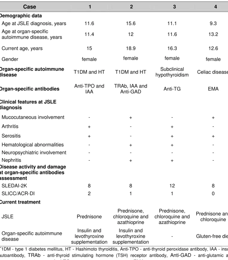
Doenças autoimunes órgão-específicas foram evidenciadas apenas em
pacientes com LESJ (24% versus 0%, p=0,13) (Tabela 1). Dois destes
pacientes preencheram critérios diagnósticos de DM1 e HT e foram tratados
com insulina e levotiroxina. Outro paciente apresentava hipotireoidismo
subclínico com positividade para anticorpo anti-Tg. O quarto paciente
preencheu diagnóstico de doença celíaca com base nas seguintes
características: anemia por deficiência crônica de ferro, presença de anticorpo
EMA, biópsia duodenal compatível com doença celíaca e resposta a dieta
sem glúten (Tabela 3). Nenhum dos 41 pacientes com DMJ apresentou
R
REESSUULLTTAADDOOSS 18
Tabela 3 – Dados demográficos, atividade da doença, outros autoanticorpos e tratamento em quatro pacientes com lúpus eritematoso sistêmico juvenil (LESJ) com doenças autoimunes órgão-específicas
Caso 1 2 3 4
Dados demográficos
Idade ao diagnóstico de LESJ,
anos 11,6 15,6 11,1 9,3
Idade ao diagnóstico da doença
autoimune órgão-específica, anos 11,4 12 11,6 13,2
Idade atual, anos 15 18,9 16,3 12,6
Sexo feminino feminino feminino feminino
Doença autoimune
órgão-específica DM1 e TH DM1 e TH
Hipotireoidismo
subclínico Doença celíaca
Anticorpos órgão-específicos Anti-TPO e IAA TRAb, IAA e
Anti-GAD Anti-Tg EMA
Características clínicas ao diagnóstico de LESJ
Envolvimento mucocutâneo - + - +
Artrite + - + -
Serosite + - + +
Anormalidades hematológicas - + + -
Envolvimento neuropsiquiátrico - - - -
Nefrite - + + -
Atividade de doença e dano na avaliação dos anticorpos órgão-específicos
SLEDAI-2K 8 8 12 8
SLICC/ACR-DI 2 1 1 0
Tratamento atual
LESJ Prednisona
Prednisona, cloroquina e azatioprina Prednisona, cloroquina e azatioprina Prednisona e cloroquina
Doença autoimune órgão-específica Insulina e reposição de levotiroxina Insulina e reposição de levotiroxina
- Dieta livre de glúten
DI S C U S S Ã O 20
Este foi o primeiro estudo que avaliou simultaneamente uma
variedade de anticorpos órgão-específicos em populações de LESJ e DMJ e
demonstrou uma alta prevalência destes anticorpos em ambas as doenças.
Outros anticorpos foram evidenciados exclusivamente em pacientes com
lúpus. Assim como, doenças autoimunes órgão-específicas foram
evidenciadas apenas em pacientes com LESJ, particularmente doenças
endócrinas e gastrintestinais, que necessitaram de tratamento específico.
O LESJ é uma doença autoimune crônica multissistêmica,
caracterizada pela presença de linfócitos auto-reativos e um risco acentuado
para o desenvolvimento de múltiplos autoanticorpos órgão-específicos e não
órgão-específicos2. No presente estudo, o perfil de outros anticorpos
específicos e não específicos foi claramente evidenciado nos pacientes com
LESJ em relação à DMJ.
Tireoidite autoimune clínica, especialmente HT, é a mais importante
doença autoimune órgão-específica no lúpus pediátrico em pacientes do
sexo feminino. A frequência de anticorpos anti-tireóide foi previamente
relatada em 14% a 26% desses pacientes2,22, como observado neste estudo.
Hipotireoidismo autoimune subclínico foi evidenciado em 0-7% da população
com LESJ, como também observado neste estudo2,22. Por outro lado,
hipertireoidismo autoimune não foi diagnosticado em nosso estudo e já foi
DI S C U S S Ã O 21
Interessantemente, nenhum estudo avaliou a freqüência de anticorpos
associados a DM1 na população de LES, e, poucos relatos descreveram
anticorpos contra o pâncreas em pacientes adultos e pediátricos com lúpus4.
Nesta população, 5% dos pacientes com LESJ tinham anticorpos e DM1
insulino-dependente controlados. Esta doença autoimune é
subdiagnosticada em pacientes com LESJ, provavelmente devido à alta
freqüência de diabetes melito induzido pelo uso de glicocorticóides.
Outro aspecto relevante deste estudo foi a avaliação da
autoimunidade gástrica e intestinal. Surpreendentemente, um paciente com
LESJ desta casuística, com anemia ferropriva crônica e sem manifestações
gastrointestinais apresentava doença celíaca. Na realidade, as
manifestações mais comuns deste doença são perda de peso e diarréia e
anemia crônica é uma das manifestações extra-intestinais encontradas nas
formas subclínicas da doença17.
O anticorpo ACP está altamente correlacionado com gastrite
autoimune crônica16. Em um paciente deste estudo com LESJ e positividade
para ACP, a ausência de manifestações gastrointestinais pode indicar que o
processo autoimune estava em uma fase inicial. De fato, a alteração atrófica
da mucosa gástrica pode evoluir para gastrite crônica autoimune em um
periodo de 20 a 30 anos16, e esse paciente requer um acompanhamento
rigoroso. Da mesma forma, neste estudo, três dos pacientes com LESJ e
DMJ apresentavam anticorpos para o fígado, e também devem ser
DI S C U S S Ã O 22
Além disso, estudos anteriores com pequenas populações de DMJ e
avaliações incompletas relataram a avaliação de anticorpos
órgão-específicos e outros autoanticorpos. Observaram-se apenas anticorpos
anti-hepáticos, endócrinos e intestinais em pacientes com DMJ, sem doenças
autoimunes, como já descrito7. Montecucco et al6 não evidenciaram
autoanticorpos hepáticos ou gástricos em 14 pacientes com DMJ, e
Martinez-Cordero et al7 descreveram apenas um paciente com DMJ e AML.
FAN e aCL IgM foram observados em quase 50% dos pacientes deste
estudo, tendo o último apresentado uma freqüência maior em comparação
com estudos anteriores6.
A atividade de doença e tratamento não foram associados com
autoanticorpos órgão-específicos nas duas populações de doenças
autoimunes estudadas. Em contraste, uma alta freqüência de anticorpos
anti-tireóide e tireoidite subclínica foi anteriormente evidenciada em
pacientes com LESJ leve2. Além disso, a flutuação destes anticorpos pode
ocorrer durante o curso da doença, como descrito no LESJ com doença
autoimune da tireóide2,12.
Em conclusão, doenças órgão-específicas foram observadas apenas
em pacientes com LESJ e necessitaram de tratamento específico. A
presença destes anticorpos recomenda a avaliação de doenças
CO N C L U S Õ E S 24
1. As prevalências de autoanticorpos órgão-específicos foram elevadas
e semelhante entre pacientes com LSEJ e DMJ, predominando
anticorpos associados a tireoidite autoimune e DM1.
2. As prevalências de outros autoanticorpos (FAN, anti-DNA, anti-Sm,
anti-RNP, anti-Ro, anti-La e aCL-IgG) foram estatisticamente mais
elevadas no LESJ em relação à DMJ.
3. Não houve associação entre dados demográficos, atividade da doença
e tratamento de pacientes com LESJ e DMJ e a presença de
autoanticorpos órgão-específicos.
4. Doenças autoimunes órgão-específicas, particularmente endócrinas e
RE F E R Ê N C I A S 26
1. Faco MM, Leone C, Campos LM, Febrônio MV, Marques HH, Silva CA.
Risk factors associated with the death of patients hospitalized for
juvenile systemic lupus erythematosus. Braz J Med Biol Res 2007; 40:
993-1002.
2. Parente Costa L, Bonfá E, Martinago CD, de Oliveira RM, Carvalho JF,
Pereira RM. Juvenile onset Systemic Lupus Erythematosus thyroid
dysfunction: a subgroup with mild disease? J Autoimmun. 2009; 33:
121-124.
3. Sallum AME, Kiss MHB, Sachetti S, et al. Juvenile Dermatomyositis:
clinical. laboratorial. histological. therapeutical and evolutive parameters
of 35 patients. Arq. Neuropsiquiatr. 2002; 60: 889-899.
4. Lidar M, Braf A, Givol N, et al. Anti-insulin antibodies and the natural
autoimmune response in systemic lupus erythematosus. Lupus 2001;
10: 81-86.
5. Deen ME, Porta G, Fiorot FJ, Campos LM, Sallum AM, Silva CA.
Autoimmune hepatitis and juvenile systemic lupus erythematosus.
Lupus. 2009; 18: 747-751.
6. Montecucco C, Ravelli A, Caporali R, et al. Autoantibodies in juvenile
dermatomyositis. Clin Exp Rheumatol. 1990; 8: 193-196.
7. Martínez-Cordero E, Martínez-Miranda E, Aguilar León DE.
Autoantibodies in juvenile dermatomyositis. Clin Rheumatol. 1993; 12:
426-428.
8. Go T, Mitsuyoshi I. Juvenile dermatomyositis associated with subclinical
hypothyroidism due to auto-immune thyroiditis. Eur J Pediatr. 2002; 161:
RE F E R Ê N C I A S 27
9. Singh R, Cuchacovich R, Gomez R, Vargas A, Espinoza LR, Gedalia A.
Simultaneous occurrence of diabetes mellitus and juvenile
dermatomyositis: report of two cases. Clin Pediatr. 2003; 42: 459-62.
10. Hochberg MC. Updating the American College of Rheumatology revised
criteria for the classification of systemic lupus erythematosus. Arthritis
and Rheumatism. 1997; 40: 1725.
11. Bohan A, Peter JB. Polymiositis and dermatomyositis. N Engl J Med.
1975; 13: 344-347.
12. Franklyn JA. Hypothyroidism. Medicine. 2005; 33: 27-29.
13. American Diabetes Association. Standards of medical care in diabetes
2007 (Positional Statement). Diabetes Care. 2007; 30: S4-S41.
14. Alvarez F, Berg PA, Bianchi FB, et al. International Autoimmune
Hepatitis Group Report: review of criteria for diagnosis of autoimmune
hepatitis. J Hepatol. 1999; 31: 929-938.
15. Kaplan MM, Gershwin ME. Primary biliary cirrhosis. N Engl J Med. 2005;
353: 1261-1273.
16. De Block CE, De Leeuw IH, Van Gaal LF. Autoimmune gastritis in type 1
diabetes: a clinically oriented review. J Clin Endocrinol Metab. 2008; 93:
363-371.
17. Catassi C, Fasano A. Celiac disease diagnosis: simple rules are better
than complicated algorithms. Am J Med. 2010; 123: 691-693.
18. Gladman DD, Ibanez D, Urowitz MB. Systemic lupus erythematosus
disease activity index 2000. J Rheumatol. 2002; 29: 288-291.
19. Brunner HI, Silverman ED, To T, Bombardier C, Feldman BM. Risk
factors for damage in childhood-onset systemic lupus erythematosus:
cumulative disease activity and medication use predict disease damage.
RE F E R Ê N C I A S 28
20. Bode RK, Klein-Gitelman MS, Miller ML, Lechman TS, Pachman LM.
Disease activity score for children with juvenile dermatomyositis:
reliability and validity evidence. Arthritis Rheum. 2003; 49: 7-15.
21. Lovell DJ, Lindsley CB, Rennebohm RM, et al. Development of validated
disease activity and damage indices for the juvenile idiopathic
inflammatory myopathies. II. The Childhood Myositis Assessment Scale
(CMAS): a quantitative tool for the evaluation of muscle function. The
Juvenile Dermatomyositis Disease Activity Collaborative Study Group.
Arthritis Rheum. 1999; 42: 2213-2219.
22. Ronchezel MV, Len CA, Spinola e Castro A, et al. Thyroid function and
serum prolactin levels in patients with juvenile systemic lupus
A N E X O S 30
Anexo I – “Organ-specific autoantibodies and autoimmune diseases
in juvenile systemic lupus erythematosus and juvenile
dermatomyositis patients”
Submetido à revista Lupus
Anexo II – “Penile and scrotum swellings in juvenile
dermatomyositis”
Submetido à revista Acta Reumatologica Portuguesa
Anexo III – “Menstrual and hormonal alterations in juvenile
dermatomyositis”
Publicado na revista Clinical and Experimental Rheumatology
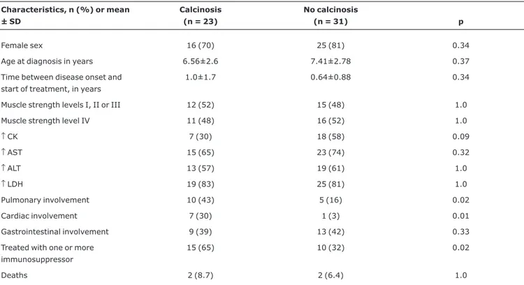
Anexo IV – “Risk factors associated with calcinosis of juvenile
dermatomyositis”
Publicado na revista Jornal de Pediatria
Anexo V – “Irreversible blindness in juvenile systemic lupus erythematosus”
Lupus - Manuscript ID LUP-11-023
1 message
editorial@lupusjournal.co.uk <editorial@lupusjournal.co.uk> Thu, Jan 13, 2011 at 6:57 PM To: nadia.aikawa@gmail.com
13-Jan-2011
Dear Dr. Aikawa:
Your manuscript entitled "ORGAN-SPECIFIC AUTOANTIBODIES AND AUTOIMMUNE DISEASES IN JUVENILE SYSTEMIC LUPUS ERYTHEMATOSUS AND JUVENILE DERMATOMYOSITIS PATIENTS" has been successfully submitted online and is being considered for processing.
Your manuscript ID is LUP-11-023.
Please quote the above manuscript ID in all future correspondence. If there are any changes in your street address or e-mail address, please log in to Manuscript Central at
http://mc.manuscriptcentral.com/lupus and edit your user information as appropriate.
You can also view the status of your manuscript at any time by checking your Author Center after logging in to http://mc.manuscriptcentral.com/lupus .
Thank you very much for submitting your manuscript to Lupus.
Kind regards.
Yours sincerely
Running title - Organ-specific diseases in JSLE and JDM
Concise Report
ORGAN-SPECIFIC AUTOANTIBODIES AND AUTOIMMUNE
DISEASES IN JUVENILE SYSTEMIC LUPUS ERYTHEMATOSUS
AND JUVENILE DERMATOMYOSITIS PATIENTS
Nádia E. Aikawa1,2, Adriana A. Jesus1, Bernadete L. Liphaus1, Clovis A. Silva1,2, Magda Carneiro-Sampaio3, Adriana M.E. Sallum1
Paediatric Rheumatology Unit1, Rheumatology Division2 and Imunology and Allergology Unit3 of Faculdade de Medicina da Universidade de São Paulo, São Paulo, Brazil
Conflicts of interest: none
Corresponding author:
Nádia Emi Aikawa
Rua Oscar Freire, 1456 - Apto 53
Jardim América – São Paulo – SP, Brazil
ZIP code: 05409-010
Summary
To our knowledge, no study has assessed simultaneously a large number of organ-specific autoantibodies, as well as the prevalence of organ-specific autoimmune diseases in juvenile systemic lupus erythematosus (JSLE) and juvenile dermatomyositis (JDM) populations. Therefore, the purpose of this study was to evaluate organ-specific autoantibodies and autoimmune diseases in JSLE and JDM patients. Forty-one JSLE and 41 JDM patients were investigated for serum autoantibodies associated with autoimmune hepatitis, primary biliary cirrhosis, type 1 diabetes mellitus (T1DM), autoimmune thyroiditis, autoimmune gastritis and celiac disease. Patients with positive organ-specific antibodies were assessed for the presence of the respective organ-specific autoimmune diseases. Mean age at diagnosis was significantly higher in JSLE compared to JDM patients (10.3±3.4 vs. 7.3±3.1years, p=0.0001), whereas the mean disease duration was similar in both groups (p=0.92). The frequencies of organ-specific autoantibodies were similar in JSLE and JDM patients (p>0.05). Of note, the high prevalence of autoantibodies related to T1DM and autoimmune thyroiditis were observed in both groups (20% vs. 15%, p=0.77 and 24% vs. 15%, p=0.41; respectively). Higher frequencies of antinuclear antibody - ANA (93% vs. 59%, p=0.0006), anti-dsDNA (61% vs. 2%, p<0.0001), anti-Ro (35% vs. 0%, p<0.0001), anti-Sm (p=0.01), anti-RNP (p=0.02), anti-La (p=0.03) and IgG aCL (p=0.001) were observed in JSLE compared to JDM patients. Organ-specific autoimmune diseases were evidenced only in JSLE patients (24% vs. 0%, p=0.13). Two JSLE patients had T1DM associated with Hashimoto thyroiditis and another had subclinical thyroiditis. Another JSLE patient had celiac disease diagnosis based on iron deficiency anaemia, presence of anti-endomysial antibody, duodenal biopsy compatible to celiac disease and response to a gluten-free diet. In conclusion, organ-specific diseases were observed solely in JSLE patients and required specific therapy. The presence of these antibodies recommends the evaluation of organ-specific diseases and a rigorous follow-up of these patients.
Introduction
Systemic lupus erythematosus (SLE) is an autoimmune multisystemic disease characterized by the presence of autoantibodies1. Juvenile dermatomyositis (JDM) is a connective tissue disease characterized by muscle and cutaneous vasculitis, which can also compromise other organs and systems2. JDM etiology is unknown. However, the presence of chronic muscle inflammation, positivity for serum autoantibodies and association with other autoimmune diseases suggest that autoimmune mechanism is involved in its pathogenesis2.
Of note, studies on organ-specific autoimmunity in juvenile SLE (JSLE) patients have shown a high prevalence of anti-thyroid antibodies and subclinical hypothyroidism1. Additionally, autoimmune hepatitis was rarely reported in our JSLE patients3. On the other hand, few studies have described organ-specific antibodies in JDM4,5, including rare cases of Hashimoto thyroiditis (HT)6 and type 1 diabetes mellitus (T1DM)7. To our knowledge, other organ-specific autoimmune diseases, such as celiac disease (CD), autoimmune gastritis and primary biliary cirrhosis, were not evaluated in both diseases.
Moreover, no study assessed simultaneously a large number of organ-specific autoantibodies, as well as the prevalence of subclinical organ-organ-specific autoimmune diseases in JSLE and JDM patients.
Patients and methods
Forty-one JSLE and 41 JDM patients regularly followed at the Pediatric Rheumatology Unit of our University Hospital were enrolled from January 2008 to January 2009. All patients fulfilled the American College of Rheumatology (ACR) criteria for JSLE8 and the Bohan and Peter criteria for JDM9. The Local Ethical Committee approved this study and an informed consent was obtained from all participants.
Organ-specific and other autoantibodies
Serum autoantibodies associated with the following organ-specific autoimmune diseases were assessed: autoimmune thyroiditis - anti-thyroid peroxidase TPO) antibody by fluoroimmunoassay, anti-thyroglobulin (anti-TG) antibody and anti-thyroid stimulating hormone (TSH) receptor antibody (TRAb) by chemiluminescence; T1DM - insulin autoantibody (IAA), anti-glutamic acid decarboxylase GAD) antibody and anti-tyrosine phosphatase (anti-IA2) antibody by radioimmunoassay; autoimmune hepatitis - anti-type I liver-kidney microsomal (anti-LKM-1) antibody and anti-smooth muscle antibody (SMA) by indirect immunofluorescence on rat liver and kidney tissue sections;
primary biliary cirrhosis - antimitochondrial antibody (AMA) by indirect immunofluorescence on rat liver, kidney and stomach parietal cells, and confirmation of the positive cases by enzyme-linked immunosorbent assay (ELISA); autoimmune gastritis - parietal cell autoantibody (PCA) by indirect immunofluorescence; celiac disease – immunoglobulin A (IgA) class anti-endomysial (EMA) antibody by indirect immunofluorescence. Patients who were positive for organ-specific autoantibodies had the test repeated for confirmation. After that, they were investigated for the presence of the organ-specific autoimmune disease.
anti-SSB/La , anti-topoisomerase 1 (anti-Scl70), anticardiolipin (aCL) isotypes IgG and IgM by ELISA and lupus anticoagulant (LAC) by the dilute Russell’s viper venom time with confirmatory testing, anti-neutrophil cytoplasm antibody (ANCA) by ELISA and direct immunofluorescence in human neutrophils fixed with ethanol and anti-Jo1 by ELISA.
All autoantibodies were assessed at Central Laboratory Division of our Hospital.
Organ-specific autoimmune diseases
Disease activity, disease damage and treatment in JSLE and JDM patients
SLE disease activity and cumulative damage were measured at the moment of organ-specific antibodies and disease evaluations in JSLE patients using the SLE Disease Activity Index 2000 (SLEDAI-2K)16 and the Systemic Lupus International Collaborating Clinics/ACR Damage Index (SLICC/ACR-DI)17.
JDM activity was assessed by disease activity score (DAS)18, and muscle strength was evaluated by childhood myositis assessment scale (CMAS)19 and manual muscle testing (MMT)19. The serum muscle enzymes performed concomitantly to organ-specific antibodies and disease assessments were creatine phosphokinase (CPK), aspartate aminotransferase (AST), alanine aminotransferase (ALT), lactate dehydrogenase (LDH) and aldolase.
Data concerning the current JSLE and JDM treatments included: prednisone, methotrexate, azathioprine, chloroquine, cyclosporine, cyclophosphamide, mycophenolate mofetil and intravenous immunoglobulin.
Statistical analysis
Results
Demographic features: The mean age at JSLE diagnosis was significantly higher compared to JDM patients (10.3 ± 3.4 vs. 7.3 ± 3.1 years, p=0.0001). However, the mean duration of disease was similar in both groups (4.4 ± 3.7 vs. 4.4 ± 3.3, p=0.92), as well as the frequency of female gender (85% vs. 71%, p=0.18).
Organ-specific and other autoantibodies
The frequencies of at least one serum organ-specific antibody were similar in JSLE and JDM patients [17 (41%) vs. 11 (27%), p=0.24]. High frequencies of autoantibodies related to autoimmune thyroiditis (TG, anti-TPO antibodies and/or TRAb) and T1DM (IAA, anti-GAD and/or anti-IA2 antibodies) were observed in both diseases (24% vs. 15%, p=0.41; 20% vs.15%, p=0.77; respectively). The frequencies of EMA and PCA were comparable in both groups (2% vs. 2%, p=1.0; 2% vs. 0%, p=1.0; respectively). Likewise, the frequencies of autoimmune hepatitis antibodies were similar: anti-LKM-1 antibody and/or SMA (2% vs. 5%, p=1.0). None of JSLE and JDM patient had AMA (Table 1).
Higher frequencies of ANA (93% vs. 59%, p=0.0006), anti-dsDNA (61% vs. 2%, p<0.0001), Ro (35% vs. 0%, p<0.0001), Sm (p=0.01), anti-RNP (p=0.02), anti-La (p=0.03) and IgG aCL (p=0.001) were observed in JSLE compared to JDM patients (Table 1).
Organ-specific autoimmune diseases
Organ-specific autoimmune diseases were evidenced only in JSLE patients (24% vs. 0%, p=0.13) (Table 1). Two of them fulfilled both T1DM and HT diagnosis criteria and were treated with insulin and levothyroxine. Another patient had subclinical hypothyroidism with presence of anti-TG antibody. The fourth patient had diagnosis of celiac disease based on the following features: chronic iron deficiency anaemia, presence of AEM antibody, duodenal biopsy compatible to celiac disease and response to a gluten-free diet (Table 3).
Discussion
As far as we know, this was the first study to evaluate simultaneously a variety of organ-specific antibodies in JSLE and JDM populations, and demonstrated a high prevalence of these antibodies in both diseases. Other antibodies were demonstrated in lupus patients, and organ-specific autoimmune diseases were evidenced exclusively in JSLE patients, particularly autoimmune endocrine and gastrointestinal illnesses that required specific treatment.
Of note, JSLE is a chronic multisystem autoimmune disease, characterized by the presence of autoreactive cells and a marked risk for the development of multiple organ and non-organ-specific autoantibodies1. The profile of other specific and non-specific antibodies was clearly evidenced in our JSLE patients compared to JDM.
Clinical autoimmune thyroiditis, specially HT, is the most important organ-specific autoimmune disease in female pediatric lupus, and the frequency of anti-thyroid antibodies were previously reported in 14% to 26% of these patients1,20, as observed in the current study. Autoimmune subclinical hypothyroidism was evidenced in 0-7% of JSLE population, as also observed herein1,20. On the other hand, autoimmune hyperthyroidism was not diagnosed in our study and has been already described in 2-3% of these patients1,20.
Interestingly, no study evaluated the frequency of T1DM-associated antibodies in JSLE population, and, to our knowledge, few reports have described pancreas autoantibodies in adult and pediatric lupus7. In our population, we found 5% of JSLE patients with these antibodies and controlled insulin-dependent T1DM. This autoimmune disease is under diagnosed in JSLE patients, probably due to a high frequency of diabetes mellitus induced by glucocorticoids use.
chronic anemia is one of the extra-intestinal manifestations found in the subclinical forms of the disease15.
The PCA antibody is highly correlated with chronic autoimmune gastritis14. The absence of gastrointestinal manifestations may indicate that the autoimmune process was at an initial stage in one of our JSLE patients with PCA. Importantly, atrophic alteration of the gastric mucosa can progress to chronic autoimmune gastritis within 20 to 30 years period14, and this patient requires a rigorous follow-up. Likewise, three of our lupus and JDM patients had liver autoantibodies, and also should be constantly monitored12.
Furthermore, previous studies were reported with small JDM populations and incomplete evaluations reported assessment of organ-specific and other antibodies. We observed only endocrine, liver and intestinal autoantibodies in JDM patients without autoimmune diseases, as previously described5. Montecucco et al4 did not evidence autoimmune liver and gastric autoantibodies in 14 JDM patients, and Martinez-Cordero et al5 described one JDM patient with SMA.ANA and aCL-IgM were observed in almost 50% of our patients, and the later had a higher frequency compared to previous studies4.
Disease activity and treatment was not associated with organ-specific autoantibodies in our two autoimmune disease populations. In contrast, a high frequency of anti-thyroid antibodies and subclinical thyroiditis were previously evidenced in mild JSLE patients1.Moreover, fluctuation of these antibodies may occur during the course of the disease, as described in JSLE with autoimmune thyroid disease1,10.
ACKNOWLEDGMENTS
REFERENCES
1. Parente Costa L, Bonfá E, Martinago CD, de Oliveira RM, Carvalho JF, Pereira RM. Juvenile onset Systemic Lupus Erythematosus thyroid dysfunction: a subgroup with mild disease? J Autoimmun 2009; 33: 121-124.
2. Sallum AME, Kiss MHB, Sachetti S, et al. Juvenile Dermatomyositis: clinical. laboratorial. histological. therapeutical and evolutive parameters of 35 patients. Arq. Neuropsiquiatr 2002; 60: 889-899.
3. Deen ME, Porta G, Fiorot FJ, Campos LM, Sallum AM, Silva CA. Autoimmune hepatitis and juvenile systemic lupus erythematosus. Lupus
2009; 18: 747-751.
4. Montecucco C, Ravelli A, Caporali R, et al. Autoantibodies in juvenile dermatomyositis. Clin Exp Rheumatol 1990; 8: 193-196.
5. Martínez-Cordero E, Martínez-Miranda E, Aguilar León DE. Autoantibodies in juvenile dermatomyositis. Clin Rheumatol 1993; 12: 426-428.
6. Go T, Mitsuyoshi I. Juvenile dermatomyositis associated with subclinical hypothyroidism due to auto-immune thyroiditis. Eur J Pediatr 2002; 161: 358-359.
7. Lidar M, Braf A, Givol N, et al. Anti-insulin antibodies and the natural autoimmune response in systemic lupus erythematosus. Lupus 2001; 10:
81-86.
8. Hochberg MC. Updating the American College of Rheumatology revised criteria for the classification of systemic lupus erythematosus. Arthritis and Rheumatism 1997; 40: 1725.
9. Bohan A, Peter JB. Polymiositis and dermatomyositis. N Engl J Med 1975;
13: 344-347.
11. American Diabetes Association. Standards of medical care in diabetes 2007 (Positional Statement). Diabetes Care 2007; 30: S4-S41.
12. Alvarez F, Berg PA, Bianchi FB, et al. International Autoimmune Hepatitis Group Report: review of criteria for diagnosis of autoimmune hepatitis. J Hepatol 1999; 31: 929-938.
13. Kaplan MM, Gershwin ME. Primary biliary cirrhosis. N Engl J Med 2005;
353: 1261-1273.
14. De Block CE, De Leeuw IH, Van Gaal LF. Autoimmune gastritis in type 1 diabetes: a clinically oriented review. J Clin Endocrinol Metab 2008; 93: 363-371.
15. Catassi C, Fasano A. Celiac disease diagnosis: simple rules are better than complicated algorithms. Am J Med 2010; 123: 691-693.
16. Gladman DD, Ibanez D, Urowitz MB. Systemic lupus erythematosus disease activity index 2000. J Rheumatol 2002; 29: 288-291.
17. Brunner HI, Silverman ED, To T, Bombardier C, Feldman BM. Risk factors for damage in childhood-onset systemic lupus erythematosus: cumulative disease activity and medication use predict disease damage. Arthritis Rheum 2002; 46: 436-444.
18. Bode RK, Klein-Gitelman MS, Miller ML, Lechman TS, Pachman LM. Disease activity score for children with juvenile dermatomyositis: reliability and validity evidence. Arthritis Rheum 2003; 49: 7-15.
Table 1 – Organ-specific autoantibodies and diseases, and other autoantibodies in juvenile systemic lupus erythematosus (JSLE) versus juvenile dermatomyositis (JDM) patients
Variables
JSLE
n=41
JDM
n=41
p
Organ-specific antibodies 17 (41) 11 (27) 0.24
Autoimmune thyroiditis
(anti-TG and/or anti-TPO and/or TRAb) 10 (24) 6 (15) 0.41
Type 1 diabetes mellitus
(IAA and/or anti-GAD and/or anti-IA2) 8 (20) 6 (15) 0.77
Celiac disease
(EMA) 1 (2) 1 (2) 1.0
Autoimmune hepatitis
(SMA and/or anti-LKM-1) 1 (2) 2 (5) 1.0
Primary biliar cirrhosis
(AMA) 0 (0) 0 (0) 1.0
Autoimune gastritis
(PCA) 1 (2) 0 (0) 1.0
Organ-specific diseases 4/17 (24) 0/11 (0) 0.13
Other autoantibodies
ANA 38 (93) 24 (59) 0.0006
RF 4 (10) 0 (0) 0.12
Anti-dsDNA 25 (61) 1 (2) < 0.0001
Anti-Sm 11 (27) 2 (5) 0.01
Anti-RNP 9 (22) 1 (2) 0.02
Anti-Ro 14 (35) 0 (0) < 0.0001
Anti-La 6 (15) 0 (0) 0.03
Anti-Scl-70 0 (0) 0 (0) 1.0
Anti-Jo1 0 (0) 2 (5) 0.49
aCL-IgM 20 (49) 17 (41) 0.66
aCL-IgG 19 (46) 5 (12) 0.001
LAC 5 (12) 1 (2) 0.2
p-ANCA 4 (10) 0 (0) 0.12
c-ANCA 1 (2) 1 (2) 1.0
Table 2 – Demographic data, disease activity, treatment and other autoantibodies in juvenile lupus erythematosus (JSLE) and juvenile dermatomyositis (JDM) patients with and without organ-specific autoantibodies
JSLE Variables
JSLE with organ-specific antibody (n=17)
JSLE without organ-specific antibody
(n=24)
p
Demographic data
Female gender 13 (76) 22 (92) 0.21
Current age, years 11 (4-13) 11 (6-17) 0.18
Disease duration, years 3.1 (0.3-7.6) 3.8 (0-12.3) 0.78
SLEDAI-2K 5 (0-12) 3 (0-14) 0.36
SLICC/ACR-DI 1 (0-2) 1 (0-2) 0.93
Current treatment
Prednisone 14 (83) 23 (96) 0.29
Immunosuppressive use 7 (41) 17 (71) 0.11
Other autoantibodies
ANA 15 (88) 23 (96) 0.56
anti-dsDNA 11 (65) 14 (58) 0.75
anti-Sm 6 (35) 5 (21) 0.48
anti-RNP 5 (29) 4 (17) 0.45
anti-Ro 8 (47) 6 (25) 0.19
anti-La 3 (18) 3 (13) 0.68
aCL-IgM 10 (59) 11 (46) 1.69
aCL-IgG 7 (41) 12 (50) 0.75
JDM Variables
JDM with organ-specific antibody
(n=11)
JDM without organ-specific antibody
(n=30)
p
Demographic data
Female gender 10 (91) 19 (63) 0.13
Current age, years 12 (5-17) 11 (6-18) 0.92
Disease duration, years 3.7 (1.1-8.1) 3.7 (0-13.5) 0.86
JDM scores and muscle enzymes
CMAS (0 - 52) 50 (44-52) 48.5 (4-52) 0.27
MMT (0 - 80) 80 (75-80) 80 (38-80) 0.47
DAS (0 - 20) 2 (0-7) 3 (0-17) 0.71
AST (10 – 36 UI/L) 23.5 (12-43) 25.5 (14-90) 0.57 ALT (24 – 49 UI/L) 34 (29-103) 33.5 (10-123) 0.76 CPK (39 – 170 UI/L) 93 (26-165) 92.5 (40-27381) 0.72 Aldolase (<7.6 UI/L) 7.05 (3.5-9.4) 7.2 (2.7-49.9) 0.95 LDH (240 – 480 UI/L) 159.5 (135-238) 200 (87-522) 0.19
Current treatment
Prednisone 6 (55) 15 (50) 1.0
Immunosuppressive use 7 (64) 17 (57) 0.74
Other autoantibodies
ANA 5 (45) 19 (63) 0.48
aCL-IgM 5 (45) 12 (40) 1.0
Table 3 – Demographic data, disease activity, other autoantibodies and treatment in four juvenile systemic lupus erythematosus (JSLE) patients with organ-specific autoimmune diseases
Case 1 2 3 4
Demographic data
Age at JSLE diagnosis, years 11.6 15.6 11.1 9.3
Age at organ-specific
autoimmune disease, years 11.4 12 11.6 13.2
Current age, years 15 18.9 16.3 12.6
Gender female female female female
Organ-specific autoimmune
disease T1DM and HT T1DM and HT
Subclinical
hypothyroidism Celiac disease
Organ-specific antibodies Anti-TPO and
IAA
TRAb, IAA and
Anti-GAD Anti-TG EMA
Clinical features at JSLE diagnosis
Mucocutaneous involvement - + - +
Arthritis + - + -
Serositis + - + +
Hematological abnormalities - + + -
Neuropsychiatric involvement - - - -
Nephritis - + + -
Disease activity and damage at organ-specific antibodies assessment
SLEDAI-2K 8 8 12 8
SLICC/ACR-DI 2 1 1 0
Current treatment
JSLE Prednisone
Prednisone, chloroquine and azathioprine Prednisone, chloroquine and azathioprine Prednisone and chloroquine Organ-specific autoimmune disease Insulin and levothyroxine supplementation Insulin and levothyroxine supplementation
- Gluten-free diet
T1DM - type 1 diabetes mellitus, HT - Hashimoto thyroiditis, Anti-TPO - anti-thyroid peroxidase antibody, IAA - insulin
autoantibody, TRAb - anti-thyroid stimulating hormone (TSH) receptor antibody, Anti-GAD - anti-glutamic acid
decarboxylase antibody, Anti-TG - anti-thyroglobulin antibody, EMA - anti-endomysial antibody, SLEDAI-2K -
Systemic Lupus Erythematosus Disease Activity Index 2000, SLICC/ACR-DI - Systemic Lupus International
1 Penile and scrotum edema in JDM
Case report
Penile and scrotum swellings in juvenile dermatomyositis
Adriana Maluf Elias Sallum
1, Marco Felipe Castro Silva
1, Cíntia Maria
Michelin
1, Ricardo Jordão Duarte
2, Ronaldo Hueb Baroni
3, Nádia Emi
Aikawa
1,4, Clovis Artur Silva
1,41
Pediatric Rheumatology Unit,
2Division of Urology,
3Radiology
Department and
4Division of Rheumatology, Faculdade de Medicina da
Universidade de São Paulo, São Paulo, Brazil.
Correspondence author
:
Dr. A. M. E. Sallum
Av Juriti 187, apto 21
São Paulo, SP, Brazil
CEP 04520-000
2
RESUMO
O edema é uma característica bem conhecida da dermatomiosite juvenil (DMJ). No entanto, para nosso conhecimento, edema simultaneamente localizados no pênis e no escroto não foi relatado. Durante um período de 27 anos, 5.506 pacientes foram acompanhados na Unidade de Reumatologia Pediátrica do nosso Hospital Universitário e 157 pacientes (2,9%) tiveram DMJ. Um deles (0,6%) desenvolveu edema localizado concomitante no pênis e no escroto. Ele apresentava atividade grave da doença, vasculite cutânea difusa, edema localizado recorrente (membros e/ou face) e um episódio de edema generalizado desde os 7 anos de idade. À admissão, ele apresentava há um mês edema proeminente e difuso do pênis e do escroto, indolor e eritema cutâneo leve. A ultra-sonografia peniana, escrotal e testicular mostrou edema cutâneo, sem envolvimento testicular, como também observado na ressonância magnética. Na ocasião, ele fazia uso de prednisona, metotrexato, ciclosporina, hidroxicloroquina e talidomida. Ele recebeu 4 doses semanais de rituximab 375 mg/m2 por dose,
com melhora das lesões cutâneas e do edema peniano e escrotal. Nenhum evento adverso foi observado após a terapia anti-CD20. Portanto, edema peniano e escroto foi uma manifestação rara da DMJ ativa, com melhora após terapia com anticorpo monoclonal anti-CD20 direcionada contra células B.
3
ABSTRACT
Edema is a well-known feature of the juvenile dermatomyositis (JDM). However, to our knowledge simultaneously localized penile and scrotum swellings were not reported. During a 27-year period, 5,506 patients were followed up at the Pediatric Rheumatology Unit of our University Hospital and 157 patients (2.9%) had JDM. One of them (0.6%) had concomitant localized penile and scrotum swellings. He had severe disease activity, diffused cutaneous vasculitis, recurrent localized edema (limbs and/or face) and one episode of generalized edema since he was 7 years old. At admission, he had one-month prominent and diffused swellings of the penis and scrotum, painless and mild skin erythema. Penis, scrotum and testicular ultrasound showed skin edema without testicular involvement, as it was observed in the magnetic resonance imaging. On that occasion, he had been taking prednisone, methotrexate, cyclosporin, hydroxychloroquine and thalidomide. He
received 4 weekly doses of rituximab at 375 mg/m2 per dose with improvements of
skin rash, penile and scrotum swellings. No adverse event was observed after anti-CD20 therapy. Therefore, penile and scrotum edema was a rare manifestation of active JDM with improvement after anti-CD20 monoclonal antibody targeting B cells treatment.
4
INTRODUCTION
Juvenile dermatomyositis (JDM) is a systemic disease characterized by nonsuppurative inflammation of the skeletal muscle and skin1,2. The disease is initially marked by the presence of vasculitis and later on by the development of calcinosis3.
Constitutional symptoms, as fever, alopecia, weight loss, fatigue, headache and irritability, are usually present at the disease onset4. Of note, edema is a
well-known feature of the disease, particularly in localized areas. The most common regions of this manifestation are eyelids, face and distal extremity regions5.
Generalized edema associated with JDM was rarely reported5-9. To our knowledge
simultaneously localized penile and scrotum swelling in JDM patient were not reported.
During a 27-year period (January 1983 to December 2010), 5,506 patients were followed up at the Pediatric Rheumatology Unit of Instituto da Criança, Faculdade de Medicina da Universidade de São Paulo and 157 patients (2.9%) had JDM. One of our pre-pubertal JDM patients (0.6%) had concomitant localized penile and scrotum swellings without testicular involvement, and was described.
CASE REPORT
A 10-year-old boy was diagnosed with JDM according to Bohan and Peter criteria due to Gottron´s papules, heliotrope rash, muscle weakness, increased muscle enzymes serum levels, inflammatory infiltrate and perifascicular atrophy at muscle histopathology and characteristic electromyographic changes10. He had severe
5 sulphate, alendronate, diltiazem, thalidomide and intravenous immunoglobulin. At admission at our University Hospital, he had one-month prominent and diffused penile and scrotum swellings, painless and mild skin erythema (Figure 1). An erythematosus rash was also observed in pubic region. At the same moment, he presented periorbital rash, erythematous maculopapular lesions on the extensor surfaces of the hands, vasculitis, photosensitivity, diffused calcinosis, symmetric proximal weakness with a grade-3 muscular strength and significant muscular atrophy. The Childhood Myositis Assessment Scale (CMAS)11 and the Disease Activity Score (DAS)12 were not performed due to knees imitation. On that occasion, he had been taking prednisone 0.12mg/kg/day, methotrexate 1.0mg/kg/week, cyclosporin 5.0mg/kg/day, hydroxychloroquine 6.2mg/kg/day, alendronate 70mg/week, diltiazen 5.6mg/kg/day and thalidomide 2.3mg/kg/day. Erythrocyte sedimentation rate was 62 mm/h (normal range 0-20 mm/h) and serum muscle enzymes showed aspartate aminotransferase 40 U/l (normal range 10-34 U/l), alanine aminotransferase 32 U/l (normal range 10-44 U/l), creatine kinase 50 U/l (normal range 24-204 U/l), lactic dehydrogenase 272 U/l (normal range 211-423 U/l), aldolase 11.8 U/l (normal range 1-7.5 U/l), albumin 4.3 g/dl (normal range 3.8-5.6 g/dl), urea 13 mg/dl (normal range 15-45 mg/dl) and creatinine 0.16 mg/dl (normal range 0.6-0.9 mg/dl). He was on pre-pubertal stage and the hormone profile was normal: follicle-stimulating hormone - FSH 4.39 IU/l (normal range 1,5-12,4 IU/l), luteinizing hormone – LH 1.09 IU/l (normal range 0,1-7,8 IU/l), and morning total testosterone 0.03 ng/dl (normal range 0,03-0,68 ng/dl). Immunological tests were positive for antinuclear antibodies (ANA) 1:640 (fine speckled pattern) and Ro 52 Kd, and negative for other serum antibodies: anti-Mi-2, anti-synthetase (anti-Jo-1, anti-PL-7 and anti-PL-12), anti-Ku, anti-PM-Scl,
anti-double stranded DNA (anti-dsDNA), anti-Sm, anti-RNP, anti-La and anti-Scl-70. Penis, scrotum and testicular ultrasound showed skin edema without testicular involvement, as it was observed in the magnetic resonance imaging. He received 4
weekly doses of rituximab at 375 mg/m2 per dose with improvements of skin rash,