Original Article
6 Arq Bras Oftalmol. 2015;78(1):6-9 http://dx.doi.org/10.5935/0004-2749.20150003
INTRODUCTION
The development of exuberant fibrinous exudation or inflamma-tion is a rare but serious complicainflamma-tion after ocular surgery. It can cause numerous sequelae including membranes, anterior or posterior synechiae, secondary glaucoma, and pupillary block. Previous stu dies showed that recombinant tissue plasminogen activator (r-TPA) is effective and safe for improving the clinical course of fibri-nous related complications in cases of traumatic hyphema(1-3), severe
post cataract fibrinous membranes in pediatric(4), fibrin formation
after cataract surgery(4-6), glaucoma surgery(7-9), subretinal hemorrhage(10),
and endophthalmitis(11).
Intracameral r-TPA has been shown to be effective for fibrin de-gradation. However, the effectiveness of topical(12), intravitreous(13),
and subconjunctival(7,9) r-TPA remains controversial. Most studies
re garding absorption or anterior chamber dosing of r-TPA were non-quan titative. To the best of our knowledge, no study has com-pared in tracameral, subconjunctival and topical application of r-TPA using the same quantitative methods.
Therefore, the purpose of this study was to quantify fibrin degra-dation products (FDP) after topical and subconjunctival administration of r-TPA in rabbits.
METHODS
This prospective double-blind experimental study was perfor-med in the Setor de Técnica Cirúrgica da Santa Casa de São Paulo under veterinary supervision. It was approved by the Animal Ethical Committee and Institutional Board of the Hospital das Clínicas and the Faculdade de Medicina da Universidade de São Paulo. All proce-dures followed proper legislation for the protection of animals (EU Directive 2010/63/EU) and adhered to the Association of Research in Vision and Ophthalmology (ARVO) statement for the Use of Animals in Ophthalmic and Vision Research.
Twenty-five New Zealand male rabbits weighing 3.5 to 4.0 kg were enrolled in this study. All rabbits underwent an ocular exami-nation and slit lamp documentation (Nikon, FS3 slit lamp, Japan). All rabbits with ocular abnormalities were excluded from the study.
Anesthesia was induced in each rabbit using intramuscular in jection of a mixture of 0.3 mL/kg tiletamine hydrochloride and zo la zepam (Zoletil 50®, Laboratoire Virbac, France). General anes-thesia was achieved with an intramuscular injection of a mixture of 0.3 mL/kg tiletamine hydrochloride and zolazepam, and a mixture of 0.4 mL/kg fentanyl and droperidol (Innovar-Vet®, MTC Pharmaceu-ticals, Ontario, Canada).
Subconjunctival and topical application of recombinant tissue plasminogen
activator in rabbits
Uso tópico e subconjuntival de ativador de plasminogênio tecidual recombinante em coelhos
José RicaRdode abReu Reggi1, RichaRd Yudi hida1,2, Milton Massato hida3, MaRia cRistina nishiwaki-dantas1, hisashi suzuki2
Submitted for publication: June 30, 2014 Accepted for publication: November 10, 2014
1 Department of Ophthalmology, Santa Casa de São Paulo, São Paulo, SP, Brazil. 2 Department of Ophthalmology, University of São Paulo (USP), São Paulo, SP, Brazil. 3 Department of Ophthalmology, Universidade Estadual Paulista “Júlio de Mesquita Filho” (UNESP),
Botucatu, SP, Brazil.
Funding: No specific financial support was available for this study.
Disclosure of potential conflicts of interest: None of the authors have any potential conflicts of interest to disclose.
Corresponding author: Richard Yudi Hida. Rua Afonso de Freitas, 488/61 - São Paulo, SP - 04006-052 - Brazil - E-mail: [email protected]
ABSTRACT
Purpose: To quantify fibrin degradation products after topical and subconjunctival administration of recombinant tissue plasminogen activator in rabbits.
Methods: Fibrin formation was induced in the anterior chamber in 25 rabbits. Subsequently, five rabbits received an injection of r-TPA (positive control) in the anterior chamber, another 10 received a subconjunctival injection of r-TPA, and the remaining 10 received instillations of topical r-TPA. Afterwards, samples of aqueous humor were collected and semi-quantitative analysis of fibrin degradation products (FDP) was performed.
Results: No statistical differences were noted between the treatment and control groups at any time point. Fibrin degradation products semi-quantification showed statistical improvement in the control group and the subconjunctival group.
Conclusion: Fibrin degradation products were observed in the anterior cham-ber after subconjunctival administration of r-TPA. However, it was probably not sufficient to cause fibrin degradation. Topical r-TPA did not effectively absorb anterior chamber fibrin.
Keywords: Tissue plasminogen activator; Anterior chamber; Hyphema; Inflamma-tion; Postoperative complications; Animals; Rabbits
RESUMO
Objetivo: Quantificar produtos de degradação de fibrina (PDF) após uso tópico e sub-conjunctival de ativador de plasminogênio tecidual recombinante (r-TPA) em coelhos.
Métodos: Formação de fibrina foi induzida na câmara anterior em 25 coelhos. Cinco coelhos foram submetidos a injeção intracameral de r-TPA (controle positivo). Dez coelhos foram submetidos a injeção subconjuntival de r-TPA e dez coelhos foram submetidos a instilação tópica de r-TPA. Amostras de humor aquoso foram coletados e uma análise quantitativa dos produtos de degradação de fibrina foi realizada.
Resultados: Não foi observado diferença estatisticamente significativa na degra-dação de fibrina em nenhum dos momentos estudados quando comparados com o controle. Porém foi observado diferença estatisticamente significante na quantificação do produtos de degradação de fibrina no grupo controle e no grupo subconjuntival.
Conclusão: Produtos de degradação de fibrina foi observado nas amostras do grupo subconjunctival, porém, provavelmente não foi suficiente para degradar a fibrin presente. r-TPA tópico não foi efetivo em absorver fibrina na câmara anterior.
Reggi JRA, et al.
7
Arq Bras Oftalmol. 2015;78(1):6-9 After appropriate anesthesia was attained, 5.0 mL of blood was
collected from the ear vein and centrifuged to isolate the plasma. Each rabbit received topical 0.5% procaine chloridrate (Anestalcon®, Alcon, São Paulo, Brazil), and then a lid speculum was used for para-centesis in the superior portion of the cornea with a 25 gauge needle. Keeping the needle in the anterior chamber, 0.1 mL of aqueous hu-mor was aspirated and 0.1 mL of plasma citrate was injected.
After 24 h, all rabbits were sedated and photographed under a slit lamp. All eyes with fibrin in the anterior chamber were graded according to the classification described by Lim et al.(14) (grade 0: no
visible fibrin; grade 1: few visible fibrin filaments and clear details of the iris; grade 2: presence of a fibrin clot and blurry details of the iris; grade 3: presence of a fibrin clot, no visible details of the iris). All eyes with fibrin levels of grade 1 and 2 of fibrin in the anterior chamber were excluded from the study. Sampling calculations were based on a previous pilot study and statistical analysis was performed using the WINPEPI program with the COMPARE2 module (Version 2.68) to avoid sampling error. The sample size was increased in 20% due to possible loss to follow-up.
Rabbits were divided into three study groups: group 1 (n=5; positive control) received intracamerular injection of r-TPA; Group 2 (n=10) received subconjunctival injection of r-TPA; Group 3(n=10) received topical r-TPA.
One eye from each rabbit in group 1 was randomly chosen (po-sitive control) to receive an injection of 0.1 mL of 0.25 µg/mL r-TPA (TPA-Alteplase®, Ophthalmos, São Paulo, Brazil) in the anterior cham-ber after instillation of topical 0.5% procaine chloridrate (Anestalcon®, Alcon, São Paulo, Brazil). The other eye from the same rabbit was in-jected with 0.1 mL balanced salt solution using the same technique.
One eye from each rabbit in group 2 was randomly chosen to receive a subconjunctival injection of 0.1 mL of 0.25 µg/mL r-TPA after instillation of topical 0.5% procaine chloridrate. The other eye was injected with 0.1 mL balanced salt solution using the same technique.
One eye from each rabbit in Group 3 was randomly chosen to receive one drop of topical r-TPA at 1 mg/mL, 9 times at intervals of every 5 min after instillation of topical 0.5% procaine chloridrate. The other eye received one drop of 0.1 mg/mL balanced salt solution in the same regimen.
Each rabbit was examined, classified, and documented under sedation using a slit lamp (Nikon, model FS3, Japan) at the moment of the procedure (M0), and 30 min (M1), 60 min (M2), 7 h (M3) and 24 h (M4) after the procedure.
After 24 h, each rabbit was euthanized with intramuscular 1 mg/kg acepromazine and intravenous 30 mg/kg thiopental sodium. All eyes underwent paracentesis and aspiration of samples of aqueous humor using a 25-gauge needle. Samples were immediately transported to our laboratory for qualitative and semi-quantitative analysis of FDP and fibrinogen using the macro-latex slide agglutination test (FDP
Plas-ma®, Diagnostica Stago Inc, France) according to the ma nufacturer`s instructions. This test involves multiple microlatex particles coated with mouse monoclonal anti-human FDP antibodies(15). Presence of
FDPs causes agglutination of the latex particles as the FDPs bind to the antibodies. Then these agglutinated particles are detected visually. The detection limit of this test is 2.5 µg/mL.
Qualitatively, the agglutination pattern was interpreted as nega-tive when transparent and posinega-tive when blurred. Quantitanega-tively, a positive agglutination pattern was interpreted as recommended in the manufacturer`s instructions.
All results were analyzed statistically with the Wilcoxon test (p<0.05). All animal care and procedures were performed under the su-pervision of a single veterinarian to avoid bias and factors that could influence the hemodynamics and neuroendocrinological conditions of the animals (anesthesia technique, stress, food, etc.). All animals were kept in individual cages and given water and food ad libitum. Light, temperature, and humidity were controlled and monitored.
RESULTS
Table 1 shows the averages and standard deviations of anterior chamber fibrin grading following Lim et al.(14) after application of r-TPA.
In group 1 (intracameral administration), no statistically signifi-cant difference was noted at M1 compared to the control group (p=0.06). However, anterior chamber fibrin grading showed statis-tically significant improvement at M2, M3, and M4 compared to the control group (p=0.0079).
In group 2 (subconjunctival administration), no statistically signi-ficant difference was noted at any time point (M1, M2, M3 and M4) compared to the control group (p>0.05).
In group 3 (topical administration), no statistically significant diffe rence was noted at any time point (M1, M2, M3 and M4) compa-red to the control group (p>0.05).
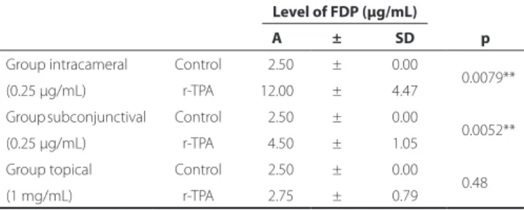
Table 2 shows the results of the semi-quantification of the FDPs following the different applications of r-TPA compared to the control group.
FDP semi-quantification showed statistically significant improve-ment in groups 1 (p=0.0079) and 2 (p=0.0052), but no statistically significant difference was observed in group 3 (p=0.48).
DISCUSSION
Fibrinous exudation is a serious complication of intraocular sur-gery and endophthalmitis. These complications can preclude fundus examination and interfere with the performance of a second inter-vention such as vitrectomy. These fibrinous membranes are usually difficult to manage with conventional steroid therapy. Typically, intrao-cular fibrin can be surgically removed, treated with an argon laser, or
Table 1. Distribution of the average and mean deviation of anterior chamber ibrin grading following criteria according to Lim et al.(14), after
applica-tion of recombinant tissue plasminogen activator
Examination time points
M1 M2 M3 M4
A ± SD p A ± SD p A ± SD p A ± SD p
Group intracameral Control 3.00 ± 0.00
0.06 3.00 ± 0.00 0.0079** 2.80 ± 0.45 0.0079** 2.00 ± 0.00 0.0079** (0.25 µg/mL) r-TPA 1.80 ± 0.84 1.60 ± 0.55 1.00 ± 0.00 0.20 ± 0.45
Group subconjunctival Control 2.90 ± 0.32
0.97 2.90 ± 0.32 1.00 2.70 ± 0.48 1.00 2.10 ± 0.32 0.80 (0.25 µg/mL) r-TPA 3.00 ± 0.00 2.90 ± 0.32 2.70 ± 0.48 2.00 ± 0.67 Group topical Control 3.00 ± 0.00
1.00 3.00 ± 0.00 1.00 2.50 ± 0.53 0.74 2.20 ± 0.42 0.74 (1 mg/mL) r-TPA 3.00 ± 0.00 3.00 ± 0.00 2.60 ± 0.52 2.30 ± 0.48
Subconjunctival and topical application of recombinant tissue plasminogen activator in rabbits
8 Arq Bras Oftalmol. 2015;78(1):6-9
occasionally fibrinolytic drugs such as streptokinase and urokinase are used(16,17). However, intraocular toxicity was often observed with
these drugs(18).
R-TPA is a genetically cloned serine protease that promotes the degradation of fibrin only at the clot surface(19). It has minimal
side effects or toxicity when applied to the eye. It is considered a clot-specific fibrinolytic agent, used for the treatment of deep vein throm bosis(20) and acute myocardial infarction(21). In ophthalmology,
it was described as effective in thrombolytic therapy in patients with branch or central vein occlusion of the retina(22), glaucoma filtration
surgery(23), or intracameral fibrinolysis following ocular surgery(5,6).
In our study using rabbit eyes, we found that intracamerular r-TPA (0.25 µg/1 mL) more effectively degraded anterior chamber fibrin compared to the control group. Other studies yielded similar re-sults(2,24,25) with similar control groups.
There was no statistically significant difference in the degradation of anterior chamber fibrin in rabbit eyes with subconjunctival r-TPA compared to the control group. Although some degradation pro-ducts were present in the anterior chamber, they were insufficient to indicate effective fibrin degradation. Additional studies are necessary to clarify whether a different concentration of subconjunctival r-TPA could actually increase fibrin degradation in the anterior chamber. The benefits and indications of r-TPA are evident especially in compli-cations of glaucoma surgery involving the conjunctiva(7,9,23).
Compared to the control group, there was also no statistically relevant difference in the degradation of anterior chamber fibrin in rabbit eyes following r-TPA injection at 1 mg/mL. Previous studies were controversial(12,26-28). The inefficacy of this method is probably
re lated to poor penetration beyond the surface of the clot. Higher concentrations of r-TPA were not administered because toxicity to the epithelium was observed during a pilot study (data not shown).
The minimum concentration necessary to promote degradation of anterior chamber fibrin varied in previous studies. Based on these stu-dies, an approximate dose of 25 µg/1 mL is suggested to effectively degrade fibrin with no intraocular toxicity(2,3). There is no evidence of
lens abnormalities in animal models following the use of this concen-tration(2,29). Other authors observed good results using 10 µg/mL r-TPA
for total hyphema in rabbit eyes(30). Subconjunctival administration of
r-TPA may be a novel method for promoting fibrin degradation. The use of r-TPA in the eye has been considered safe with very few complications. The potential complications related to the use of r-TPA are corneal edema and anterior chamber turbidity, which results from immediate fibrinolysis products, prostaglandins, and leu-kocyte hydrolytic enzymes(31). Previous studies did not show clinically
relevant abnormalities in the cornea, lens, or retina(2,29), intraocular
pressure changes, or an inflammatory response or rebleeding when intracameral or vitreous r-TPA was used at doses under 25 µg/1 mL. However, some studies suggested that it can increase the risk of in-traocular rebleeding, ocular toxicity, and corneal abnormalities. Our study showed promising results with a lower dose of intracameral r-TPA. However, further studies are required to determine whether higher doses could be more effective.
FDPs were observed in the anterior chamber after 0.25 µg/mL subconjunctival r-TPA; however, this dose was probably insufficient to cause fibrin degradation. Topical 1 mg/mL r-TPA did not effective degrade anterior chamber fibrin.
ACKNOWLEDGMENT
Drs. Abreu Reggi and Hida contributed equally to this work and request acknowledgement as co-first authors.
REFERENCES
1. Laatikainen L, Mattila J. The use of tissue plasminogen activator in post-traumatic total hyphaema. Graefes Arch Clin Exp Ophthalmol. 1996;234(1):67-8.
2. Lambrou FH, Snyder RW, Williams GA. Use of tissue plasminogen activator in experi-mental hyphema. Arch Ophthalmol. 1987;105(7):995-7.
3. Moon J, Chung S, Myong Y, Chung S, Park C, Baek N, et al. Treatment of postcataract fibrinous membranes with tissue plasminogen activator. Ophthalmology. 1992;99(8): 1256-9.
4. Klais CM, Hattenbach LO, Steinkamp GW, Zubcov AA, Kohnen T. Intraocular recombi-nant tissue-plasminogen activator fibrinolysis of fibrin formation after cataract surgery in children. J Cataract Refract Surg. 1999;25(3):357-62. Comment in: J Cataract Refract Surg. 1999;25(7):880; J Cataract Refract Surg. 2000;26(1):4-5; J Cataract Refract Surg. 2000;26(1):4; author reply 5.
5. Wedrich A, Menapace R, Muhlbauer-Ries E. The use of recombinant tissue plasmino-gen activator for intracameral fibrinolysis following cataract surgery. Int Ophthalmol. 1994-1995;18(5):277-80.
6. Wedrich A, Menapace R, Ries E, Polzer I. Intracameral tissue plasminogen activator to treat severe fibrinous effusion after cataract surgery. J Cataract Refract Surg. 1997; 23(6):873-7.Comment in: J Cataract Refract Surg. 1998;24(5):575.
7. Fourman S, Vaid K. Effects of tissue plasminogen activator on glaucoma filter blebs in rabbits. Ophthalmic Surg. 1989;20(9):663-7.
8. Lundy DC, Sidoti P, Winarko T, Minckler D, Heuer DK. Intracameral tissue plasminogen activator after glaucoma surgery. Indications, effectiveness, and complications. Ophthalmology. 1996;103(2):274-82.
9. Ortiz JR, Walker SD, McManus PE, Martinez LA, Brown RH, Jaffe GJ. Filtering bleb thrombolysis with tissue plasminogen activator. Am J Ophthalmol. 1988;106(5):624-5. 10. Hattenbach LO, Klais C, Koch FH, Gumbel HO. Intravitreous injection of tissue plasmi-nogen activator and gas in the treatment of submacular hemorrhage under various conditions. Ophthalmology. 2001;108(8):1485-92.Comment in: Ophthalmology. 2002; 109(5):824; author reply 825.
11. Wu TT, Wang HH. Intracameral recombinant tissue plasminogen activator for the treat ment of severe fibrin reaction in endophthalmitis. Eye (Lond). 2009;23(1):101-7. 12. Gerding PA, Jr., Hamor RE, Ramsey DT, Vasaune S, Schaeffer DJ. Evaluation of topically administered tissue plasminogen activator for intraocular fibrinolysis in dogs. Am J Vet Res. 1994;55(10):1368-70.
13. Johnson MW, Olsen KR, Hernandez E, Irvine WD, Johnson RN. Retinal toxicity of re-combinant tissue plasminogen activator in the rabbit. Arch Ophthalmol. 1990;108(2): 259-63.
14. Lim JI, Maguire AM, John G, Mohler MA, Fiscella RG. Intraocular tissue plasminogen activator concentrations after subconjunctival delivery. Ophthalmology. 1993;100(3): 373-6.
15. Mirshahi M, Soria J, Soria C, Perrot JY, Boucheix C. A latex immunoassay of fibrin/fibrino-gen degradation products in plasma using a monoclonal antibody. Thromb Res. 1986; 44(6):715-28.
16. WuDunn D. Intracameral urokinase for dissolution of fibrin or blood clots after glauco-ma surgery. Am J Ophthalmol. 1997;124(5):693-5.
17. Bernatchez SF, Tabatabay C, Belin D. Urokinase-type plasminogen activator in human aqueous humor. Invest Ophthalmol Vis Sci. 1992;33(9):2687-92.
18. Ramsby ML, Kreutzer DL. Fibrin induction of tissue plasminogen activator expression in corneal endothelial cells in vitro. Invest Ophthalmol Vis Sci. 1993;34(11):3207-19. 19. Pennica D, Holmes WE, Kohr WJ, Harkins RN, Vehar GA, Ward CA, et al. Cloning and
expression of human tissue-type plasminogen activator cDNA in E. coli. Nature. 1983; 301(5897):214-21.
20. Milligan KS. Tissue-type plasminogen activator: a new fibrinolytic agent. Heart Lung. 1987;16(1):69-74.
21. Collen D, Bounameaux H, De Cock F, Lijnen HR, Verstraete M. Analysis of coagulation and fibrinolysis during intravenous infusion of recombinant human tissue-type plas-minogen activator in patients with acute myocardial infarction. Circulation. 1986;73(3): 511-7.
Table 2. Distribution of average and mean deviation of the dilution of a positive agglutination pattern of ibrin degradation products as determined by semi-quantiication analysis using the macro-latex slide agglutination test (FDP Plasma®, Diagnostica Stago Inc., France). Semi-quantitatively, a positive agglutination pattern was interpreted as positive when the examiner visually detected agglutinated particles
Level of FDP (µg/mL)
A ± SD p
Group intracameral Control 02.50 ± 0.00
0.0079** (0.25 µg/mL) r-TPA 12.00 ± 4.47
Group subconjunctival Control 02.50 ± 0.00
0.0052** (0.25 µg/mL) r-TPA 04.50 ± 1.05
Group topical Control 02.50 ± 0.00 0.48 (1 mg/mL) r-TPA 02.75 ± 0.79
Reggi JRA, et al.
9
Arq Bras Oftalmol. 2015;78(1):6-9
22. Kreutzer A, Brunner R, Schafer HJ, Sickel W, Auel H, Hossmann V. [Thrombolytic the-rapy with recombinant tissue-type plasminogen activator in patients with branch or central vein occlusion of the retina]. Fortschr Ophthalmol. 1988;85(5):511-3.German. 23. Ozment RR, Laiw ZC, Latina MA. The use of tissue plasminogen activator in
experimen-tal filtration surgery. Ophthalmic Surg. 1992;23(1):22-30.
24. Johnson RN, Olsen K, Hernandez E. Tissue plasminogen activator treatment of pos-toperative intraocular fibrin. Ophthalmology. 1988;95(5):592-6.
25. Moon J, Chung S, Myong Y, Park C, Baek N, Rhee S. Treatment of postcataract fibrinous membranes with tissue plasminogen activator. Ophthalmology. 1992;99(8):1256-9. 26. Lim JI, Fiscella R, Tessler H, Gagliano DA, Chaques-Alepuz V, Mohler MA. Intraocular
penetration of topical tissue plasminogen activator. Arch Ophthalmol. 1991;109(5):714-7.
27. Cellini M, Baldi A, Possati GL. Topical treatment of postvitrectomy fibrin formation with tissue plasminogen activator. Int Ophthalmol. 1994;18(6):351-3.
28. Zwaan J, Latimer WB. Topical tissue plasminogen activator appears ineffective for the clearance of intraocular fibrin. Ophthalmic Surg Lasers. 1998;29(6):476-83. 29. Snyder RW, Sherman MD, Allinson RW. Intracameral tissue plasminogen activator for
treatment of excessive fibrin response after penetrating keratoplasty. Am J Ophthalmol. 1990;109(4):483-4.
30. Kim MH, Koo TH, Sah WJ, Chung SM. Treatment of total hyphema with relatively low-do se tissue plasminogen activator. Ophthalmic Surg Lasers. 1998;29(9):762-6. 31. McDermott ML, Edelhauser HF, Hyndiuk RA, Koenig SB. Tissue plasminogen activator
and the corneal endothelium. Am J Ophthalmol. 1989;108(1):91-2.