Apolipoprotein E and PAI-1 gene polymorphisms
and no association with arterial ischemic stroke and
peripheral arterial disease manifestations
Polimorfismos nos genes da apolipoproteína E e do PAI-1 e não associação com as
manifestações de acidente vascular cerebral e doença arterial periférica
Fernanda Cristina G. Evangelista1; Danyelle R. A. Rios2; Daniel D. Ribeiro1; Maria das Graças Carvalho1; Luci Maria S. Dusse1; Ana Paula L. Mota1; Ana Paula S. M. Fernandes1; Adriano de Paula Sabino1
1. Universidade Federal de Minas Gerais (UFMG), Minas Gerais, Brazil. 2. Universidade Federal São João del-Rei (UFSJ), Minas Gerais, Brazil.
First submission on 07/25/17; last submission on 05/04/18; accepted for publication on 05/08/18; published on 06/20/18
ABSTRACT
Introduction: Arterial thrombosis is considered a multifactorial disease, resulting from the interaction of genetic and acquired risk
factors. Objectives: The aim of this study was to investigate the presence of the polymorphism in inhibitor of plasminogen activator type 1 (PAI-1) and apolipoprotein E (ApoE) genes and its interactions with PAI-1 levels and lipids and apolipoprotein profiles, respectively, as well as the frequencies of these polymorphisms and their association with thrombosis. Methods: Ninety-seven patients [48 with arterial
ischemic stroke (IS) and 49 with peripheral arterial disease (PAD)], treated at the hematology medical service were included in this study. Polymorphisms were also investigated in 201 control subjects. Polymorphisms were investigated by polymerase chain reaction-restriction fragment length polymorphism (PCR-RFLP). Results: For the PAI-1 polymorphism, there were 54.2% heterozygous (HT) genotypes and 12.5% homozygous (HM) genotypes in the patients’ group, and 52.7% HT genotypes and 21.3% HM genotypes in the controls. For the ApoE polymorphism, there were 56.3% (ε3ε3), 6.3% (ε4ε4), 8.3% (ε2ε3), 4.2% (ε2ε4) and 24.9% (ε3ε4) in the patients, and 61.2%
(ε3ε3), 4.5% (ε4ε4), 8% (ε2ε3), 4.5% (ε2ε4) and 21.8% (ε3ε4) in the controls. Conclusion: No significant difference was observed by comparing patients and controls. In this study, no association was found between the presence of the evaluated polymorphisms and the occurrence of thrombotic events.
Key words: thrombosis; apoenzymes; polymorphism genetic.
INTRODUCTION
The discovery of hereditary conditions that affect coagulation has a major impact on our understanding of thrombosis, which is now viewed as a complex disease in which interactions between environmental and genetic components contribute to clinical evaluation(1). As thrombus formation and dissolution are crucial events in the pathogenesis of ischemic stroke, a number of candidate genes involved in both coagulation and fibrinolytic pathways have been studied as potential risk factors for ischemic stroke in young adults. Among them, apolipoprotein E (ApoE) and the inhibitor of plasminogen activator type 1 (PAI-1) gene polymorphisms(2, 3).
The ApoE gene is located on chromosome 19 and encodes a protein of 299 amino acids, being polymorphic, consisting of three common alleles (ε2, ε3 and ε4) and six different homozygous (ε2/ε2, ε3/ε3, ε4/ε4) and heterozygous (ε2/ε3,
ε2/ε4, ε3/ε4)(4, 5) genotypes. The elevated level of plasma ApoE is an important risk factor for stroke. ApoE alleles have been linked to early development of atherosclerosis or cerebrovascular disease(4, 6). There is substantial evidence of association between Apo ɛ4 allele and elevated low-density-lipoprotein cholesterol (LDL-C) levels, thereby increasing the risk of cardiovascular diseases(1). The ɛ2 isoform has been associated to carotid artery atherosclerosis, atherothrombosis, cardioembolism and intracerebral hemorrhage(7). Although the
mechanism of this association is unknown, it is probably due to the effects of ɛ2 allele in slower “clearance” of triglyceride-rich lipoproteins(8). Individuals with ɛ4/ɛ4 and ɛ3/ɛ4 genotypes present higher levels of LDL-C and total cholesterol compared to the ɛ3/ɛ3 individuals, being particularly susceptible to early development of coronary artery disease(9, 10). PAI-1 plays a role of primary inhibitor of fibrinolysis. PAI-1, a member of the serpin superfamily with molecular masses in the range of 48 kb, consists of 379 amino acids and lacks cysteines, but contains multiple methionines(11). The human PAI-1 gene is ~ 12.2 kb long, contains nine exons and eight introns and is located on chromosome 7q22(12).
The plasma and tissue concentrations of PAI-1 are extremely decreased under normal circumstances, but increased under pathological conditions(13). Increased PAI-1 activity is associated to increased risk of ischemic cardiovascular events and tissue
fibrosis(14). PAI-1 is a remarkable molecule, which modulates the development of atherosclerosis and cardiovascular disease. Interrelated mechanisms comprise an altered fibrinolysis towards a prothrombotic state, changes in vascular remodeling and the association with other risk factors(15). PAI-1 gene polymorphisms may influence PAI-1 levels, but their influence on PAI-1 activity is unknown(11). Both G and A alleles of the -844 G⁄A polymorphism affect binding of nuclear factors, which may influence the regulation of the PAI-1 gene transcription(16). PAI-1 gene -844 A/G polymorphism has been associated to both coronary heart disease and venous thrombosis, resulting from decreased fibrinolysis(12, 17). Genetic abnormalities, in association with acquired factors that compromise production, activity, bioavailability or metabolism of specific factors, can alter the physiological balance and predispose to early thromboembolic and atherothrombotic events. Thus, in this study, we investigated the ApoE and PAI-1 polymorphism in patients with arterial and venous thrombosis in an attempt to identify additional insights into the underlying thrombotic processes.
METHODS
Patients and control subjects
A total of 298 individuals participated in this cross-sectional study. Forty-eight survival patients with diagnosis of ischemic stroke (IS) and forty-nine with peripheral arterial disease (PAD) were consecutively selected by physicians at the Hematology Unit of the University Hospital [Universidade Federal de Minas Gerais (UFMG), Belo Horizonte, Minas Gerais, Brazil] to participate in the present study between July 2007 and
December 2010. Diagnosis of arterial thrombosis was confirmed by magnetic resonance, brain computed tomography, and/or arteriography. Patients with major systemic diseases that are known to predispose to thrombosis, such as cancer, infections, hepatic diseases, or autoimmune disorders, as well as those with coagulation disorders, were excluded. Baseline information on smoking habits, medication use, and personal history of disease were gathered by trained medical staff during a standardized interview. In addition, all participants underwent an extensive standardized medical examination. The control group comprised 201 subjects with no previous history of arterial or venous thrombosis from the same demographic area as the patients, but with no familial relationship to them. The institutional Ethics Committee of UFMG approved this study, and informed consent was obtained from all participants.
Samples
Venous blood was collected from all subjects into ethylenediaminetetraacetic acid (EDTA) (5 ml) tubes using the Vacutainer® System (Becton, Dickinson and Company, Franklin Lakes, NJ, USA), and it was subjected to genomic deoxyribonucleic acid (DNA) extraction using the Wizard purification system (Promega Inc., Madison, WI, USA), according to instructions by the manufacturer. The DNA samples were prepared and stored at -80°C for future analysis.
Investigation of polymorphism in the ApoE and
PAI-1 genes
The investigation of ApoE and PAI-1 polymorphisms was conducted with the polymerase chain reaction (PCR), followed by digestion with restriction endonucleases for the analysis of restriction fragment length polymorphism (RFLP). ApoE and PAI-1 polymorphisms were investigated using sequences previously described: sense 5’-GCACGGCTGTCCAAGGAGCTGCAGGC-3’ and anti-sense 5’- CTCAGCGC CATCCGCGAGCGCC-3’, sense 5’-CAGGCTCCCACTGATTCTAC-3’ and anti-sense 5’- AGGGCTCTCTTGTGTCAAC-3’, respectively(18, 19). PCRs were performed in a PT100 PCR thermocycler (MJ Research, Waltham, Massachusetts, USA). The PCR products were subjected to endonuclease digestion for 5 h at 37°C with
enzyme HhaI(CfoI) (Promega Inc.) in the case of ApoE; for 4 h at 37°C with enzyme XhoI (Promega Inc.) in the case of PAI-1.
RESULTS
Baseline characteristics of subjects
According to the anatomical site of the arterial thrombotic event, the 97 patients included in the study were distributed into two groups: patients with IS (n = 48) and patients with PAD
(n = 49). The IS group was composed by 22 (45.8%) men and 26
(54.2%) women with a mean age of 36 years, and the PAD group was composed by 20 (40.8%) men and 29 (59.2%) women, also with a mean age of 36 years. The average age of the patients did not change significantly compared with the control group (p > 0.05). The early occurrence of thrombotic event was a striking feature observed in patients, including children aged between 8 and 15
years (n = 13). Among patients with PAD, the most affected sites were the left lower limb (26.6%), the right lower limb (22.4%) and retinal occlusion (22.4 %) also occurred. Other sites observed were the left arm (8.2%), the right arm (6.1%), with carotid artery occlusion (6.1%) mesenteric ischemia (4.1%) occurring, and both lower limbs (4.1%) being affected.
Genotyping
No significant differences were observed for ApoE polymorphism when comparing controls and IS and PAD patients considering the genotypes found. For PAI-1 genotyping, again no differences in heterozygous or homozygous genotypes were found between patients and controls. No significant differences were observed for ApoE and PAI-1 allelic frequencies (Tables 1 to 4).
In a previous study our research group reported no significant association between any of the PAI-1 4G5G polymorphism genotypes (4G4G, 4G5G or 5G5G) and increased PAI-1 plasma levels. After adjustment for sex, age, smoking and hypertension using a multivariate regression model, only PAI-1 levels were independently associated with risk for IS [OR 3.40, 95% confidence interval (CI) 1.49-7.74, p = 0.001](21). It was also shown that the presence of an unfavorable balance 500
300
200
100
1 2 3 4 5 6 7 8 9 10
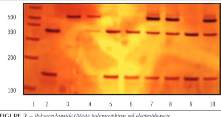
FIGURE 2 − Polyacrylamide G844A polymorphism gel electrophoresis
Lane 1: molecular weight standard; lanes 2, 5, 6 and 9: homozygous normal DNA (146 and 314 bp); lane 3: individual DNA homozygous mutant (510 bp); lanes 4, 7, 8 and 10: DNA heterozygotes (146, 314 and 510 bp).
DNA: deoxyribonucleic acid. 91
74
72
64
48
1 2 3 4 5
FIGURE 1 − Polyacrylamide ApoE gel electrophoresis
Lane 1: ε2ε4profile (91, 74, 72 and 48 bp); lanes 2 and 4: ε4ε4(72, 64 and 48 bp); lane 3: ε3ε3(91, 64 and 48 bp); lane 5: ε2ε3(91, 74, 64 and 48 bp).
ApoE: apolipoprotein E.
TABLE 1 − Frequency of ApoE gene polymorphisms in patients with stroke and individuals in the control group
Frequency of genotypes Allele frequencies
Polymorphism ε3ε3 ε4ε4 ε2ε3 ε2ε4 ε3ε4 ε2 ε3 ε4
Control (n = 201) 123 (61.2%) 9 (4.5%) 16 (8%) 9 (4.5%) 44 (21.8%) 0.06 0.76 0.18
Stroke (n = 48) 27 (56.3%) 3 (6.3%) 4 (8.3%) 2 (4.2%) 12 (24.9%) 0.06 0.73 0.21
OR 0.82 1.42 1.05 0.93 1.19 1.01 0.84 1.44
95% CI 0.41-1.62 0.29-6.06 0.28-3.58 0-4.84 0.53-2.61 0.36-2.68 0.49-1.43 0.79-2.62
p-value 0.64 0.71 1 1 0.79 0.82 0.57 0.26
ApoE: apolipoprotein E; OR: odds ratio; CI: confidence interval.
Statistical analysis
TABLE 2 − Frequency of G844A polymorphism in the promoter region of the PAI-1 gene among stroke patients and individuals in the control group
Polymorphism Frequency of genotypes Allele frequencies
G844A Not detected (GG) Heterozygous (GA) Homozygous (AA) G A
Control (n = 201) 52 (26%) 106 (52.7%) 43 (21.3%) 0.52 0.48
Stroke (n = 48) 16 (33.3%) 26 (54.2%) 6 (12.5%) 0.59 0.41
OR 1.06 0.52 1.4 0.72
95% CI 0.54-2.09 0.19-1.4 0.87-2.25 0.44-1.15
p-value 0.99 0.23 0.18 0.18
PAI-1: plasminogen activator type 1; OR: odds ratio; CI: confidence interval.
TABLE 3 − Frequency of ApoE gene polymorphisms in patients with PAD and individuals in the control group
Frequency of genotypes Allele frequencies
Polymorphism ε3ε3 ε4ε4 ε3ε4 ε2 ε3 ε4
Control (n = 201) 123 (61.2%) 9 (4.5%) 44 (21.8%) 0.06 0.76 0.18
PAD (n = 49) 31 (63.3%) 2 (4.1%) 10 (20.4%) 0.06 0.8 0.14
OR 1.09 0.91 0.91 1.01 1.22 0.91
95% CI 0.55-2.19 0-4.73 0.39-2.09 0.36-2.68 0.69-2.18 0.46-1.78
p-value 0.92 1 0.97 0.82 0.55 0.9
ApoE: apolipoprotein E; PAD: peripheral arterial disease; OR: odds ratio; CI: confidence interval.
TABLE 4 − Frequency of G844A polymorphism in the promoter region of the PAI-1 gene among patients with PAD and individuals in the control group
Polymorphism Frequency of genotypes Allele frequencies
G844A Not detected (GG) Heterozygous (GA) Homozygous (AA) G A
Control (n = 201) 52 (26%) 106 (52.7%) 43 (21.3%) 0.52 0.48
PAD (n = 49) 19 (38.8%) 19 (38.8%) 11 (22.4%) 0.58 0.42
OR 0.57 1.06 1.34 0.75
95% CI 0.29-1.12 0.47-2.38 0.83-2.15 0.46-1.2
p-value 0.11 0.97 0.25 0.25
PAI-1: plasminogen activator type 1; OR: odds ratio; CI: confidence interval.
between potentially atherogenic particles and antiatherogenic ones was independently associated with the development of atherothrombotic disease in young IS and PAD patients, and that the ApoB/ApoA-I ratio represents a significant marker of these alterations(22). Continuing this approach we investigated the association of PAI-1 G844A polymorphism and PAI-1 plasma levels previously determined in these patients. No statistically significant differences were observed when plasma levels were compared to the observed genotype.
No significant differences were observed when the evaluated patients were grouped according to the presence of the allele in heterozygotes or homozygotes. No correlation was observed
between plasma levels of PAI-1 and the G and A alleles, when Spearman’s correlation test was applied. With the aim of verifying the relationship between ApoE genotypes and plasma levels of the biochemical and hemostatic parameters previously
determined(22), patients and controls were allocated into three groups: group 1, consisting of the ε2ε3 and ε2ε4 genotypes; group 2, of the ε3ε3 genotype; and group 3, of the ε3ε4 and
ε4ε4 genotypes, but no significant difference was observed
(Table 5).
Likewise, no significant differences were observed when the evaluated patients were grouped according to age (> 37 years,
DISCUSSION
Several studies have reported an association between the presence
of alleles ε2, ε3 and ε4 and the occurrence of arterial thrombotic events as well as variations in plasma lipid concentrations. However, after molecular characterization of patients and controls, in this study significant difference was not observed in the distribution of ApoE genotypes (homozygous or heterozygous)(1, 2). Data from similar genotypic frequencies to those observed in this study were described by Wiegman et al. (2003)(23) when evaluating a group of 450 children with familial hypercholesterolemia and 2018 control subjects: they did not find significant differences between patients and controls for the genotypes. Similarly, association between the
ε4 allele and plasma levels of LDL-C, high-density-lipoprotein cholesterol (HDL-C) or triglyceride was not observed, suggesting a low influence of ApoE genotype on these parameters. Cerrato et al.
(2005)(24) also reported no association between the alleles of the ApoE, specifically the ε4 allele and stroke in assessing a group of 302 Italian patients, with ischemic cerebrovascular disease (ICVD). Lin et al. (2011)(25) evaluated Taiwanese patients, with the same profile and reported no association between the ε4 allele and ICVD, but indicated a protective role for the ε2 allele and the occurrence of these events(26, 27).
Considering the biochemical parameters evaluated in our previous studies, significant differences were observed between patients with IS or PAD in levels of total cholesterol, HDL-C, LDL-C, triglycerides, ApoA-I, ApoB, including ApoB/ApoA-I and PCR index, compared with the control group(22). However, even though plasma levels of these biochemical parameters were increased compared to the control group, there was no association between changes
in these parameters and ApoE alleles, showing that, in the present study, there was no effect of ApoE genotype on changing lipid profile and apolipoprotein.
The studies have associated increased levels of PAI-1 with the occurrence of coronary artery disease, insulin resistance, dyslipidemia in adults as well as cerebrovascular diseases(28). In a previous study, we evaluated the plasma levels of PAI-1 as well as the influence of 4G/5G polymorphism in the promoter region of the gene(21). Here we investigated the influence of PAI-1 G844A polymorphism and the increased levels of PAI-1; however, no association was observed. Although elevated plasma levels of PAI-1 have been considered, until recently, a potential risk factor for stroke, due to its antifibrinolytic activity, this association should be considered carefully because of the complex biological variations in the levels of PAI-1 dependent on estrogen levels, lipids and proinflammatory cytokines(29). Studies with transgenic mice show PAI-1 has a role both as a risk factor and as a protector in the development of arterial
disease(30). Furthermore, PAI-1 is required for protection against tissue plasminogen activator (tPA), which causes local damage in nervous tissue after an IS(16, 31). No significant association was observed between the levels of PAI-1 and G844A polymorphism among patients in the present study. Although this polymorphism has been studied extensively, there is still controversy about the association between the development of IS and PAD and the possible genotypes, especially in the Brazilian population. Studies were not found in the literature reporting the frequency or correlation of these polymorphisms with the occurrence of atherothrombotic events in Brazil.
The differential regulation or function of PAI-1 in plasma levels and tissue may explain the different findings for the
TABLE 5 − Biochemical, haemostatic parameters and ApoE genotypes: comparison between groups
Controls Patients
Groups 1 (ε2ε3, ε2ε4) 2 (ε3ε3) 3 (ε3ε4, ε4ε4) p 1 (ε2ε3, ε2ε4) 2 (ε3ε3) 3 (ε3ε4, ε4ε4) p
TC (mg/dl) 168.6 ± 31.7 179.4 ± 38.2 167.1 ± 31.6 > 0.05 179.1 ± 68.5 205.1 ± 54.6 194.6 ± 45.2 > 0.05
HDL-C (mg/dl) 56.6 ± 23 57.7 ± 15.9 52.5 ± 13 > 0.05 46.1 ± 17.6 50.5 ± 13.5 46.4 ± 13.4 > 0.05
LDL-C f (mg/dl) 87.6 ± 32.7 101 ± 33.7 92.2 ± 30.5 > 0.05 121 ± 55.4 129.4 ± 47.1 119.8 ± 39.5 > 0.05
TG (mg/dl) 121.5 ± 54.2 99.7 ± 56.7 93.6 ± 42.3 > 0.05 101.3 ± 43.8 133.4 ± 77.7 142.4 ± 78.6 > 0.05
ApoA-I (mg/dl) 162.3 ± 8.1 159.8 162.1 ± 16 > 0.05 140.7 ± 9.8 149.6 ± 9.3 144.6 ± 13.1 > 0.05
ApoB (mg/dl) 60.9 ± 11.3 66.2 ± 17.1 63.6 ± 13.9 > 0.05 75.2 ± 26.9 87.5 ± 22.5 87 ± 20.5 > 0.05
ApoB/ApoA-I 0.37 ± 0.1 0.4 0.39 ± 0.1 > 0.05 0.54 ± 0.21 1.12 ± 4 0.6 ± 0.12 > 0.05
hs-CRP (mg/l) 3.1 ± 5.2 0.9** 1.3** > 0.05 7.9 ± 10.9 8.8 ± 15.8 4.5 ± 4.8 > 0.05
PAI-1 (ng/ml) 37.3 ± 14.1 36.9 ± 12.4 37.1 ± 11.8 > 0.05 49 ± 26.7 60 ± 21 51.2 ± 22.1 > 0.05
polymorphism and levels of PAI-1 found in this and other studies. The local cellular production of PAI-1 and tPA and a definite role of this proteases system in the tissues of animals have been
demonstrated(11, 16, 32, 33). A localized inflammation process plays a major role in atherosclerosis and in plaque rupture present in brain tissue. Note that the plasma levels of PAI-1 can be modulated by several factors acting in tandem, wherein this impact modifier is not associated with the genetic profile of the individual. Thus, the role of PAI-1 and the G844A polymorphism as risk factors for the development of IS and PAD needs to be fully understood and requires further studies.
In this study, an aspect that draws attention is the fact that patients were aged below 37 years, featuring a distinguished group of individuals who had early arterial thrombotic events. Noteworthy is also the fact that the majority of patients were not under the influence of classic risk factors for the vascular obstruction, such as hypertension, diabetes mellitus, obesity and smoking, but showed significant change related to the deposition of lipids, inflammation and hypercoagulability. Thus, one can consider that the biochemical and molecular parameters evaluated in this study provided important data on the occurrence of arterial thrombotic events in young individuals in our population compared to multifactorial aspects.
CONCLUSION
No association was observed between the polymorphism of PAI-1 gene and IS and PAD or between the polymorphism and increased levels of PAI-1. There was no association between ApoE gene polymorphisms and changes in lipid profile and the occurrence of IS and PAD.
ACKNOWLEDGMENTS
The authors are grateful to Conselho Nacional de Desenvolvimento Científico e Tecnológico (CNPq), Fundação de Amparo à Pesquisa do Estado de Minas Gerais (Fapemig) and Coordenação de Aperfeiçoamento de Pessoal de Nível Superior (Capes) for sponsoring this investigation.
CONFLICT OF INTEREST
The authors declare that they have no conflict of interest.
RESUMO
Introdução: A trombose arterial é considerada uma doença multifatorial, resultante da interação de fatores de risco genéticos
e adquiridos. Objetivos: O objetivo deste estudo foi investigar a presença dos polimorfismos nos genes do inibidor da ativação do plasminogênio tipo 1 (PAI-1) e da apolipoproteína E (ApoE), bem como suas interações com níveis de PAI-1 e lipídios e perfis de apolipoproteína, respectivamente, além das frequências desses polimorfismos e sua associação com trombose.
Métodos: Noventa e sete pacientes [48 com acidente vascular cerebral isquêmico arterial (AVC) e 49 com doença arterial
periférica (DAP)], tratados no serviço médico de hematologia, foram incluídos neste estudo. Os polimorfismos também foram investigados em 201 indivíduos-controle. Os polimorfismos foram investigados por reação em cadeia da polimerase-fragmento de restrição polimorfismo (PCR-RFLP). Resultados: Para o polimorfismo PAI-1, havia 54,2% genótipos heterozigotos (HT) e 12,5% genótipos de homozigoto (HM) no grupo dos pacientes, e 52,7% genótipos HT e 21,3% genótipos HM nos grupos-controle. Para o polimorfismo da ApoE, havia 56,3% (ε3ε3), 6,3% (ε4ε4), 8,3% (ε2ε3), 4,2% (ε2ε4) e 24,9% (ε3ε4) nos pacientes, e 61,2% (ε3ε3), 4,5% (ε4ε4), 8% (ε2ε3), 4,5% (ε2ε4) e 21,8% (ε3ε4) nos controles. Conclusão: Nenhuma diferença significativa foi observada comparando pacientes e controles. Neste estudo, não foi encontrada associação entre a presença dos polimorfismos avaliados e a ocorrência de eventos trombóticos.
REFERENCES
1. Moriarty PM, Varvel SA, Gordts PLSM, McConnell JP, Tsimikas S. Lipoprotein(a) mass levels increase significantly according to APOE genotype: an analysis of 431 239 patients. Arterioscler Thromb Vasc Biol. 2017; 37(3): 580-8.
2. Ranellou K, Paraskeva A, Kyriazopoulos P, et al. Polymorphisms in prothrombotic genes in young stroke patients in Greece: a case-controlled study. Blood Coagul Fibrinolysis. 2015; 26(4): 430-5.
3. Zhu S, Wang Z, Wu X, Shu Y, Lu D. Apolipoprotein E polymorphism is associated with lower extremity deep venous thrombosis: color-flow Doppler ultrasound evaluation. Lipids Health Dis. 2014; 13: 21. 4. Kumar A, Sagar R, Kumar P, et al. Identification of genetic contribution to ischemic stroke by screening of single nucleotide polymorphisms in stroke patients by using a case control study design. BMC Neurol. 2013; 13(1): 136.
5. Tudorache IF, Trusca VG, Gafencu AV. Apolipoprotein E - a multifunctional protein with implications in various pathologies as a result of its structural features. Comput Struct Biotechnol J. 2017; 15: 359-65.
6. Liu G, Liu X, Yu P, et al. APOE gene polymorphism in long-lived individuals from a central China population. Sci Rep. 2017; 7(1): 3292. 7. Nagato LC, de Souza Pinhel MA, de Godoy JM, Souza DR. Association of ApoE genetic polymorphisms with proximal deep venous thrombosis. J Thromb Thrombolysis. 2011: 116-9.
8. Henriques AD, Tonet-Furioso AC, Machado-Silva W, et al. Apolipoprotein E genotype is associated with apolipoprotein B plasma levels but not with coronary calcium score in very elderly individuals in primary care setting. Gene. 2014; 539(2): 275-8.
9. Kulminski A, Ukraintseva SV, Arbeev KG, et al. Association between APOE ε2/ε3/ε4 polymorphism and disability severity in a national long-term care survey sample. Age Ageing. 2008; 37: 288-293.
10. Heiman M, Gupta S, Khan SS, Vaughan DE, Shapiro AD. In: Adam AP, Ardinger HH, Pagon RA, et al., editors. Complete plasminogen activator inhibitor 1 deficiency. Seattle (WA): August 2017.University of Washington, Seattle; 1993-2018.
11. Ye Y, Vattai A, Zhang X, et al. Role of plasminogen activator inhibitor type 1 in pathologies of female reproductive diseases. Int J Mol Sci. 2017; 18(8).
12. De la Cruz-Mosso U, Munoz-Valle JF, Salgado-Goytia L, et al. Relationship of metabolic syndrome and its components with 844 G/A and HindIII C/G PAI-1 gene polymorphisms in Mexican children. BMC Pediatr. 2012; 12(1): 41.
13. Liguori R, Quaranta S, Di Fiore R, Elce A, Castaldo G, Amato F. A novel polymorphism in the PAI-1 gene promoter enhances gene expression. A novel pro-thrombotic risk factor? Thromb Res. 2014; 134(6): 1229-33. 14. Munoz SA, Aranda F, Allievi A, et al. 4G/5G plasminogen activator inhibitor-1 and -308 A/G tumor necrosis factor-alpha promoter gene
polymorphisms in Argentinean lupus patients: focus on lupus nephritis. Clin Exp Med. 2014; 14(1): 83-9.
15. Padilla-Gutiérrez JR, Palafox-Sánchez CA, Valle Y, et al. Plasminogen activator inhibitor-1 polymorphisms (-844 G>A and HindIII C>G) in systemic lupus erythematosus: association with clinical variables. Clin Exp Med. 2011; 11(1): 11-7.
16. García-González IJ, Valle Y, Sandoval-Pinto E, et al. The -844 G>A PAI-1 polymorphism is associated with acute coronary syndrome in Mexican population. Dis Markers. 2015; 2015: 460974.
17. Ozolina A, Strike E, Nikitina-Zake L, et al. Polymorphisms on PAI-1 and ACE genes in association with fibrinolytic bleeding after on-pump cardiac surgery. BMC Anesthesiol. 2015; 15(1): 122.
18. Souza GMA, Romano NL, Whittle MR. Efficient mispriming during apolipoprotein E genotyping. J Bras Patol Med Lab. 2005; 41(1): 25-8. 19. Morange PE, Henry M, Tregouet D, et al. The A -844G polymorphism in the PAI-1 gene is associated with a higher risk of venous thrombosis in factor V Leiden carriers. Arterioscler Thromb Vasc Biol. 2000; 20(5): 1387-91.
20. Dean Andrew G, Dean Jeffrey A CD, Coulombier D, et al. Epi Info, version 6.04: a word processing database and statistics program for epidemiology on microcomputers. Cent Dis Control Prev. 1997. 21. de Paula Sabino A, Ribeiro DD, Domingueti CP, et al. Plasminogen activator inhibitor-1 4G/5G promoter polymorphism and PAI-1 plasma levels in young patients with ischemic stroke. Mol Biol Rep. 2011; 38(8): 5355-60.
22. Sabino AP, de Oliveira Sousa M, Moreira Lima L, et al. ApoB/ApoA-I ratio in young patients with ischemic cerebral stroke or peripheral arterial disease. Transl Res. 2008; 152(3): 113-8.
23. Wiegman A, Sijbrands EJG, Rodenburg J, et al. The apolipoprotein ε4 allele confers additional risk in children with familial hypercholesterolemia. Pediatr Res. 2003; 53(6): 1008-12.
24. Cerrato P, Baima C, Grasso M, et al. Apolipoprotein E polymorphism and stroke subtypes in an Italian cohort. Cerebrovasc Dis. 2005; 20(4): 264-9.
25. Lin HF, Lai CL, Tai CT, Lin RT, Liu CK. Apolipoprotein E polymorphism in ischemic cerebrovascular diseases and vascular dementia patients in Taiwan. Neuroepidemiology. 2011; 23(3): 129-34.
26. Satrupa Das, Subhash Kaul, Akka Jyothy, and Anjana Munshi. Association of APOE (E2, E3 and E4) gene variants and lipid levels in ischemic stroke, its subtypes and hemorrhagic stroke in a South Indian population. Neuroscience Letters. 2016; 628: 136-41.
27. Konialis C, Spengos K, Iliopoulos P, et al. The APOE E4 allele confers increased risk of ischemic stroke among Greek carriers. Adv Clin Exp Med. 2016; 25(3): 471-8.
29. Tang J, Zhu W, Mei X, Zhang Z. Plasminogen activator inhibitor-1: a risk factor for deep vein thrombosis after total hip arthroplasty. J Orthop Surg Res. 2018; 13(1): 8.
30. Prabhudesai A, Shetty S, Ghosh K, Kulkarni B. Investigation of plasminogen activator inhibitor-1 (PAI-1) 4G/5G promoter polymorphism in Indian venous thrombosis patients: a case-control study. Eur J Haematol. 2017; 99(3): 249-54.
31. Song C, Burgess S, Eicher JD, O’Donnell CJ, Johnson AD. Causal effect
of plasminogen activator inhibitor type 1 on coronary heart disease. 2017; 86(1): 138-43.
32. Vaughan DE, Rai R, Khan SS, Eren M, Ghosh AK. Plasminogen activator inhibitor-1 is a marker and a mediator of senescence. Arterioscler Thromb Vasc Biol. 2017; 37(8): 1446-52.
33. Chang ML, Lin Y, Pao LH, Huang HC, Chiu CT. Link between plasminogen activator inhibitor-1 and cardiovascular risk in chronic hepatitis C after viral clearance. Sci Rep. 2017; 7: 42503.
CORRESPONDING AUTHOR
Adriano de Paula Sabino
Rua Indiana, 173, apto 202; Nova Suíça; CEP: 30421-322; Belo Horizonte-MG, Brasil; e-mail: [email protected].