Oral Delivery of a Novel Recombinant
Streptococcus mitis
Vector Elicits Robust
Vaccine Antigen-Specific Oral Mucosal and
Systemic Antibody Responses and T Cell
Tolerance
Emily Xie1, Abhiroop Kotha1, Tracy Biaco1, Nikita Sedani1, Jonathan Zou1,
Phillip Stashenko1,3, Margaret J. Duncan2, Antonio Campos-Neto1,3, Mark J. Cayabyab1,3*
1Global Infectious Disease Research Center and the Department of Immunology and Infectious Diseases, The Forsyth Institute, 245 First Street, Cambridge, Massachusetts, United States of America,2Department of Microbiology, The Forsyth Institute, 245 First Street, Cambridge, Massachusetts, United States of America,3Department of Oral Medicine, Infection and Immunity, Harvard School of Dental Medicine, Boston, Massachusetts, United States of America
Abstract
The pioneer human oral commensal bacteriumStreptococcus mitishas unique biologic features that make it an attractive mucosal vaccine or therapeutic delivery vector.S.mitisis safe as a natural persistent colonizer of the mouth, throat and nasopharynx and the oral commensal bacterium is capable of inducing mucosal antibody responses. A recombinant
S.mitis(rS.mitis) that stably expresses HIV envelope protein was generated and tested in the germ-free mouse model to evaluate the potential usefulness of this vector as a mucosal vaccine against HIV. Oral vaccination led to the efficient and persistent bacterial coloniza-tion of the mouth and the induccoloniza-tion of both salivary and systemic antibody responses. Inter-estingly, persistently colonized animals developed antigen-specific systemic T cell
tolerance. Based on these findings we propose the use ofrS.mitisvaccine vector for the induction of mucosal antibodies that will prevent the penetration of the mucosa by patho-gens such as HIV. Moreover, the first demonstration ofrS.mitishaving the ability to elicit T cell tolerance suggest the potential use ofrS.mitisas an immunotherapeutic vector to treat inflammatory, allergic and autoimmune diseases.
Introduction
Streptococcus mitisis a commensal species of Gram-positive oral streptococci that inhabits the human mouth.S.mitisand other oral commensal streptococci includingS.salivariusandS.
oralisare among the first colonizers of the human mouth; hence, they are called pioneer oral bacteria.S.mitiscolonizes hard surfaces in the oral cavity such as dental hard tissues as well as OPEN ACCESS
Citation:Xie E, Kotha A, Biaco T, Sedani N, Zou J, Stashenko P, et al. (2015) Oral Delivery of a Novel RecombinantStreptococcus mitisVector Elicits Robust Vaccine Antigen-Specific Oral Mucosal and Systemic Antibody Responses and T Cell Tolerance. PLoS ONE 10(11): e0143422. doi:10.1371/journal. pone.0143422
Editor:John S Tregoning, Imperial College London, UNITED KINGDOM
Received:August 29, 2015
Accepted:November 4, 2015
Published:November 30, 2015
Copyright:© 2015 Xie et al. This is an open access article distributed under the terms of theCreative Commons Attribution License, which permits unrestricted use, distribution, and reproduction in any medium, provided the original author and source are credited.
Data Availability Statement:All relevant data are within the paper and its Supporting Information file.
Funding:This work was supported by a grant (RO3 DE022525) from the National Institute of Dental and Craniofacial Research. The funder had no role in study design, data collection and analysis, decision to publish, or preparation of the manuscript.
mucous membranes andS.mitisis also found in the throat and nasopharynx.S.mitistakes res-idence in the human oral cavity as early as 1–3 days postpartum [1] and continue to persist until other oral bacteria co-inhabit the mouth. An established adult human oral microbiota is comprised of over 600 prevalent taxa [2–4]. An extensive microbiota study showed that during infancy and adult life,S.mitiscan predominate, both in prevalence and proportion of oral streptococci recovered in the mouth [5] andS.mitiswas the most predominant bacterial spe-cies colonizing all oral surfaces of adults [6]. The success ofS.mitislikely hinges on its ability to adapt to the oral microenvironment and evade host defenses. For example, some strains ofS.
mitisand other oral streptococci express IgA1 protease that cleaves host neutralizing IgA anti-bodies [3,4].
Mucosal antibody responses toS.mitisare well-documented. In adult human, salivary IgA toS.mitisis present in high titers [7] and in infants, salivary antibodies are generated soon after birth. The nature of the T cell response toS.mitisis not fully understood, although,S.
mitis-specific T cell clones have been isolated from human peripheral blood [8]. Certain com-mensal bacteria especially those found in the gut induce T cell tolerance [9] and it is possible thatS.mitisand perhaps other oral commensal bacteria are also capable of inducing T cell tolerance.
The ability to safely and efficiently colonize and persist in the human mouth and other mucosal surfaces as well as the ability to induce oral mucosal antibodies are unique biologic features ofS.mitisthat make this pioneer bacterium a potential antigen vaccine or therapeutic delivery vehicle. The genome ofS.mitishas been fully sequenced and the oral bacterium is genetically tractable for the expression of foreign antigens.
In this study, we exploredS.mitis’potential as a mucosal vaccine vector for the delivery of antigens against HIV. Mucosal HIV transmission is responsible for the majority of the current estimated 34 million HIV infection globally and current vaccine strategies for HIV and other mucosal pathogens efficiently induce systemic immune responses but many do not efficiently induce mucosal immunity. Mucosal immunity is likely to play a critical role in vaccine-medi-ated protection against HIV. In HIV infection, the virus primarily penetrates across mucosal surfaces and viral replication in the intestinal mucosa contributes significantly to pathogenesis in adult HIV infection [10–13]. In mother-to-child transmission (MTCT), studies in an impor-tant breast milk AIDS transmission model in infant rhesus macaques suggest that HIV-1 likely penetrates oral and gut mucosal surfaces of the infant, infects intraepithelial dendritic cells and CCR5+ CD4 T cells, then disseminates to the oral buccal mucosa, tonsils, esophageal and intes-tinal mucosa [10]. It is argued that a preventive vaccine must induce protective mucosal anti-bodies at the time of transmission to prevent pathogen adherence and penetration of the epithelial surface and/or induce pathogen-specific mucosal T responses that will destroy the pathogen in infected tissues [11,13].
We constructed a recombinantS.mitis(rS.mitis) vector prototype expressing HIV antigens and tested its ability to induce B and T cell responses in the germ-free mouse model. We found that oral inoculation withrS.mitisresulted in efficient and persistent colonization of the oral cavity of mice. Furthermore,rS.mitismarkedly induced systemic and salivary vaccine antigen-specific antibody responses. Interestingly, persistent colonization byrS.mitisinduced T cell non-responsiveness to antigen stimulation, which is likely due to the induction of oral T cell tolerance. These findings suggest that anrS.mitisvector prototype is capable of colonizing oral mucosal surfaces and inducing mucosal and systemic antibody responses, which argue in favor of further development ofrS.mitisvaccine strategy to elicit protective mucosal and systemic antibodies against HIV and other mucosal pathogens. In addition, the ability ofrS.mitisto induce systemic T cell tolerance also has clinical implication for the development ofrS.mitis -based therapy to treat inflammatory, allergic and autoimmune diseases.
Materials and Methods
Generation of recombinant
Streptococcus mitis
RecombinantS.mitis(rS.mitis) was generated by homologous recombination. The p5E3 sui-cide plasmid vector was created by inserting HIV-1 HXBc2 Env gp120-His-tag (HIV Env) [14] (codon-optimized forS.mitisexpression by BlueHeron Technology, Bothel, WA) in-frame with the 250bp 5’end of the pullulanase gene (pulA/Smt0163) followed by Ermrgene [15] and the 250bp 3’end of the pulA gene into pCR2.1 (Invitrogen, Carlsbad, CA) for transformation and integration into the Smt0163 locus (Fig 1A).S.mitiswas transformed by electroporation with p5E3 to generate homologous recombinantS.mitisexpressing HIV-1 Env or with pCR2.1 containing Ermrflanked by 250bp pulA 5’and 3’fragments to generate controlS.mitisempty vector.rS.mitisclones were selected on Todd Hewitt Broth (THB) plates containing 50μg/ml erythromycin (Fisher Scientific, Pittsburgh, PA).
Expression of the HIV Env gp120 protein was assessed by Western blotting. Single colonies were grown in THB medium containing 50μg/ml of erythromycin and grown 3–5 days until an optical density at 600 nm (OD600) approximately equal to 1.rS.mitiscells were then har-vested and washed twice in ice-cold phosphate-buffered saline (PBS). Cell pellets were collected and bacterial lysates were generated by vortexing at top speed with 106 micron glass beads (Sigma, St. Louis, MO) for 15 minutes. Proteins from 10 ml culture supernatants were filtered through a 0.2-μm-pore-size filter to remove cell material and either concentrated 5x using a 10-kDa membrane filter (Amicon) or precipitated with either acetone or 10% trichloroacetic acid. Expression of the viral gp120 protein was assessed by Western blotting of bacterial lysates, concentrated or TCA- or Acetone-precipitated supernatants (1μg of total protein) using Penta-His HRP (Qiagen, Hilden, Germany). Expression of HIV gp120 in concentrated super-natant was also assessed by binding with human patient sera recognizing HIV-1 gp120, which were pooled from HIV-infected individuals followed by a secondary goat anti-human IgG con-jugated with HRP (ThermoFisher, Waltham, MA). For detection a chemiluminescence ECL kit was used according to the manufacturer's protocol (Invitrogen, Carlsbad, CA).
Production of HIV-1 HXBc2 Env and
S
.
mitis
antigens
The HXBc2 Env gp120 gene [14] was subcloned into pET14b expression vector (Novagen-EMD Chemicals, Gibbstown, NJ), expressed in BL21(DE3) pLysSE.colihost (Invitrogen, Carlsbad, CA) and the over-expressed recombinant protein was purified by affinity chromatog-raphy under denaturing condition as we have previously described [16]. To produceS.mitis
antigens,S.mitiswas grown in THB media to OD value of 1.0.S.mitiscells were washed twice and resuspended in PBS. Bacterial lysate was generated by vortexing at top speed with 106 micron glass beads (Sigma, St. Louis, MO) for 15 minutes and filter sterilized.
Mice, immunizations, blood and saliva collection
6–8 week old female Balb/c mice were purchased from Charles River Laboratories (Wilming-ton, MA) and kept under specific pathogen-free conditions at the Forsyth Institute Animal Facility. 6–8 week old germ-free Balb/c were housed at the Gnotobiotic and Microbiology Core (CHB) Facility, Brigham Women’s Hospital and Harvard Medical School, Boston, MA. All animal procedures were carried out under the guidelines of the Institutional Animal Care and Use Committee at the Forsyth Institute and the Animal Care and Use Committee at Har-vard Medical School.
phosphate-buffered saline (PBS) and inoculated into mice orally at 109cfu or intraperitoneally at 2x10⁸cfu in 200μl PBS.
For saliva and blood collection, mice first received ketamine (100 mg/kg of body weight) with xylazine (12 mg/kg) anesthesia intraperitoneally. 100–200μl blood was collected retro-orbitally from anesthetized mice using a sterile beveled tip. Saliva secretion was stimulated by subcutaneous injection of 0.1 ml 0.1mg/ml carbachol and approximately 100–200μl saliva was collected from each animal.
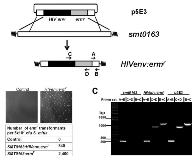
Fig 1. Construction of recombinantS.mitisexpressing HIV Env gp120.(A) RecombinantS.mitis(rS.mitis) was generated by homologous
recombination. The p5E3 suicide plasmid was created by inserting theS.mitiscodon-optimized HIV-1 HXBc2 Env gp120-His-tag (HIV Env) in-frame with the 250bp 5’end of the pullulanase gene (pulA/Smt0163) followed by ermr gene and the 250bp 3’end of the pulA gene into pCR2.1 for transformation and integration into the Smt0163 locus. (B)S.mitiswas transformed with water (control), p5E3 (Smt0163:HIVenv:ermr) or pCR2.1 containing ermr flanked by 250bp pulA 5’and 3’fragments (Smt0163:ermr
). Growth of transformants on THB plates containing 50μg/ml erythromycin and transformation frequency are shown. (C) Integration was confirmed by PCR usingS.mitis-specific primers A/B, HIV-specific primer C and ermr-specific primer D.
Colonization study
rS.mitiscolonization of mice was determined by the presence of the oral bacterium in the mouth and feces. The upper right buccal cheek of each mouse was scrubbed with calcium algi-nate oral swab (Fisher Scientific, Pittsburgh, PA), which was then immersed in 1 ml THB media and vortexed thoroughly. Two fecal pellets per mouse were collected and resuspended vigorously in 1 ml THB media by vortexing. Undiluted and diluted oral and fecal samples were spread on THB agar plates, containing 50μg/ml erythromycin to determine the total number of colony-forming-units (cfu) present in the collected feces. The cfu counts were normalized against the wet weight of the fecal pellets.
Measurement of Antibody Responses
High-binding 96-well microplates (Costar, Lowell, MA) were coated with the purified HIV-1 HXBc2 Env protein (2μg/ml) orS.mitislysate antigens (2μg/ml) prepared in 0.2 M sodium carbonate/bicarbonate buffer (pH 9.6) and incubated overnight at 4°C. Wells were washed with PBS containing 0.05% Tween-20 (PBS-T) and blocked with 1% BSA in PBS-T for 2 hr. Serum or saliva samples were diluted 1:3 and added at 2- to 3-fold serial dilutions in PBS-T containing 0.1% BSA and plates were incubated for 1 hr at room temperature. After another washing step, horseradish peroxidase, HRP-conjugated rat anti-mouse IgA (clone 11-44-2), IgG1 (clone SB77e) or IgG2a (clone SB84a) (Southern Biotech, Birmingham, AL) was added at 1:4000, 1:5000, 1:5000 dilution, respectively and incubated for 1 hr. A substrate solution con-taining tetramethylbenzidine (KPL, Gaithersburg, MD) was added and colour development was stopped using 1 m HCl. Optical density data were recorded as absorbance at 450 nm. OD values twice above the background were considered positive and values between 0.01 and 1.0 were used to determine the amount of antibody produced and adjusted according to the dilu-tion factor to determine the OD value of the undiluted saliva and serum samples.
Intracellular cytokine staining
Blood collected from anesthetized mice was mixed with PBS containing 10mM EDTA to pre-vent coagulation and peripheral blood mononuclear cells (PBMCs) were purified using lym-pholyte-M (Cedarlane, Burlington, NC). 1–2 x 10⁵PBMCs were cultured at 37°C in a 5% CO2 environment for 6 hr in the presence of RPMI-1640/10% fetal calf serum alone or with either 10μg/mlS.mitislysate or 10μg/ml HIV HXBc2 envelope protein. All cultures contained Mon-ensin (GolgiStop; BD Biosciences, San Jose, CA). The cultured cells were cell-surface stained with the following monoclonal antibodies purchased from BD Biosciences: anti-CD3-FITC (145-2C11), anti-CD4-allophycocyanin-Cychrome7 (GK1.5), anti-CD8α-perdinin chlorophyll protein-Cychrome 55 (53–6.7). After fixing with Cytofix/Cytoperm solution (BD Biosciences),
Multiplex Luminex Assay
Cytokines present in culture supernatants of stimulated cells were quantitated using MILLI-PLEX1MAPmultiplex cytokine biomarker magnetic bead panel for detection of mouse IL-2, IL-4, IL-6, IL-10, IL-13, IL-17A, TGFβ, TNFαand IFNγ(EMD Millipore, Darmstadt, Ger-many). Samples were analyzed in a microplate well using Luminex1200™(Luminex, Austin, TX).
Statistical analysis
Data were expressed as means ± standard error of the means (SEM). Statistical tests were per-formed using Student’sttest. APvalue of less than 0.05 was considered significant.
Results
Construction of the prototype recombinant
S
.
mitis
HIV vaccine vector
To generate recombinantS.mitis (rS.mitis), strain NCTC 12261 was transformed with DNA fragment p5E3 containing the HIV Env gene to allow homologous recombination and integra-tion into the pullulanase (pulA/Smt0163) gene (Fig 1A). ThepulA/Smt0163) gene was selected because it is non-essential and highly expressed [17]. The p5E3 DNA fragment consists of the pCR2.1 plasmid containing theS.mitiscodon-optimized HIV-1 HXBc2 gp120env-His-tag (HIV Env) in-frame with the 250bp 5’end of the pullulanase gene (pulA/Smt0163), followed by an Ermr gene and the 250bp 3’end of the Smt0163 gene (Fig 1A). Transformation frequen-cies on erythromycin-containing plates were 840 and 2,400 transformants per 5 x10⁷cfu bacte-ria added for plasmids containing the HIVenv:ermror ermrgene only, respectively (Fig 1B). Integration of the HIV gene was confirmed by PCR and sequencing. PCR analysis showed that theS.mitis-specific primers A/B generated the expected 250 bpsmt0163PCR product, while the HIV-specific primer C andermr-specific primer D generated the expected 1 kb product (Fig 1C). Sequencing of the PCR products further showed the integration of the HIV Env gene inS.mitis(data not shown). Thus, we successfully constructedrS.mitiscontaining an HIV gene integrated at thepulAgene locus of theS.mitisgenome.
Recombinant
S
.
mitis
expresses HIV-1 envelope protein
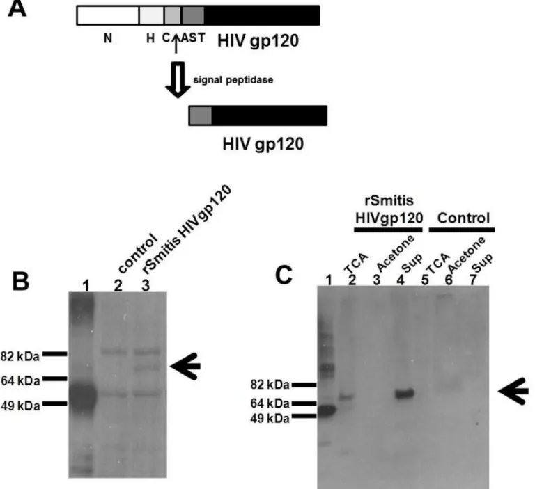
Our strategy was to express foreign vaccine antigens in the secreted form, which enhances the immunogenicity of bacterial antigens [14]. The constructedrS.mitiscontains the integratedS.
mitiscodon-optimized HIV-1 HXBc2env-His-tag (HIV Env) in-frame with the 250bp 5’end of the pullulanase gene (pulA/Smt0163) encoding a signal peptide that allows processing and secretion of the HIV antigen (Figs1Aand2A) [18,19]. The signal peptide has a predicted amino-terminal region (N), a hydrophobic core (H), a signal peptidase cleavage site (C), and an accessory Sec transport motif (AST).
Fig 2. RecombinantS.mitisexpresses HIV envelope protein.rS.mitiswith the integrated HIV HXBc2 Env gp120 was designed to secrete HIV Env by ligating the HIV Env in frame with 250bp 5’end of the pullulanase gene (pulA/Smt0163) encoding a signal peptide that allows processing and secretion of the HIV antigen. (A) The signal peptide has an amino-terminal region (N), a hydrophobic core (H), a signal peptidase cleavage site (C), and an accessory Sec transport motif (AST). Expression of HIV Env containing a C-terminal His tag was assessed by Western blotting using Penta-His-HRP from a representative recombinant clone inS.mitislysates (B) and in culture supernatants (C) by TCA-precipitation (TCA), acetone precipitation (Acetone) and Amicon filter-concentration (Sup). HIV Env expression in lysates (B) and supernatants of controlS.mitisvector (control) (C) is shown. The arrow denotes expression of the Env Ag band. 100 ng of His-taggedM.tuberculosisprotein (MT0401) was used as a positive control (B and C, lane 1). (D) The expression of HIV-1 gp120 in
rS.mitiscontaining the HIV Env gene (lane 2) in Amicon filter-concentrated supernatant was detected using human HIV patient sera.rS.mitiscontaining the empty plasmid was used as a negative control (lane 1).
(Fig 2B, lane 2) and supernatants of the controlS.mitisempty vector (Fig 2C, lanes 5–7). Addi-tionally, we determined the expression of HIV-1 gp120 in the concentrated supernatant using human HIV patient sera and found that therS.mitiscontaining the Env gp120 but not control
rS.mitisexpresses HIV g120 (Fig 2D). The apparent molecular weight of gp120 was similar to the expected molecular weight (of approximately 70 kDa), suggesting that Env likely was not glycosylated inS.mitis.
Expression of HIV Env in
rS
.
mitis
is stable
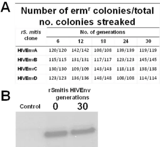
To determine the stability of the integrated HIV-1 Env gp120 gene inrS.mitis, four clones were picked at random and grown anaerobically for 30 generations. Loss of erythromycin-resistance was taken as an indication of the loss of the Env transgene. The progeny from all four clones were stable, with no loss of erythromycin resistance after ten subcultures or 30 gen-erations of replication without antibiotic (Fig 3A). In addition, we analyzed the expression of Env in the same daughter clones. Western blot analysis revealed that they secreted the same amount of Env antigen as the originalrS.mitisclones; Env production in a representative
Fig 3.rS.mitisexpressing HIV Env is stable.To determine the stability of the integrated Env gp120 gene in
rS.mitis, four clones (HIVEnvA, B, C, and D) were picked at random and grown anaerobically for 24 hours which represents approximately three generations (7.1 hours per generation) in THB media without erythromycin. Cultures were grown for approximately 30 generations without erythromycin. From each generation 100 and 150 colonies were picked at random and streaked to THB plates with and without erythromycin. For eachS.mitisHIV Env clone, the number of Ermrcolonies/total number of colonies streaked after 6, 12, 18, 24, and 30 generations was determined (Fig 3A). Expression of Env in the same daughter clones was analyzed by Western blot analysis. Env production in a representative daughter clone (after 30 generations) and the original HIVgp120A clone is shown (Fig 3B).
daughter clone after 30 generations and the original HIVgp120A clone is shown (Fig 3B). These results indicate that ourrS.mitisis stable, a vaccine property that makes it a good candi-date for further preclinical testing.
Recombinant
S
.
mitis
vaccine vector abundantly and persistently
colonized germ-free mice but not conventional mice
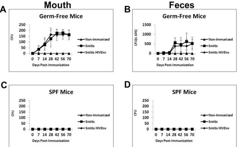
Oral pioneer commensal bacteria such asS.mitis,S.salivariusandS.oralisare excellent colo-nizers of the mouth. Therefore, a suitable animal model to test the preclinical efficacy of recom-binantS.mitismust be able to be effectively colonized. A prior study showed that gnotobiotic or germ-free mice were colonized by human oral bacteria, includingS.mitis[21]. To assess whether germ-free mice are a suitable animal model, germ-free mice as well as conventional mice were inoculated orally with 109cfurS.mitisexpressing HIV Env gp120 (Smitis HIVEnv) and controlS.mitis(Smitis). Following oral vaccination, colonization of the mouth was observed at day 7 and the number of bacteria continued to increase over time, peaking on day 30 and remained persistently high thereafter (Fig 4A). BothrS.mitisHIV Env and controlS.
mitiscolony-forming-units were significantly higher in immunized compared to
non-Fig 4. RecombinantS.mitiscolonizes germ-free mice efficiently and persistently.Germ-free Balb/c mice and conventional SPF mice were inoculated orally with 109cfurS.mitisexpressing HIV Env gp120 (Smitis HIV Env),rS.mitiscontaining an integrated Ermrgene without Env (Smitis) or PBS (Non-Immunized). Following inoculation the upper right buccal cheek was swabbed and two pellets of feces were collected from each mouse at various time points.rS.mitiscolonization was assessed by growth on THB plates containing 50μg/ml erythromycin. The mean colony-forming-units, cfu (±SEM) from 3 mice/group at various timepoints, present in the mouth (A) and feces (B) of germ-free mice and in the mouth (C) and feces (D) of conventional mice inoculated withrS.mitisare shown.
immunized mice on day 7, 14, 28, 42, 56 and 70 (p<0.05 at each timepoint). On those same
days,rS.mitiswas also found abundantly in the feces of the immunized mice but not in non-immunized mice (Fig 4B). Conventional SPF mice were not colonized byrS.mitis(Fig 4C and 4D). These results demonstrate that germ-free mice are efficiently and persistently colonized byS.mitisand therefore are a viable animal model for the preclinical testing ofrS.mitisvaccine vectors.
Recombinant
S
.
mitis
vaccine vector induces antigen-specific salivary
and systemic antibody responses in colonized mice
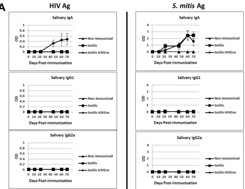
Infants, children and adults colonized byS.mitisdevelop salivary antibodies to the pioneer bacterium [7,22], and therefore we determined whether the vaccinated germ-free mice also develop antibody responses torS.mitis. Salivary IgA specific to HIV Env protein as well asS.
mitislysate antigens were initially detected three weeks post-inoculation and the antibody responses increased concomitant withS.mitispersistence in the oral cavity (Figs4Aand5A). The amount of HIV-specific salivary IgA inrS.mitisHIV Env-vaccinated mice was signifi-cantly higher than in controlS.mitis-vaccinated or in non-immunized mice on day 22, 45, 60 and 70 (p<0.05 at each timepoint). As expected, on those same days, similar amounts ofS. mitisantigen-specific salivary IgA were found in eitherrS.mitisHIV Env or controlS.mitis -immunized mice, which were both significantly higher than in non--immunized mice (p<0.05
at each timepoint). Salivary IgG1 and IgG2a antibodies specific torS.mitiswere undetectable. Systemic antibody responses were also generated in the vaccinated mice. TherS.mitis vac-cine-elicited serum IgA, IgG1 and IgG2a specific to HIV inrS.mitisHIV Env colonized but not in mice colonized by controlS.mitis(Fig 5B). The amount of HIV-specific serum IgA and IgG1 were significantly higher inrS.mitisHIV Env-vaccinated mice than in controlS.mitis -vaccinated or in non-immunized mice on day 16, 31, and 58 (p<0.05 at each timepoint),
while the HIV-specific IgG2a concentrations were significantly higher on day 31 and 51 in mice immunized withrS.mitisHIV Env than in mice immunized with controlS.mitisand non-immunized mice (p<0.05 at each timepoint). As expected, serum IgA, IgG1 and IgG2a
responses specific toS.mitislysate antigens were found in bothS.mitis-vaccinated groups (Fig 5B). Compared to the levels of antibodies found in non-immunized mice, the amount ofS.
mitisantigen-specific serum IgA and IgG1 in eitherS.mitis-vaccinated group were significantly higher on days 16, 31, and 56, while the antigen-specific serum IgG2a were significantly higher on day 31 and 56 (p<0.05 at each timepoint). Taken together, these results show thatrS.mitis
is capable of eliciting robust mucosal and systemic antibody responses in vaccinated animals.
Recombinant
S
.
mitis
vaccination induces systemic T cell
non-responsiveness, likely due to oral tolerance
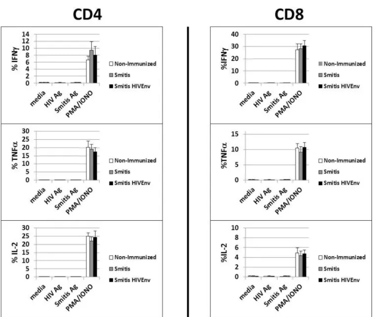
The nature of the T cell response toS.mitisin humans is not clearly known. We investigated whetherrS.mitisinduced T cell responses in the vaccinated germ-free mice. Vaccinated mice that harboredrS.mitispersistently for three months developed no vaccine-specific T cell-responses systemically. Intracellular cytokine staining analysis revealed that peripheral blood T cells isolated from vaccinated mice stimulated with either HIV orS.mitisantigens did not pro-duce IFNγ, TNFαand IL-2 (Fig 6A). In contrast, T cells expressed large amounts of IFNγ, TNFαand IL-2 in response to PMA and Ionomycin stimulation (Fig 6A).
(colonized) or non-vaccinated and three months later, they were injected intraperitoneally (IP) with 2x10⁸cfurS.mitisHIV Env. Two weeks after IP injection, splenocytes and mesenteric lymph node lymphocytes were isolated and proliferative and cytokine responses to HIV andS.
mitislysate antigens were assessed. In mice previously vaccinated and colonized withrS.mitis
followed by systemic IP injection ofrS.mitisthree months later (Balb/c (colonized) IP group), in vitro proliferative and cytokine responses in the spleen and MLN to HIV andS.mitis anti-gens were not seen (Fig 6B and 6C). In contrast, lymphocytes isolated from non-orally colo-nized germ-free mice that received IP injection withrS.mitis(Balb/c IP group) showed significant proliferative responses (p<0.05 for spleen and MLN) to HIV andS.mitisantigens
compared to media only control as well as to lymphocytes isolated from those mice that were previously colonized byrS.mitis(Balb/c (colonized) IP group). In addition, the lymphocytes Fig 5. RecombinantS.mitisinduces mucosal and systemic antibody responses.Germ-free Balb/c mice were inoculated orally with 109cfurS.mitis expressing HIV Env gp120 (Smitis HIV Env),rS.mitiscontaining an integrated Ermrgene without Env gp120 (Smitis), or PBS (Non-Immunized). Following immunization, the presence of IgA, IgG1, and IgG2a antibodies specific to the HIV Env gp120 protein (HIV Ag) orS.mitislysate antigens (S.mitisAg) was measured by ELISA in the saliva (A) and serum (B) of mice. The mean optical density (OD) (±SEM) values of the undiluted samples from 3 mice/group at various timepoints post-immunization are shown. The saliva and serum antibody dilution factor was 1:3 or indicated otherwise.
from therS.mitisIP-injected mice produced various cytokines in response to in vitro stimula-tion with HIV andS.mitisantigens. Using multiplex Luminex assay, the stimulated splenocytes and MLN cells were found to produce IL-2, IL-4, IL-6, IL-10, IL-13, IL-17, TNFαand IFNγ (Fig 6C). As expected, spleen and MLN cells from mice that were neither orally vaccinated nor IP injected withrS.mitis(Non-immunized group) did not proliferate and produced little or no Fig 6.rS.mitisinduces systemic T cell tolerance.(A) Germ-free mice were vaccinated orally with 109cfurS.mitisexpressing HIV Env gp120 (Smitis HIV Env) orrS.mitiscontaining an integrated Ermrgene without Env gp120 (Smitis). Ten weeks later, peripheral blood monuclear cells were isolated from vaccinated mice and stimulated with media alone (media),S.mitislysate antigens (S.mitisAg), the HIV Env gp120 protein (HIV Ag), or PMA and Ionomycin (PMA/IONO). Production of IFNγ, TNFα, and IL-2 by the stimulated cells measured by % IFNγ+, TNFα+, and IL-2+ CD4 or CD8 T cells from each group (3 mice/group) is shown. (B) To assess tolerance induction, germ-free mice were inoculated orally with 109cfu to allow colonization and three months later, these mice were injected intraperitoneally (IP) with 2x10⁸cfu rS.mitisHIV Env (Balb/c (colonized) IP group). Non-colonized mice were either non-immunized (Non-immunized group) or inoculated IP with 2x10⁸cfu rS.mitisHIV Env (Balb/c IP group). Two weeks after intraperitoneal immunization of the colonized and non-colonized mice, the spleen and mesenteric lymph nodes (MLN) were harvested and stimulated in vitro with media, 10μg/ml purified HIV Env gp120 protein (HIV Ag), 10μg/ml ofS.mitisantigens (Smitis Ag), Concanavalin A (ConA), or PMA/IONO. T cell proliferative responses were measured by using 3H-Thymidine incorporation assay. Mean CPM from 3 mice/group is shown. The production of IL-2, IL-4, IL-6, IL-10, IL-13, IL-17A, TGFβ, TNFαand IFNγby cells stimulated with media, HIV antigen, orS.mitisantigens was measured using multiplex luminex assay (C). The assay was performed in duplicates and the mean concentration (pg/ml) for each cytokine from pooled supernatants samples (3 mice/group) is shown.
cytokines (Fig 6B and 6C). Altogether, these results suggest that oral immunization and persis-tent colonization byrS.mitisinduces systemic T cell non-responsiveness likely due to oral T cell tolerance.
Discussion
Streptococcus mitishas unique biologic features that make this oral bacterium an attractive vac-cine vector against HIV/AIDS.S.mitisis a commensal nonpathogenic bacterium in pediatric and adult populations and therefore should be safe clinically. The sequence of theS.mitis
genome is available and the organism is genetically tractable for expression of HIV immuno-gens.S.mitisis most abundant in the mouth and persistently colonizes oral surfaces and the nasopharynx of infants and adults, and this microbe has been shown to induce durable muco-sal immunity. We constructed recombinantStreptococcus mitisstably expressing the model HIV HXBc2 envelope antigen and performed preclinical immunogenicity studies in mice to evaluate this delivery system as a possible mucosal AIDS vaccine. Germ-free mice were found to be a good animal model for the preclinical evaluation ofrS.mitis. Germ-free mice supported the growth and colonization of the vaccine vector.rS.mitisefficiently and persistently colo-nized the mouth of germ-free mice, similar to what is seen in humans. Conventional SPF mice, on the other hand, were not colonized byrS.mitis, perhaps due to the inability ofS.mitisto compete against resident microbes, or due to the presence of cross-reactive mucosal immune responses elicited by resident microbes that preventsS.mitiscolonization.
Vaccine immunogenicity studies in the germ-free mouse model reveal the ability ofrS.mitis
to elicit preferentially antigen-specific mucosal and systemic immune responses. Salivary IgA responses were generated three weeks after vaccination. Serum IgA and IgG1 were detected two weeks after vaccination while IgG2a responses were seen approximately 4 weeks after vac-cination. Antibody responses specific to the HIV Env vaccine and vector antigens subsequently increased over time asrS.mitispersisted in the host. Consistent with these findings, we previ-ously found thatrS.mitisexpressing the Mtb 85b also induced systemic and mucosal antibod-ies in gnotobiotic pigs which were also colonized by the oral vaccine vector [24]. Conventional SPF mice, despite not capable of being colonized, also developed HIV-specific antibody responses torS.mitisbut only after repeated immunizations (data inS1 Fig). Collectively, these findings suggest that persistence and continuous exposure to antigen is necessary for the gener-ation of mucosal and systemic antibody responses torS.mitis.
Since HIV-1 Env is not glycosylated inS.mitis, expression of neutralizing Env gp120/gp41 epitopes independent of glycosylation will be necessary. Another viable approach is to prime withrS.mitisfollowed by boost with relevant Env immunogens may be needed to elicit broadly neutralizing antibodies. A possible boosting Env immunogen may include improved glycosy-lated HIV Env gp120/gp41 trimer constructs that will elicit broadly neutralizing antibodies [25]. These vaccine approaches will be explored and antibodies generated will be assessed for their ability to neutralize HIV. Mucosal antibody responses in the gut and vagina ofrS.mitis -vaccinated germ-free mice are currently being assessed, since these mucosal surfaces are likely portals of HIV entry.
The ability of oralS.mitisto elicit mucosal immunity early (within the first month after birth) may help prevent breast milk transmission of HIV because babies are estimated to become infected between 2 and 28 weeks of age [27]. With no intervention, the estimated cumulative risk of infant HIV-1 infection or death between 2 and 28 weeks is 7.0%. In light of these studies, anS.mitisvaccine vector can be developed to induce anti-HIV mucosal immunity soon after birth that will protect infants from contracting HIV from their infected mothers via breastfeed-ing. How soon after birth anti-HIV antibodies actually develop afterrS.mitisvaccination in humans will be critical in that it may be necessary to treat infants with anti-retroviral drugs in the first few weeks to prevent HIV infection until protective antibodies against HIV are generated.
T cell responses toS.mitisin humans have not been fully investigated. Our immunogenicity study showed that vaccinated mice, despite being continuously exposed to a high dose ofS.
mitisantigens as a result of being efficiently and persistently colonized byrS.mitis, failed to develop systemic T cell responses. We began to explore the possibility that the systemic T cell non-responsiveness seen inS.mitiscolonized germ-free mice could likely be due to oral toler-ance. Oral tolerance studies show that systemic T cells from mice tolerized orally fail to respond to antigenic challenge in vivo [23]. Indeed, T cells from germ-free mice previously oral vaccinated and colonized withrS.mitisdid not respond to intraperitoneal injection withrS.
mitis. In contrast, germ-free mice injected systemically withrS.mitisgenerated HIV andS.
mitisantigen-specific T cell responses. These results suggest thatrS.mitisvaccination and per-sistent colonization induces systemic T cell tolerance. Human T cell clones specific toS.mitis
antigens have previously been isolated in culture suggesting that systemic T cells are not toler-ant toS.mitisantigens. However, stimulation with mitogen and IL-2 for the expansion of the T cell clones from highly purified memory T cells could have led to the reversal of tolerant T helper cells induced naturally byS.mitisresident in the human oral cavity [8].
Persistent and efficient colonization byS.mitislikely leads to high dose and persistent stim-ulation of the oral mucosa as well as the gut immune system since a large number ofrS.mitis
bacteria was found both in the mouth and in the feces. As a result, systemic T cell tolerance is induced concomitant with the generation of strong mucosal and systemic antibody responses torS.mitis. Similar to systemic tolerance induced through oral administration of large dose of antigen,rS.mitisoral colonization could induce early T cell activation and TCR modulation, followed by an intermediate stage of anergy and subsequent deletion ofrS.mitis-specific T cells after prolonged and persistent colonization by the oral bacterium [28]. If systemic T cells were tolerant, then why were there systemic antibody responses in the vaccinated animals? One likely explanation is that during the initial period of T cell activation prior to tolerance induc-tion, T helper cells likely stimulated antigen-specific B cell responses that were seen inrS.mitis
colonized mice.
role in protection against HIV [29]. Taken together, the possible efficacy of rS.mitis-based vac-cines needs to be explored further.
Ongoing studies include delineating the mechanism(s) involved in therS.mitis-induced T cell tolerance, which may involve suppression by T regulatory cells, T cell exhaustion, anergy, deletion, or IgA-mediated suppression [30–36]. In addition, the kinetics of persistence relative to development of systemic T cell tolerance is also being investigated to develop a vaccine strat-egy that would either promote immune activation or tolerance. One possible stratstrat-egy to pro-mote mucosal T cell effector and memory immune responses is to conjugaterS.mitisto charge-switching synthetic adjuvant nanoparticles (cSAPs). cSAP conjugated to inactivated
Chlamydia trachomatisvaccine was recently shown to induce protective memory T cells and prevent Chlamydia infection in mice [37]. Combining rS.mitisantibody-based vaccines with T cell vaccines to achieve both antibody and T cell immunity in mucosal and systemic compart-ments would also be an attractive vaccine regimen.
It is entirely possible that the T cell non-responsiveness observed inrS.mitisorally vacci-nated mice was not be due to the mechanism of immunologic tolerance. A plausible alternative explanation is that mucosal surface colonization byrS.mitispromotes the trafficking of anti-gen-specific T cells from the periphery to the oral and gut mucosa [38]. This possibility is now being explored experimentally.
We have generatedrS.mitisvaccine vector as a potential vaccine against mucosal pathogens such as HIV-1. As a pioneer oral commensal bacterium,S.mitisshould be safe as a mucosal vaccine vector for infants, children and adults. The ability ofrS.mitisto induce systemic and mucosal antibody responses warrants further development ofrS.mitisstrategy against HIV and other mucosal pathogens. IfS.mitisis proven to be capable of inducing T cell tolerance,S.
mitis-based immunotherapy could also be developed to help prevent the progression of inflam-matory diseases as well as allergic and autoimmune diseases.
Supporting Information
S1 Fig. Anti-HIV antibody response in conventional mice after repeated immunization
with recombinantS.mitis.Conventional specific pathogen free (SPF) mice were inoculated
with 109cfu recombinantS.mitis3 consecutive days on a weekly basis for a total of 4 weeks. IgA, IgG1, and IgG2a antibody responses in the saliva and serum were assessed on the indi-cated days following the last inoculation. The mean optical density (O.D) values ± SEM of the antibody produce in the undiluted samples are shown.
(PPTX)
Acknowledgments
This work was supported by grant (RO3 DE022525) from the National Institute of Dental and Craniofacial Research. We thank Daniel Nguyen for flow cytometry assistance, Danielle Ste-phens for performing the multiplex Luminex assay, Rakesh Dihman and Nada Daifalla for technical assistance.
Author Contributions
References
1. Fitzsimmons S, Evans M, Pearce C, Sheridan MJ, Wientzen R, Bowden G, et al. Clonal diversity of Streptococcus mitis biovar 1 isolates from the oral cavity of human neonates. Clin Diagn Lab Immunol. 1996; 3(5):517–22. PMID:8877128; PubMed Central PMCID: PMCPMC170399.
2. Dewhirst FE, Chen T, Izard J, Paster BJ, Tanner AC, Yu WH, et al. The human oral microbiome. Jour-nal of bacteriology. 2010; 192(19):5002–17. Epub 2010/07/27. doi:10.1128/jb.00542-10PMID:
20656903; PubMed Central PMCID: PMCPmc2944498.
3. Plaut AG, Genco RJ, Tomasi TB Jr. Isolation of an enzyme from Streptococcus sanguis which specifi-cally cleaves IgA. J Immunol. 1974; 113(1):589–91. PMID:4832315.
4. Kilian M, Holmgren K. Ecology and nature of immunoglobulin A1 protease-producing streptococci in the human oral cavity and pharynx. Infect Immun. 1981; 31(3):868–73. PMID:7014463; PubMed Cen-tral PMCID: PMCPMC351399.
5. Smith DJ, Anderson JM, King WF, van Houte J, Taubman MA. Oral streptococcal colonization of infants. Oral Microbiol Immunol. 1993; 8(1):1–4. PMID:8510978.
6. Aas JA, Paster BJ, Stokes LN, Olsen I, Dewhirst FE. Defining the normal bacterial flora of the oral cav-ity. J Clin Microbiol. 2005; 43(11):5721–32. doi:10.1128/JCM.43.11.5721-5732.2005PMID:
16272510; PubMed Central PMCID: PMCPMC1287824.
7. Wirth KA, Bowden GH, Richmond DA, Sheridan MJ, Cole MF. Antibody binding to Streptococcus mitis and Streptococcus oralis cell fractions. Arch Oral Biol. 2008; 53(2):141–9. doi:10.1016/j.archoralbio. 2007.08.003PMID:17904095; PubMed Central PMCID: PMCPMC2519026.
8. Engen SA, Valen Rukke H, Becattini S, Jarrossay D, Blix IJ, Petersen FC, et al. The oral commensal Streptococcus mitis shows a mixed memory Th cell signature that is similar to and cross-reactive with Streptococcus pneumoniae. PLoS One. 2014; 9(8):e104306. doi:10.1371/journal.pone.0104306
PMID:25119879; PubMed Central PMCID: PMCPMC4131883.
9. Nutsch KM, Hsieh CS. T cell tolerance and immunity to commensal bacteria. Curr Opin Immunol. 2012; 24(4):385–91. doi:10.1016/j.coi.2012.04.009PMID:22613090; PubMed Central PMCID:
PMCPMC3423487.
10. Brenchley JM, Schacker TW, Ruff LE, Price DA, Taylor JH, Beilman GJ, et al. CD4+ T cell depletion during all stages of HIV disease occurs predominantly in the gastrointestinal tract. J Exp Med. 2004; 200(6):749–59. doi:10.1084/jem.20040874PMID:15365096; PubMed Central PMCID:
PMCPMC2211962.
11. Li Q, Duan L, Estes JD, Ma ZM, Rourke T, Wang Y, et al. Peak SIV replication in resting memory CD4+ T cells depletes gut lamina propria CD4+ T cells. Nature. 2005; 434(7037):1148–52. doi:10.1038/ nature03513PMID:15793562.
12. Mattapallil JJ, Douek DC, Hill B, Nishimura Y, Martin M, Roederer M. Massive infection and loss of memory CD4+ T cells in multiple tissues during acute SIV infection. Nature. 2005; 434(7037):1093–7. doi:10.1038/nature03501PMID:15793563.
13. Veazey RS, DeMaria M, Chalifoux LV, Shvetz DE, Pauley DR, Knight HL, et al. Gastrointestinal tract as a major site of CD4+ T cell depletion and viral replication in SIV infection. Science. 1998; 280
(5362):427–31. PMID:9545219.
14. Cayabyab MJ, Hovav AH, Hsu T, Krivulka GR, Lifton MA, Gorgone DA, et al. Generation of CD8+ T-cell responses by a recombinant nonpathogenic Mycobacterium smegmatis vaccine vector expressing human immunodeficiency virus type 1 Env. J Virol. 2006; 80(4):1645–52. doi: 10.1128/JVI.80.4.1645-1652.2006PMID:16439521; PubMed Central PMCID: PMCPMC1367151.
15. Tobian JA, Macrina FL. Helper plasmid cloning in Streptococcus sanguis: cloning of a tetracycline resistance determinant from the Streptococcus mutans chromosome. Journal of bacteriology. 1982; 152(1):215–22. PMID:6288658; PubMed Central PMCID: PMCPMC221394.
16. Mukherjee S, Kashino SS, Zhang Y, Daifalla N, Rodrigues V Jr., Reed SG, et al. Cloning of the gene encoding a protective Mycobacterium tuberculosis secreted protein detected in vivo during the initial phases of the infectious process. J Immunol. 2005; 175(8):5298–305. PMID:16210635.
17. Santi I, Pezzicoli A, Bosello M, Berti F, Mariani M, Telford JL, et al. Functional characterization of a newly identified group B Streptococcus pullulanase eliciting antibodies able to prevent alpha-glucans degradation. PLoS One. 2008; 3(11):e3787. doi:10.1371/journal.pone.0003787PMID:19023424; PubMed Central PMCID: PMCPMC2582482.
19. Hytonen J, Haataja S, Finne J. Streptococcus pyogenes glycoprotein-binding strepadhesin activity is mediated by a surface-associated carbohydrate-degrading enzyme, pullulanase. Infect Immun. 2003; 71(2):784–93. PMID:12540558; PubMed Central PMCID: PMCPMC145387.
20. Cayabyab MJ, Qin L, Kashino SS, Izzo A, Campos-Neto A. An unbiased peptide-wide discovery approach to select Mycobacterium tuberculosis antigens that target CD8+ T cell response during infec-tion. Vaccine. 2013; 31(42):4834–40. doi:10.1016/j.vaccine.2013.07.077PMID:23933335; PubMed Central PMCID: PMCPMC3778375.
21. Gibbons RJ, Socransky SS, Kapsimalis B. Establishment of Human Indigenous Bacteria in Germ-Free Mice. Journal of bacteriology. 1964; 88:1316–23. PMID:14234787; PubMed Central PMCID: PMCPMC277410.
22. Smith DJ, King WF, Taubman MA. Salivary IgA antibody to oral streptococcal antigens in predentate infants. Oral Microbiol Immunol. 1990; 5(2):57–62. PMID:2087350.
23. Weiner HL, da Cunha AP, Quintana F, Wu H. Oral tolerance. Immunol Rev. 2011; 241(1):241–59. doi:
10.1111/j.1600-065X.2011.01017.xPMID:21488901; PubMed Central PMCID: PMCPMC3296283. 24. Daifalla N, Cayabyab MJ, Xie E, Kim HB, Tzipori S, Stashenko P, et al. Commensal Streptococcus
mitis is a unique vector for oral mucosal vaccination. Microbes Infect. 2015; 17(3):237–42. doi:10. 1016/j.micinf.2014.11.002PMID:25522856; PubMed Central PMCID: PMCPMC4346494.
25. Haynes BF, Moody MA, Alam M, Bonsignori M, Verkoczy L, Ferrari G, et al. Progress in HIV-1 vaccine development. J Allergy Clin Immunol. 2014; 134(1):3–10; quiz 1. doi:10.1016/j.jaci.2014.04.025PMID:
25117798; PubMed Central PMCID: PMCPMC4133697.
26. Smith DJ, Taubman MA. Ontogeny of immunity to oral microbiota in humans. Crit Rev Oral Biol Med. 1992; 3(1–2):109–33. PMID:1730067.
27. Chasela CS, Hudgens MG, Jamieson DJ, Kayira D, Hosseinipour MC, Kourtis AP, et al. Maternal or infant antiretroviral drugs to reduce HIV-1 transmission. N Engl J Med. 2010; 362(24):2271–81. doi:10. 1056/NEJMoa0911486PMID:20554982; PubMed Central PMCID: PMCPMC3440865.
28. Benson JM, Campbell KA, Guan Z, Gienapp IE, Stuckman SS, Forsthuber T, et al. T-cell activation and receptor downmodulation precede deletion induced by mucosally administered antigen. J Clin Invest. 2000; 106(8):1031–8. doi:10.1172/JCI10738PMID:11032863; PubMed Central PMCID:
PMCPMC314345.
29. Tomaras GD, Haynes BF. Advancing Toward HIV-1 Vaccine Efficacy through the Intersections of Immune Correlates. Vaccines (Basel). 2014; 2(1):15–35. doi:10.3390/vaccines2010015PMID:
24932411; PubMed Central PMCID: PMCPMC4053939.
30. Chen Y, Inobe J, Marks R, Gonnella P, Kuchroo VK, Weiner HL. Peripheral deletion of antigen-reactive T cells in oral tolerance. Nature. 1995; 376(6536):177–80. doi:10.1038/376177a0PMID:7603570. 31. Chen Y, Kuchroo VK, Inobe J, Hafler DA, Weiner HL. Regulatory T cell clones induced by oral toler-ance: suppression of autoimmune encephalomyelitis. Science. 1994; 265(5176):1237–40. PMID:
7520605.
32. Fishman-Lobell J, Friedman A, Weiner HL. Different kinetic patterns of cytokine gene expression in vivo in orally tolerant mice. Eur J Immunol. 1994; 24(11):2720–4. doi:10.1002/eji.1830241122PMID:
7957564.
33. Marth T, Strober W, Kelsall BL. High dose oral tolerance in ovalbumin TCR-transgenic mice: systemic neutralization of IL-12 augments TGF-beta secretion and T cell apoptosis. J Immunol. 1996; 157 (6):2348–57. PMID:8805632.
34. Mkaddem SB, Christou I, Rossato E, Berthelot L, Lehuen A, Monteiro RC. IgA, IgA receptors, and their anti-inflammatory properties. Curr Top Microbiol Immunol. 2014; 382:221–35. doi: 10.1007/978-3-319-07911-0_10PMID:25116102.
35. Pape KA, Merica R, Mondino A, Khoruts A, Jenkins MK. Direct evidence that functionally impaired CD4 + T cells persist in vivo following induction of peripheral tolerance. J Immunol. 1998; 160(10):4719–29. PMID:9590217.
36. Van Houten N, Blake SF. Direct measurement of anergy of antigen-specific T cells following oral toler-ance induction. J Immunol. 1996; 157(4):1337–41. PMID:8759712.
37. Stary G, Olive A, Radovic-Moreno AF, Gondek D, Alvarez D, Basto PA, et al. VACCINES. A mucosal vaccine against Chlamydia trachomatis generates two waves of protective memory T cells. Science. 2015; 348(6241):aaa8205. doi:10.1126/science.aaa8205PMID:26089520; PubMed Central PMCID: PMCPMC4605428.