Development in Starfish
Filip Vasilev, Jong T. Chun, Giovanni Gragnaniello, Ezio Garante, Luigia Santella*
Laboratory of Cellular and Developmental Biology, Stazione Zoologica Anton Dohrn, Villa Comunale, Napoli, Italy
Abstract
Ionomycin is a Ca2+-selective ionophore that is widely used to increase intracellular Ca2+levels in cell biology laboratories. It
is also occasionally used to activate eggs in the clinics practicingin vitrofertilization. However, neither the precise molecular action of ionomycin nor its secondary effects on the eggs’ structure and function is well known. In this communication we have studied the effects of ionomycin on starfish oocytes and zygotes. By use of confocal microscopy, calcium imaging, as well as light and transmission electron microscopy, we have demonstrated that immature oocytes exposed to ionomycin instantly increase intracellular Ca2+levels and undergo structural changes in the cortex. Surprisingly, when microinjected
into the cells, ionomycin produced no Ca2+increase. The ionomycin-induced Ca2+rise was followed by fast alteration of the
actin cytoskeleton displaying conspicuous depolymerization at the oocyte surface and in microvilli with concomitant polymerization in the cytoplasm. In addition, cortical granules were disrupted or fused with white vesicles few minutes after the addition of ionomycin. These structural changes prevented cortical maturation of the eggs despite the normal progression of nuclear envelope breakdown. At fertilization, the ionomycin-pretreated eggs displayed reduced Ca2+
response, no elevation of the fertilization envelope, and the lack of orderly centripetal translocation of actin fibers. These alterations led to difficulties in cell cleavage in the monospermic zygotes and eventually to a higher rate of abnormal development. In conclusion, ionomycin has various deleterious impacts on egg activation and the subsequent embryonic development in starfish. Although direct comparison is difficult to make between our findings and the use of the ionophore in thein vitrofertilization clinics, our results call for more defining investigations on the issue of a potential risk in artificial egg activation.
Citation:Vasilev F, Chun JT, Gragnaniello G, Garante E, Santella L (2012) Effects of Ionomycin on Egg Activation and Early Development in Starfish. PLoS ONE 7(6): e39231. doi:10.1371/journal.pone.0039231
Editor:Robert Alan Arkowitz, Institute of Developmental Biology and Cancer Research, France
ReceivedFebruary 8, 2012;AcceptedMay 21, 2012;PublishedJune 18, 2012
Copyright:ß2012 Vasilev et al. This is an open-access article distributed under the terms of the Creative Commons Attribution License, which permits unrestricted use, distribution, and reproduction in any medium, provided the original author and source are credited.
Funding:This work has been supported by Stazione Zoologica Napoli institutional funds. The funders had no role in study design, data collection and analysis, decision to publish, or preparation of the manuscript.
Competing Interests:The authors have declared that no competing interests exist.
* E-mail: [email protected]
Introduction
Fertilized eggs undergo a series of rapid changes such as rearrangement of the cytoskeleton, alteration of the electrical property of the plasma membrane, coordinated exocytosis of cortical granules, and initiation of DNA replication and protein synthesis [1,2]. In virtually all animal species, these metabolic and cytological changes, collectively termed ‘egg activation,’ are accompanied by a substantial increase of the intracellular Ca2+
that propagates through the cytoplasm as a single or oscillating wave [3–5]. The demonstration that nearly all aspects of egg activation can be recapitulated by artificially increasing the intracellular Ca2+levels has led to the prevailing view that Ca2+
serves as a master key to initiate all these cytological changes in the fertilized eggs [6,7], although Ca2+-independent pathways might
also exist and contribute to egg activation [8].
In physiological conditions, intracellular Ca2+ level can be
increased either by influx from the extracellular space or by release from the intracellular stores. It has been demonstrated that Ca2+
-linked second messengers such as InsP3 (inositol 1,4,5-trispho-sphate), cADPr (cyclic ADP ribose) and NAADP (nicotinic acid adenine dinucleotide phosphate) are the mediators of the intracellular Ca2+
release in response to various stimuli [9–11], and these second messengers may have distinct roles in creating
and propagating Ca2+
waves inside fertilized eggs [12]. However, intracellular Ca2+
levels can also be conveniently elevated by use of ionophores. Ionomycin is a widely used Ca2+-selective ionophore
that has been isolated from the bacteriumStreptomyces conglobatus
[13]. This mobile ion carrier binds Ca2+
in one-to-one stoichiom-etry and promotes mostly electrically neutral exchange of Ca2+for
2H+
or other divalent cations such as Mg2+
, and thereby transports Ca2+
ions across the vesicle membranes or through the water-lipid interface [14,15]. As a Ca2+
-selective ionophore, ionomycin is known to be more specific and potent than A23187 [16], but the exact mechanism by which it raises the Ca2+
levels inside an integral living cell is not fully understood and still remains controversial [17]. Extending the findings from the vesicle membranes, ionomycin may directly target the plasma membrane and induce Ca2+
influx, or act on the intracellular organelles to release Ca2+
. Alternatively, it may take an indirect route either to potentiate the existing Ca2+-mobilizing mechanisms or to activate
other enzymes such as phospholipase C (PLC) that in turn produces the Ca2+
-mobilizing second messenger InsP3. The alternative actions of ionomycin might depend on its concentra-tion, as ionomycin has predominantly ionophoretic effects at .1mM [17,18].
meiotic division. These immature oocytes characteristically containing a large nucleus termed ‘germinal vesicle (GV)’ can be induced to reenter the cell cycle (meiotic maturation) by exposure to the maturation hormone, 1-methyladenine (1-MA). At the onset of meiotic maturation, starfish oocytes readily (,2 min after 1-MA addition) releases Ca2+
from internal stores [19–21] and slowly relocate cortical granules toward the plasma membrane by an actin-dependent mechanism, as was also demonstrated in sea urchin [22,23]. However, the physiological significance of this Ca2+
mobilization has not been fully established because artificial elevation of intracellular Ca2+levels with A23187 did not induce
maturation by itself [24]. Nonetheless, inhibition of the natural occurrence of the Ca2+
signals with calcium chelators blocked GV breakdown and the meiotic progress in starfish oocytes [20]. A possible explanation reconciling these two seemingly conflictive results would be that, unlike egg activation, triggering the adequate cytological changes at meiotic maturation might require highly delicate intracellular Ca2+
signaling that cannot be mimicked by the Ca2+ ionophore, as was exemplified in the
Ca2+
-dependent cytoskeletal changes in neuronal growth cones [25–27]. In addition, an imprecise Ca2+
increase by ionophores might have caused other structural and functional changes in cells. In this communication, we have studied the effects of ionomycin on egg activation and subsequent development in starfish. Since ionomycin by itself activates mature eggs, we have used the GV stage oocytes briefly exposed to the ionophore, and followed the consequences in meiotic maturation, fertilization, and early development. We observed that artificial elevation of intracellular Ca2+levels with ionomycin immediately led to drastic alteration of
the actin cytoskeleton in the immature oocytes, which exhibited depolymerization of the actin meshwork in the subplasmalemmal regions along with highly enhanced polymerization and bundling of the actin filaments in the inner cytoplasm. These cytoskeletal changes were accompanied by retraction of the microvilli at the oocyte surface and the loss of cortical granules that were either engulfed by other vesicles or exocytosed to the perivitelline space, which mimicked the changes at egg activation. Ionomycin neither provoked nor inhibited breakdown of the nuclear envelope, which is the hallmark of the meiotic maturation. However, the oocytes’ history of being briefly exposed to ionomycin prevented comple-tion of the other processes of meiotic maturacomple-tion: the structural changes in the ectoplasm of the oocytes [28,29]. As a result, the mature eggs that had been briefly exposed to ionomycin at the GV stage responded to fertilizing sperm and InsP3with no elevation of the fertilization envelope. The monospermic zygotes derived from these eggs tended to have problems in cell cleavage and showed a higher rate of abnormal development. The detrimental effect of ionomycin on the early stage of development was also confirmed in the monospermic zygotes briefly exposed to ionomycin.
Results
Ionomycin increases intracellular Ca2+levels in the
immature oocytes of starfish by mobilizing Ca2+from
intracellular stores and by Ca2+influx
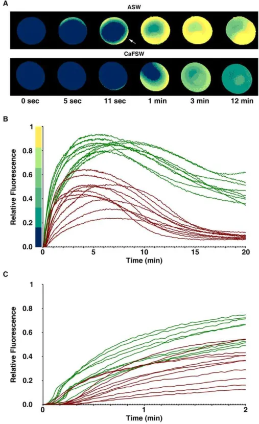
Live oocytes of Astropecten aranciacus were microinjected with Calcium Green and Rhodamine Red and examined with the CCD camera to monitor the changes of cytosolic Ca2+levels in response to
5mM ionomycin. In the artificial seawater containing 10 mM Ca2+
(ASW), oocytes promptly started to increase the intracellular Ca2+
level after the addition of ionomycin. The Ca2+
signal surpassed half the maximal level within 90 sec and arrived at the plateau by 5 min (Fig. 1B). The Ca2+
signals were initially prominent in the cortical area before spreading to the center of oocytes (Fig. 1A), and eight out
of nine oocytes displayed a sharp rise and fall of Ca2+
signals in the entire cortical regions subjacent to the plasma membrane (cortical flashes) at the initial stage of the Ca2+
rise (10.562.9 sec) (Fig. 1A, arrow). In the Ca2+
-free seawater (CaFSW), however, the Ca2+
response came out with a significantly longer latent period after the addition of ionomycin (16.265.3 sec, n = 11) in comparison with the oocytes in ASW (6.762.9 sec; n = 9,p,0.0001), as shown in Fig. 1C. In line with the idea that the short-lived (,2 sec) cortical flash represents a Ca2+
influx from outside [30,31], the same concentra-tion of ionomycin (5mM) produced no cortical flash (0 out of 11) in the CaFSW (Fig. 1A). In the absence of extracellular Ca2+
, the ionomycin-exposed oocytes released Ca2+
from the internal stores, but the amplitude of the Ca2+
peak was significantly lower (0.4660.12 RFU, n = 11) than that in the Ca2+
-containing seawater (0.8660.04 RFU, n = 9;p,0.0001). In addition, the Ca2+
level inside the oocytes exposed to 5mM ionomycin in CaFSW fell to the basal level within 15 min (Fig. 1B), whereas the oocytes in the ASW containing 5mM ionomycin displayed the declining Ca2+
signals still being maintained at much elevated levels (Fig. 1B). Hence, while both external and internal Ca2+
stores contribute to the Ca2+
rise in response to ionomycin, full-fledged and prolonged elevation of cytosolic Ca2+
requires external Ca2+
.
Ionomycin induces retraction of microvilli and fusion of cortical vesicles in the immature oocytes and eggs of starfish
Figure 1. Changes of intracellular Ca2+levels in the starfish oocytes exposed to ionomycin.A. aranciacusoocytes at the GV stage were
microinjected with Calcium Green/Rhodamine Red and subsequently exposed to 5mM ionomycin in artificial seawater in the presence (ASW) or
absence (CaFSW) of 10 mM Ca2+
. Ca2+
images were then captured with epifluorescence microscopy as described in Materials and Methods. (A) The pseudocolored images of Ca2+changes within the representative oocytes at several key time points. Indicated by an arrow is the cortical flash. (B) The
trajectories of the Ca2+
responses quantified at the entire cytoplasmic field. The Ca2+
responses in ASW and CaFSW are represented in green and brown curves, respectively. To compare the kinetics of the Ca2+
rises in ASW and CaFSW, the moment of the first detectable Ca2+
signal was set to t = 0 in panels A and B. (C) The initial response of the oocytes to ionomycin in ASW (green curves) and CaFSW (brown curves). To better illustrate the difference in the time lag before the first detectable Ca2+
Ionomycin induces drastic rearrangement of the actin cytoskeleton in the immature oocytes and eggs of starfish
As demolition of microvilli on the oocyte surface implied rapid depolymerization of microfilament bundles within, we examined the structural changes of the actin cytoskeleton inside a living oocyte after the ionomycin treatment. Under the confocal microscope, the GV stage oocytes microinjected with the Alexa Fluor 488-conjugated phalloidin revealed dramatic alteration of the actin-cytoskeleton after 5 minutes’ exposure to 5mM ionomy-cin (Fig. 3A), whereas F-actin structures of the untreated oocytes remained virtually unchanged within the same timeframe (not shown). Thus, during the period of ionomycin-induced Ca2+
rise, the actin cytoskeleton underwent highly accelerated rearrange-ment. Specifically, in the inner cytoplasm, the actin cytoskeleton became extensively polymerized, forming much longer and thicker microfilament bundles and patches. In the subplasmalemmal region, however, the typical actin filaments that are intimately associated with the plasma membrane (Fig. 3A, white arrow at t = 0) conspicuously disappeared by 5 min, indicating active depolymerization of the actin filaments in this subcellular domain. This striking structural rearrangement of the actin filaments in the
subplasmalemmal area and the inner cytoplasm were maintained for more than 20 min as long as ionomycin was kept in the media (data not shown). Hence, ionomycin had dual effects on the actin cytoskeleton remodeling: fast and extensive depolymerization in the subplasmalemmal region and the simultaneous polymerization in the inner cytoplasm. When the GV stage oocytes briefly (5 min) exposed to ionomycin were washed and switched to normal seawater for .1 h in the presence of 1-MA (10mM), the actin cytoskeletal structure was, to some extent, restored to the state of the control mature eggs (Fig. 3B). However, in these eggs, the organization of the F-actin bundles that are perpendicular to the plasma membrane in the control eggs (Fig. 3B arrowheads) were largely absent despite the restoration of the dense actin meshwork in the region. Interestingly, the borders of the large vesicles formed by ionomycin treatment were vested with a thick layer of actin fibers (Fig. 3B, arrows). Despite these structural changes in the ectoplasm, the eggs that had been pretreated with ionomycin at the GV stage underwent apparently normal meiotic maturation in the nucleus, displaying GV breakdown in the same time schedule (1 h) after 1-MA addition.
Brief exposure of the GV stage oocytes to ionomycin induces disruption and exocytosis of cortical granules
In parallel with the general propensity for actin depolymeriza-tion in the subplasmalemmal region (Fig. 3), the oocytes briefly (3– 5 min) exposed to 5mM ionomycin before the addition of
Figure 2. Morphological changes in the cortex of the starfish oocytes exposed to ionomycin.A. aranciacus oocytes at the GV stage were fixed in glutaraldehyde after 5 min incubation with 5mM
ionomycin in natural seawater. (A) Bright field view in the light microscope. GV = germinal vesicle. Scale bar = 50mm. (B) The magnified
views of the dot-lined rectangular areas in panel A. The same large vesicles in panel A were marked with yellow arrowheads. Note that cortical granules that appear as dark vesicles sized about 1mm (arrow)
had largely disappeared in the oocytes briefly exposed to ionomycin. Scale bar = 10mm. (C) TEM image of the same batch of oocytes
incubated in the absence (left) or present of 5mM ionomycin for 5 min.
Blue arrowheads indicate microvilli in cross-section. Red arrows indicate the white vesicles engulfing electron-dense cortical granules. Blue arrows, white vesicles at fusion; Scale bar = 10mm.
doi:10.1371/journal.pone.0039231.g002
Figure 3. Ionomycin induces rapid rearrangement of the actin cytoskeleton.(A) A living oocyte (A. aranciacus) microinjected with Alexa Fluor 488-conjugated phalloidin was exposed to 5mM ionomycin
and monitored under the confocal microscope. Note the continuous layer of the subplasmalemmal actin network delineating the plasma membrane before the ionomycin treatment (arrow, t = 0) had mostly disappeared within 5 min in the same oocytes. In contrast, the actin filaments in the inner cytoplasm formed bundles and became much thicker and longer. (B) After the brief exposure (5 min) to 5mM
ionomycin, the oocytes were switched to normal seawater without ionomycin and induced to undergo meiotic maturation in the presence of 10mM 1-MA for 1 hour. The orderly arranged actin filaments seen in
the control eggs (arrowheads) are mostly lost in the eggs briefly exposed to ionomycin at the GV stage. Instead, a thick layer of actin fibers surrounded the big fused white vesicles (arrows).
maturation hormone 1-MA displayed signs of cortical granules exocytosis and disruption. Whereas the control eggs displayed typical arrangement of cortical granules closely attached to the plasma membrane at the end of maturation (Fig. 4A, arrowheads), the eggs that had been briefly exposed to ionomycin before undergoing maturation process showed much reduced number of cortical granules. It appears that those cortical granules that were already at the vicinity of the plasma membrane underwent exocytosis (Fig. 4A, blue arrows), while the other ones that had not been yet translocated toward the plasma membrane were engulfed by white vesicles (Fig. 4A, red arrows). This is in sharp contrast with the control eggs in which cortical granules remain intact despite the presence of vicinal white vesicles. It is also noteworthy that the ultrastructure of the cortices of the oocytes exposed to 5mM ionomycin were not altered by further exposure to 1-MA
(compare the TEM images of the ionomycin-exposed oocyte and egg in Fig. 2C and 4A). Hence, the brief exposure to ionomycin inflicted largely irreversible structural changes in the subplasma-lemmal region. Indeed, despite the fact that the depolymerized actin reassembled the actin meshwork, albeit irregular, subjacent to the plasma membrane (Fig. 3B), the microvilli were still totally absent in these eggs (Fig. 4A). Interestingly, when exposed again to ionomycin after maturation, these eggs no longer produced intracellular Ca2+
release (Fig. 4B, brown curves) unlike the control eggs that had not been pretreated with 5mM ionomycin at
the GV stage (Fig. 4B, green curves). The same result was also obtained with the oocytes of a different starfish species, Patiria miniata(also known asAsterina miniata). As shown in Fig. 5A, a brief exposure to 5mM ionomycin at the GV stage prevented the
mature eggs from responding to ionomycin with intracellular Ca2+
increase. The failure of the second dose of ionomycin to evoke a Ca2+response is not due to the potential acidification of the eggs
and the reported inefficacy of ionomycin at low pH; unlike ionomycin that has no Ca2+
-complexing activity below pH 7.0, A23187 maintains its Ca2+ionophore activity at pH 5 to 10 [14].
With 40mM A23187, which induced intracellular Ca2+increase in
control eggs, the ionomycin-pretreated eggs at the GV stage did not mobilize Ca2+
(Fig. 5B). Taken together, these results suggest that the structural changes inflicted by the brief exposure to ionomycin, i.e. elimination of microfilaments-filled microvilli and disruption of the cortical granules and the fusion of white vesicles, are linked to the depletion of the ionomycin-sensitive Ca2+stores.
Ionomycin-sensitive Ca2+pool in the cortex of starfish eggs
When ionomycin-pretreated oocytes were switched back to normal seawater and induced to mature for 1 h in the presence of 1-MA, the cytosolic Ca2+returned to the basal level of the control
mature eggs that had not been exposed to ionomycin (data not shown). Thus, the mobilized Ca2+
had mostly returned or was at least en route to its original stores. Although not responding to the second exposure to ionomycin nor A23187 with Ca2+
release (Fig. 4 and 5), these ionomycin-pretreated eggs still responded to InsP3with a substantial release of Ca2+
(Fig. 6). The amplitude of the Ca2+ peak in these eggs (0.78
60.23 RFU, n = 10) was consistently lower (by 43%) than that of the control mature eggs (1.3860.20 RFU, n = 9;p,0.0001). Thus, the alteration of the egg cortex, e.g. retraction of microvilli and disruption of cortical granules (Fig. 4A), was associated with the reduction of the amplitude of the intracellular Ca2+
release in response to InsP3. However, the kinetics of the Ca2+
rise was not changed by the ionomycin treatment, as the time required to reach the Ca2+peak
remained virtually the same in the control (12.264.8 sec, n = 9) and the ionomycin-pretreated eggs (12.563.1 sec, n = 10;
p= 0.8681) (Fig. 6B, right panel). It is noteworthy that the eggs that had been briefly exposed to ionomycin at the GV stage did not elevate the vitelline layer despite the substantial increase of intracellular Ca2+
in all cases (Fig. 6C). In addition, for these eggs to produce the characteristic fertilization Ca2+
wave, repeated insemination was required in the majority of cases (Fig. 7A). Even when the ionomycin-pretreated eggs responded at once to a single sperm, the onset of the Ca2+
wave was much delayed (68.7664.3 sec after sperm addition, n = 3) in comparison with the control eggs in the same batch of experiment (19.269.2 sec, n = 10; p,0.05). Similar to the results obtained with uncaged InsP3, the peak amplitude of the sperm-induced Ca2+
transient in the eggs that had been exposed to ionomycin at the GV stage was about 35.9% lower (0.6760.23 RFU, n = 20) than that of the control eggs (1.0560.13 RFU n = 23;p,0.0001) (Fig. 7A and B). Again, however, the kinetics of the Ca2+
rise was not significantly altered, as the time required for reaching the peak in the ionomycin-pretreated eggs (148.4652.4, n = 20) was not signifi-cantly different from that in the control (128.0626.1 sec, n = 23;
p= 0.1061), implying that the intracellular mechanism that sustain the propagation of the Ca2+wave remained intact in these eggs
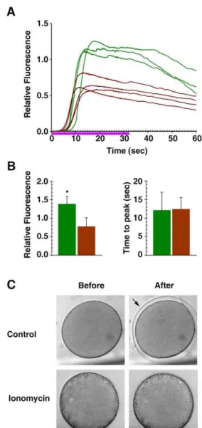
(Fig. 7B). Another conspicuous feature of the fertilization in the ionomycin-pretreated eggs is that the cortical flash that normally occurs at the very beginning of fertilization in this species [10] is often missing or substantially reduced in its amplitude. In five independent experiments, the incidence of the cortical flash in the ionomycin-pretreated eggs (30624%, n = 5) was greatly reduced from the average values in the corresponding control eggs (7662%, n = 5;p,0.05) (Fig. 7E and F). The amplitude of the cortical flash displayed by the ionomycin-pretreated eggs was also significantly reduced, corresponding to merely 47.8631.2% (n = 7) of the values in the control (100627.6%, n = 17; p= 0.0005) (Fig. 7D and E). Hence, the results on the cortical flash again indicated that the subcellular events highly restricted to the egg surface were substantially influenced by the pretreatment of the oocytes with ionomycin.
Ionomycin-pretreated eggs responding to InsP3 and fertilizing sperm (Fig. 6 and 7) but not to the second dose of ionomycin (Fig. 4 and 5) raises a possibility that the cation-complexing agent does not reach deep down to the inner cytoplasm of big cells like starfish eggs in bath incubation. To test if the lack of Ca2+
response is due to this, we have microinjected the eggs with ionomycin. To our surprise, not only the ionomycin-pretreated eggs but also the control eggs responded to the microinjected ionomycin with no significant Ca2+
increase with 50mM of ionomycin in the injection pipette. This is not due to a technical problem related to the simultaneous microinjection and Ca2+
detection because the parallel experiments with InsP3 microinjection (5mM in the injection pipette) using the same method produced high level of Ca2+response as was expected (Fig. 8). Microinjected ionomycin
did not induce any increase of Ca2+
even with 10 times higher dose (500mM in the injection pipette), and the same negative result was observed with the immature oocytes (not shown).
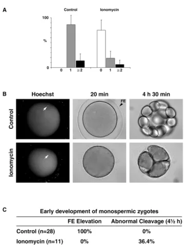
Fertilization and early development of the starfish eggs exposed to ionomycin at the GV stage
sperm that entered each egg following fertilization. In the given sperm concentration at fertilization, the control eggs responded mostly with monospermic fertilization (86.4618.8%, n = 4 inde-pendent experiments), but the ionomycin-pretreated eggs were mainly with no sperm entry (75.1620.5%, n = 4) (Fig. 9A). Thus, ionomycin-pretreated eggs appeared to have much less efficient gametes interaction that leads to sperm incorporation into the egg. For the control eggs, the frequency of monospermic fertilization far exceeds (6.4 fold) the frequency of polyspermic fertilization (13.6618.9%, P,0.001, Turkey’s test in one-way ANOVA). On the other hand, the ionomycin-pretreated eggs also displayed 3.1 fold higher rate of monospermy (18.5614.0%) over polyspermy (6.168.8%), but the difference was not statistically significant. Hence, a mechanism that favors monospermic fertilization is at work in the control eggs, but the same cannot be assuredly said for the ionomycin-pretreated eggs. However, the frequency of
polyspermy in the total egg population that displayed sperm entry was not significantly increased either by the ionomycin-pretreat-ment (20.8619.1%, n = 3), when compared with the control (16.1619.6%, n = 4; p= 0.7614). To test the effect of the ionomycin-pretreatment on early development, we followed the fate of the monospermic zygotes. Firstly, all the zygotes from the ionomycin-pretreated eggs underwent their developmental process without being shielded by the thick fertilization envelope that is normally seen in the control (Fig. 9B). Secondly, four out of eleven zygotes from the ionomycin-pretreated eggs, which had clearly displayed monospermy at fertilization, failed to develop normally at the early stages of the cell cleavage, whereas the zygotes from the monospermic control eggs all developed normally to the 16 cell stage (Fig. 9B and C). In particular, the failing zygotes from the ionomycin-pretreated eggs appeared to have a problem in creating a clear-cut cleavage furrow and thereby tended to form
Figure 4. Disruption of cortical granules and microvilli by the brief exposure to ionomycin leads to depletion of the ionomycin-sensitive Ca2+stores.A. aranciacusoocytes at the GV stage were exposed to 5
mM ionomycin for 3 min before switched to the media containing
1-MA. (A) After 1 h incubation, the mature eggs were fixed with glutaraldehyde and analyzed by TEM. Blue arrows indicate the remnant of the cortical granules that were extruded in the perivitelline space. Red arrows indicate fragments of cortical granules being engulfed by white vesicles. Scale bar = 10mm. (B) The same batch of oocytes were exposed to 5mM ionomycin for 3 min and switched to the fresh media containing 1-MA. After GV
breakdown, the mature eggs were microinjected with Calcium Green/Rhodamine Red and subsequently re-exposed to 5mM ionomycin (t = 0) to
monitor the Ca2+
response. The trajectory of intracellular Ca2+
levels in the eggs with or without (control) ionomycin pretreatment were depicted in brown and green curves, respectively.
amorphous cell clusters (Fig. 9B). On the other hand, all polyspermic zygotes in both cases failed to develop normally (not shown).
Ionomycin pretreatment disrupts the functionality of the cortical actin cytoskeleton
Actin is highly implicated in the process of egg activation at fertilization. Besides Ca2+, the actin cytoskeleton is a decisive factor
controlling exocytosis of vesicles, as has been suggested in various cell types [21,32–36]. Despite the substantial increase of intracel-lular Ca2+in response to InsP
3and fertilizing sperm, the ionomycin-pretreated eggs did not show any sign of vitelline layer elevation (Fig. 6 and 8), implying that the cortical changes induced by ionomycin at the GV stage of the oocytes interfered with the normal proceeding of egg activation at fertilization. In echinoderm eggs, it has been known that the subplasmalemmal actin fibers readily migrate centripetally at fertilization in concert with the elevation of the fertilization envelope [35–37]. In pursuit of a potential cause of the ineffective sperm entry and the tendency toward failing cleavage in the ionomycin-pretreated eggs, we have surveyed how the normal mobilization of the actin cytoskeleton is affected by the ionomycin pretreatment (Fig. 10). Whereas the control eggs at fertilization exhibited the orderly movement of the cortical actin fibers toward
the center, the eggs that had been pretreated with ionomycin at the GV stage displayed neither such coordinated translocation of the ectoplasmic actin fibers nor the elevation of the fertilization envelope (Fig. 10). Hence, the brief ionomycin treatment of the oocytes at the
Figure 5. The mature eggs pretreated with ionomycin at the GV stage respond to the second dose of ionomycin or A23187 with no intracellular Ca2+increase.P. miniataoocytes at the GV
stage were briefly exposed to 5mM ionomycin and switched to the
normal seawater containing 10mM 1-MA for 1 h and subsequently
challenged with the second dose of 5mM ionomycin (A) or 40mM
A23187 (B). In both cases, the green curves depict the Ca2+
response in the control eggs, and the brown ones the response of the eggs that had been briefly exposed to 5mM ionomycin at the GV stage.
doi:10.1371/journal.pone.0039231.g005
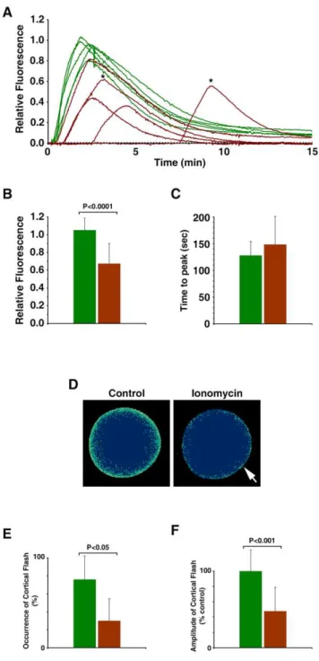
Figure 6. Ionomycin-exposed eggs with cortical granule disruption still respond to InsP3 with an intracellular Ca2+
release, but to a reduced extent. A. aranciacus oocytes were exposed to 5mM ionomycin for 5 min at the GV stage and
microinjected with caged InsP3and Calcium Green. The oocytes were
matured in the fresh seawater containing 10mM 1-MA and then
irradiated with UV to photoactivate the Ca2+
-mobilizing second messenger. (A) Results of one of the three independent experiments showing the trajectories of the quantified Ca2+
responses at the entire cytoplasmic field. Ca2+ responses in the control eggs and the eggs
briefly exposed to 5mM ionomycin at the GV stage were shown in
green and brown curves, respectively. Violet line indicates the duration of UV irradiation. (B) Summary of the data pooled from three independent batches of experiments comprising 3 or 4 microinjected eggs with (brown bars, n = 10) or without (control, green bars; n = 9) ionomycin pretreatment at the GV stage. The average amplitude (mean 6standard deviation, left histogram) and the time interval between the onset and the peak of the Ca2+
signals (right histogram) were depicted separately. Asterisk indicates a significant difference between the control and the ionomycin-pretreated eggs (p,0.0001). (C) Despite the substantial amount of Ca2+released, the ionomycin-pretreated eggs did
GV stage affected not only the structure of the actin cytoskeleton but also its functionality.
Deleterious effects of ionomycin on development To test if ionomycin affects egg activation and development of the early embryos, we have screened the fertilized eggs to select the monospermic zygotes that clearly demonstrated the entry of a single sperm and full elevation of the fertilization envelope. The monospermic zygotes were then exposed to 5mM ionomycin for
10 min, which is close to the dose generally used on a special occasion to activate the human or animal eggs in the practice of intracytoplsmic sperm injection (ICSI) [38,39]. The data pooled from three independent experiments indicated that, whereas 88.361.4% of the control zygotes developed normally 4 h after fertilization, only 24.8610.9% of the monospermic zygotes exposed to ionomycin developed normally at 4 h (p,0.001) (Fig. 11). In the latter case, the majority of the zygotes was either blocked at the first cell division or displayed a problem in cell cleavage at the later stages (Fig. 11A). After 3 days, in the given experimental condition where 68.3616.1% of the control embryos developed normally, merely 16.2619.3% of the monospermic zygotes exposed to ionomycin after fertilization displayed normal development (p,0.05) (Fig. 11).
Figure 7. Fertilization of the ionomycin-pretreated eggs with altered cortical structure. A. aranciacus oocytes were briefly exposed to ionomycin (5mM for 5 min) at the GV stage and
subsequently incubated in fresh seawater containing 10mM 1-MA.
The mature eggs were then inseminated. (A) Results of one of the five independent experiments. The trajectories of the quantified Ca2+
responses at the entire cytoplasmic field. Ca2+
responses in the control eggs and the eggs briefly exposed to 5mM ionomycin at the GV stage
were shown in green and brown curves, respectively. To illustrate the difference in the latent period before the Ca2+response, the moment of
the fertilizing sperm addition was set to t = 0. Asterisks indicate the Ca2+
peaks of the eggs that required a second addition of sperm (5 min after the first insemination). (B–F) Summary of the data pooled from five independent batches of experiments comprising 4 to 8 eggs with (brown bars, n = 20) or without (control, green bars; n = 23) ionomycin pretreatment at the GV stage. The average amplitude (mean 6 standard deviation) of the Ca2+
peaks and the time interval between the onset and the peak of the signals were plotted in panels BandC, respectively. (D) Pseudocolor images of the representative cortical
flashes in the control and the ionomycin-pretreated eggs (arrow) from the same batch of experiment. Ca2+ images were captured with
epifluorescence microscopy as described in the Materials and Methods. (E) Frequency of the detectable cortical flashes in the same five independent experiments. (F) Comparison of the amplitude of the cortical flashes. Data were normalized in reference to the average value of the control eggs in each batch of experiment.
doi:10.1371/journal.pone.0039231.g007
Figure 8. Microinjected ionomycin does not induce Ca2+
increase inside the starfish eggs. P. miniata oocytes were microinjected with Calcium Green and induced to mature in 10mM
1-MA for 1 h. Under the CCD camera, the mature eggs were microinjected with InsP3(without caging, 5mM in pipette tip), ionomycin (50mM), or
the injection buffer only. Results of one of the three independent experiments are shown. (A) Transmission views of the eggs 10 min after microinjection. (B) Quantified Ca2+
signals for InsP3 (blue curve),
Discussion
Since its first discovery in the 1970s, ionomycin has been widely used to increase intracellular Ca2+levels in cell biology
laborato-ries. On more rare occasions, it has also been used as a part of the standard protocol to activate the eggs that do not respond to ICSI in thein vitrofertilization clinics [8,38,39]. In this communication, by use of starfish oocytes and eggs, we have studied how egg activation and the early development are affected by the exposure to ionomycin at the GV stage or immediately after fertilization. We have found that ionomycin had detrimental effects on both egg activation and early development. First, we have demonstrated that 5mM ionomycin readily mobilizes intracellular Ca2+in the
oocytes. Owing to the large cell size, we were able to visualize the spatiotemporal changes of the intracellular Ca2+levels following
ionomycin treatment. With the removal of the external Ca2+in the
media, the peak of the Ca2+response by the immature oocytes
were merely 53.4% of the level in the Ca2+-containing seawater,
implying a significant contribution of the influx to the Ca2+
rise (Fig. 1). This discretion over the amplitude of Ca2+
response in the presence or absence of the external Ca2+
is largely alleviated in mature eggs, as the intracellular Ca2+
rise in CaFSW was as high as 79.0% (0.6060.18 RFU, n = 11) of that in the ASW (0.7660.12 RFU, n = 11) containing 10 mM Ca2+
. Hence, with meiotic maturation, mobilization of Ca2+
from internal stores seems to make more contribution to the net intracellular Ca2+
increase by ionomycin. We have then shown that the rapid increase of intracellular Ca2+was accompanied by accelerated rearrangement
of the actin cytoskeleton, which exhibited depolymerization near the plasma membrane with concomitant massive polymerization and bundling in the inner cytoplasm (Fig. 3A). This is one of the most striking examples of the actin cytoskeleton being rapidly and differentially rearranged by Ca2+increase in the distinct
subcel-lular domains, but the molecular mechanisms underlying the cytoskeletal changes and their physiological significance are yet to be known. Since a certain class of actin-binding protein, e.g. gelsolin, serves as a Ca2+sensor to transduce the Ca2+signals into
actin cytoskeletal remodeling [40], it is conceivable that similar pathway is at work to sever actin filaments or increase the barbed ends of actin filaments. Considering that actin is the most abundant (up to 300mM) Ca2+
-binding protein in the cytosol with unusually high binding affinity (Kd = 2–861029M for ATP-bound G-actin) [41,42], and that the Ca2+ion, once bound and
buried in the groove of F-actin, is virtually inaccessible for exchange [43], such an extensive hyperpolymerization of actin might serve as a mechanism to alleviate the cytosolic Ca2+
increase at least in part [44,45] and thereby function as a cell’s adaptive defense mechanism against the potential toxicity of prolonged Ca2+
upheaval. On the other hand, depolymerization of actin in the subplasmalemmal region might be involved in the regulation of store-operated Ca2+
entry that was reported to take place in ionomycin-exposed cells [17,46].
We have also shown that the ionomycin-induced massive Ca2+
increase and the drastic reorganization of the actin cytoskeleton are accompanied by the formation of the large white vesicles, which are often vested with actin filaments (Fig. 3B). This type of large (4–8mm) vesicle was not present in the TEM of the control eggs, but appeared with the concomitant loss of smaller vesicles of the same morphology which were always present in the cortex of control oocytes. Based on the intermediate twined structure resembling two white vesicles at fusion (Fig. 2C) found in the ionomycin-pretreated oocytes but not in the control ones, we concluded that the large white vesicles were formed by fusion with other vesicles. These vesicles often contained electron-dense materials that are likely to be fragments of cortical granules, sometimes multiple in numbers (Fig. 4A, red arrow on the left). The physiological role of this type of large vesicle is unknown, although an extreme case of growing autophagic vacuoles that fuse with other vesicles or engulf cortical granules were observed in the starfish oocytes (Pisaster ochraceus) undergoing atresia to give room to other growing oocytes [47]. The morphology of the ‘large white vesicles’ that we observed in ionomycin-pretreated oocytes are reminiscent of the ‘clear granules’ containing residual electron-dense materials in the normal eggs ofArbacia punctulata[48]. These clear granules (0.83mm in diameter) in the sea urchin eggs were
identified as acidocalcisome-like organelles containing large amount of Ca2+
and polyphosphate. While the large white vesicles in the ionomycin-pretreated oocytes might be indicative of a cell stress after cytoskeletal rearrangement and the prolonged elevation of intracellular Ca2+levels, yet to be resolved is the question about
the physiological role of the ‘small’ white vesicles seen in the control oocytes.
Figure 9. Fertilization and the early development of the ionomycin-pretreated eggs. (A) Astropecten aranciacus oocytes were pretreated with 5mM ionomycin at the GV stage and matured
with 1-MA for 1 h. Subsequently, eggs with or without (control) ionomycin pretreatment were fertilized with Hoechst 33342-stained sperm (see Materials and Methods). After 20 min, the number of the internalized sperm in each egg was counted, and the frequencies of monospermy (gray bars), polyspermy (black bars, sperm count.2), or the case with no evident sperm entry (white bar) were calculated in four independent experiments. (B) Developmental progress of the repre-sentative control and the ionomycin-pretreated eggs that clearly established monospermic sperm entry (arrows). (C) Summary of the fertilization envelope (FE) formation and the rate of abnormal development in the control and the ionomycin-pretreated eggs that established monospermic zygotes.
During the brief ionomycin-induced intracellular Ca2+
rise, cortical granules are either exocytosed or engulfed by large white vesicles (Fig. 2 and 4). Interestingly, these eggs did not respond to a second dose of ionomycin 1 h after 1-MA addition (Fig. 4B), raising the possibility that these lost vesicles are mainly responsible for ionomycin-sensitive Ca2+
increase. Our result is in line with the early studies in which Xenopus oocytes did not produce Ca2+
-dependent Cl2 currents in response to the second dose of ionomycin if the oocytes had been previously exposed to the suprathreshold (1mM) level of ionomycin [49]. This refractory period in Xenopus oocytes could be overcome by 50 min of washing, but our starfish eggs did not show any sign of recovery from the refractory phase 70 min after the removal of ionomycin in the media. Thus, we were not able to establish whether the refractory phase of ionomycin is reversible in starfish eggs. In contrast to the apparent loss of the responsiveness to ionomycin, the eggs of the same treatment (5 min exposure to ionomycin at the GV stage) still responded to InsP3and fertilizing sperm with substantial increase of intracellular Ca2+
(Fig. 6 and 7). However, the amplitude of the Ca2+
responses was consistently lower than in the ionomycin-untreated eggs. One possible explanation for the results is that the intracellular stores that directly contribute to the Ca2+
release in response to InsP3 or sperm were not fully recharged after the ionomycin treatment. Alternatively, we cannot rule out the possibility that the optimization of the Ca2+
-releasing mechanisms at the intracellular stores that take place during oocyte maturation [50,51] might have been interfered with as a result of the ionomycin pretreatment. Finally, considering that these eggs failed to respond to ionomycin and A23187 (Fig. 5), the reduction of the Ca2+
response might represent the fraction of the Ca2+
stores that are sensitive to the ionophores and InsP3but had been destroyed by the ionomycin pretreatment. However, it is difficult to pinpoint which structure represents the ionomycin-disrupted InsP3-sensitive Ca2+
stores. While the conventional Ca2+
stores such as the endoplasmic reticulum may be accountable for them, it is also possible that the loss of the microvilli, cortical granules, and white vesicles might be linked to the reduction of the InsP3-dependent Ca2+
rise. In theory, the reduction in
InsP3-dependent Ca2+
release may be ascribed to the loss of cortical granules or their associated structures. According to the early studies, cortical granules represent the major cortical Ca2+
stores, accounting for nearly 10% of total Ca2+
storage in sea urchin eggs [52]. However, the luminal Ca2+
of cortical granules is not likely to contribute to the intracellular Ca2+
increase at fertilization. Secretory vesicles of any types seem to lack InsP3receptors [53], and if cortical granules possess no Ca2+
-releasing channels, the luminal Ca2+
will be mostly extruded to the extracellular space at exocytosis. Nonetheless, early studies indicated that cortical granules of sea urchin eggs are tightly associated with fine network of endoplasmic reticulum that is endowed with ryanodine receptors [54]. If it also contain InsP3 receptors, it would be conceivable that the ionomycin-disrupted InsP3-sensitive Ca2+
store that contributes to Ca2+
signaling at fertilization may be the fine cisternae of endoplasmic reticulum that ensheaths the cortical granules, and not the cortical granules proper. However, the endoplasmic reticulum-enriched microsomal fraction of sea urchin egg cortices sometimes does not respond to InsP3with detectable Ca2+
release [55]. It would be interesting to know if this fraction corresponds to the stores operated by NAADP and two-pore channels on the acidic vesicles [56]. Thus, the contribution of the Ca2+
pool in the cortical granules or in their associated structures still remains an open question. Alternatively, the diminished Ca2+
response to InsP3 and fertilizing sperm might reflect a reduced efficacy of the eggs’ InsP3-dependent Ca2+
-releasing mechanism following the extensive rearrangement of the actin cytoskeleton, as has been repeatedly suggested in the starfish oocytes and eggs [21,35,57,58].
Our results in this communication raise a question about the way how the Ca2+
-selective ionophore ionomycin works inside the cell. Intuitively, ionomycin might infiltrate the cell as a mobile ion carrier and target all the Ca2+
stores to transport Ca2+
across the membrane dissipating the concentration gradient. However, ionomycin appeared to have specific targets in exerting its impact. Firstly, the membrane fusion was apparently restricted to cortical granules and white vesicles, while yolk platelets and other organelles at the same profundity of the egg cytoplasm were not
Figure 10. Ionomycin pretreatment disrupts the functionality of the cortical actin cytoskeleton.P. miniataoocytes were exposed to ionomycin (5mM for 8 min) at the GV stage and subsequently incubated in fresh seawater containing 10mM 1-MA. The mature eggs were then
microinjected with Alexa Fluor 488-phalloidin and inseminated to monitor with confocal microscopy the real-time changes of the actin cytoskeleton. In the control eggs, the orderly arranged subplasmalemmal actin filaments migrated centripetally (arrow), which was synchronized with the elevation of the fertilization envelope. In contrast, mature eggs previously exposed to 5mM at the GV stage failed to show such migration. At the right side of
each panel, the fluorescence image of F-actin in confocal microscopy was merged with the transmission view of the same specimen. Images of the eggs before and after fertilization (13 min post-insemination) were taken from the same individual eggs.
disrupted or modified by ionomycin. One possible explanation is that vesicles specializing in secretion, e.g. cortical granules and white vesicles that are normally exocytosed during fertilization (Data S1A), might be specifically vested with Ca2+
-sensing proteins that interact with the cytoskeleton and facilitate membrane fusion, as was exemplified by synaptotagmin in synaptic vesicles [59]. Implying a role of actin cytoskeleton in the process of membrane fusion, the large white vesicles are often delineated by fluorescent probes for F-actin (Fig. 3B). Hence, it is conceivable that prolonged elevation of intracellular Ca2+
by ionomycin may have induced deregulated membrane fusion between these vesicles in a mechanism that involves actin. Alternatively, since the action of ionomycin is highly dependent on pH [14], such specificity might be related to the subtle difference in the luminal pH of these vesicles and organelles [60]. Secondly, the majority of InsP3-sensitive stores in the cortex were neither destroyed by the first
exposure to 5mM ionomycin nor responsive to the second dose of
ionomycin with Ca2+
release. While we could not rule out that this might reflect the potential difficulties of ionomycin in diffusing inside the inner cytoplasm, we have found that ionomycin does not work when it is microinjected into the eggs (Fig. 8). Being permeant to cell membrane, ionomycin is virtually always added to the media to induce intracellular Ca2+
increase. Since cytosolic Ca2+
concentration is extremely low (1027M), our result might suggest that, to transport Ca2+
ions across the membrane, ionomycin should be added to the side of higher Ca2+
concentration. Alternatively, Ca2+
is transported into the cell from outside by ionomycin and this priming Ca2+
increase may ignite further Ca2+
release in an indirect mechanism either by inducing calcium-induced calcium release (CICR) or activating Ca2+-dependent enzymes such as phospholipase C. However, we
have seen no evidence of PIP2 hydrolysis after ionomycin-induced Ca2+
increase inside starfish oocytes in an assay using PH-GFP (data not shown), which is at variance with the results obtained with smaller cells such as fibroblasts [61] and starfish eggs at fertilization [36]. Thus, this intriguing fundamental issue of how the ionophore ionomycin works inside the cells merits further investigation also in other experimental systems.
Finally, the results of our study indicate that the Ca2+
ionophore ionomycin might have a detrimental effect on both egg activation and the early embryonic development. Starfish eggs that had been briefly exposed to ionomycin at the GV stage may still undergo meiotic maturation process at least in the nucleus, but the resulting eggs displayed several problems at fertilization. First, these eggs had difficulties in producing effective sperm interaction at fertilization. While generation of the initial Ca2+
spot underneath the egg plasma membrane is considered as one of the indices for a meaningful sperm-egg interaction at fertilization [10], the onset of the Ca2+
waves in the ionomycin-pretreated eggs required more than three times longer latent period than in the control (Fig. 7A). In line with that, we have shown that the eggs pretreated with ionomycin largely lack microvilli on their surface (Fig. 2 and 4), which are known to play important roles in gametes interaction [62]. Having already lost cortical granules and other secretory vesicles (Fig. 2 and 4), the zygotes from the ionomycin-pretreated eggs failed to elevate the fertilization envelope in all cases (Fig. 9B). Furthermore, these fertilized eggs did not exhibit the characteristic centripetal migration of the cortical actin fibers that is typically seen during egg activation (Fig. 10). Hence, the ionomycin-pretreated eggs at the GV stage lacked several important hallmarks of the echinoderm egg activation. Upon sperm incorporation, the monospermic zygotes that derived from these eggs displayed further abnormality during development. Unlike the control zygotes that all normally progressed beyond the 4–16 egg stages by 4.5 h after insemination, the monospermic zygotes from ionomycin-pretreated eggs exhibited signs of failure in cell cleavage (Fig. 9B and C). Whether this abnormal development in the embryos deriving from the ionomycin-pretreated eggs is simply due to the lack of the fertilization envelope, which is thought to be protective of the developing embryo, or ascribable to the drastic alteration of the egg ectoplasm (Fig. 2 and 4) is a matter of dispute. However, it should be reminded that the monospermic zygotes that had been exposed to the same dose of ionomycin after fertilization (rather than at the GV stage) had a fully elevated fertilization envelope like the control eggs but displayed a comparable failure rate for cell cleavage and development (Fig. 11). Thus, the fertilization envelope does not seem to act as a chemical barrier to protect the embryo, and the detrimental effect of ionomycin is likely to be caused by the changes in the egg ectoplasm. There might be several different contributing factors to
Figure 11. Deleterious effects of ionomycin on development. Mature eggs of Astropecten aranciacus were fertilized with Hoechst 33342-prestained sperm. After 20 min, zygotes displaying clear signs of monospermy and fully elevated fertilization envelope were exposed either to 5mM ionomycin or to 0.1% DMSO (control, vehicle) for 10 min
this, but we have focused on the actin cytoskeleton. Being dynamically regulated upon cell signals, the actin cytoskeleton is indeed implicated in many aspects of fertilization and early development [45]. As demonstrated in sea urchin, the cell cleavage of zygotes heavily depends on the regulation of the actin cytoskeleton [63]. The actin filaments in the egg cortex even extend through the perivitelline space of the fertilization envelope [36]. Hence, actin is intimately implicated in fertilization and in the early stages of development. We have shown that the elevated rate of aberrant development in the ionomycin-treated eggs is preceded by the drastic alteration of the actin cytoskeleton in the egg cortex and the retraction of microvilli (Fig. 2–4), as well as the failure of the centripetal migration of the cortical actin filaments (Fig. 10). Finally, it bears an emphasis that it is difficult to extrapolate our findings to make a direct comparison with the similar use of ionomycin in the rare case of the medical practice of ICSI in thein vitrofertilization clinics [64]. The assessment on the safety of such procedure awaits more detailed studies using the zygotes being produced with the same procedure, e.g. intracyto-plasmic sperm injection.
Materials and Methods
Ethics statements
No specific permits were required for the described field studies for starfish. Field studies did not involve endangered or protected species nor private territory for sample collection.
Preparation of oocytes and reagents
Captured starfish (Astropecten aranciacusandPisaster miniata) were maintained in circulating cold seawater (16uC). The female gonads were dissected from the central dorsal area near the arms and transferred to the filter-sterilized cold seawater. Free individual oocytes were isolated by repeatedly passing through gauze and rinsing in cold filtered seawater. Oocytes were collected by low speed (,1,000 rpm) sedimentation in each step. Immature oocytes obtained in this way were marked by the presence of the large nucleus (germinal vesicles). Ionomycin and InsP3were purchased from Invitrogen. All other chemicals were purchased from Sigma-Aldrich unless stated otherwise, and all solutions were prepared following the manufacturers’ instruction. Certain experiments were performed in artificial seawater (ASW: 490 mM NaCl, 8 mM KCl, 10 mM CaCl2, 12 mM MgCl2, 2.5 mM NaHCO3, pH 8.0) or Ca2+-free seawater (500 mM NaCl, 8 mM KCl,
12 mM MgCl2, 2.5 mM NaHCO3, 2 mM EGTA, pH 8.0). Hoechst 33342 was purchased from Sigma and the final concentration of 5mM was used to stain sperm for 1 min before
insemination. The number of egg-incorporated sperm was counted in epifluorescence microscopy.
Microinjection, caged compounds, and video imaging Microinjection of the oocytes was performed with an air-pressure Transjector (Eppendorf), following the procedure that was previously described in detail [36]. Fluorescent calcium dye (Calcium Green) conjugated with 10 kDa dextran (Molecular Probes) was used in 500mM (pipette concentration). The injection
buffer contained 10 mM Hepes and 100 mM L-Asp at pH 7.0. To activate the caged InsP3(Molecular Probes), microinjected eggs were irradiated with 330 nm UV light by using the computer-controlled shutter system Lambda 10-2 (Sutter Instruments, Co., Novato, CA). Alexa Fluor 488-phalloidin (Molecular Probes) was injected as described previously [35]. All Ca2+ imaging work in
this study was done by use of epifluorescence microscopy. Cytosolic Ca2+
changes were detected using a cooled CCD
camera (MicroMax, Princeton Instruments, Inc., Trenton, NJ) mounted on a Zeiss Axiovert 200 microscope with a Plan-Neofluar 20x/0.50 objective. The quantified Ca2+
signal at a given time point was normalized to the baseline fluorescence (F0) following the formula Frel= [F2F0]/F0, where F represents the average fluorescence level of the entire oocyte. Fluorescence of Ca2+
images were analyzed with the MetaMorph Imaging System software (Universal Imaging Corporation, West Chester, PA).
Statistical analysis
The numerical MetaMorph data were compiled and analyzed with Excel of Microsoft Office 2003. The average and variation of the data were reported as ‘mean6standard deviation (SD)’ in all cases. The paired t-test and the one-way ANOVA were performed by use of Prism 3.0 (GraphPad Software, La Jolla, CA, USA), and the P-values smaller than 0.05 (P,0.05) were considered statistically significant.
Light and Transmission electron microscopy (TEM) Starfish oocytes and eggs to be analyzed following the ionomycin and 1-MA treatment were fixed together with proper control samples in filtered seawater containing 1% glutaraldehyde (pH 8.0) for 1 h at room temperature and then post-fixed with 1% osmium tetroxide for 1 h. Specimens were dehydrated in increasing concentrations of alcohol and embedded in EPON 812. The polymerized resin containing the fixed material was sectioned in two ways. Semi-thin (1 micron) sections were stained with toluidine blue and examined by light microscopy with a Zeiss Axiovert 200 microscope, and the images were captured by the CCD camera. For TEM, ultrathin sections were stained with 2% uranyl acetate and 0.2% lead citrate, and examined with a LEO 912 AB energy filter transmission electron microscope.
Confocal Microscopy
GV stage oocytes and eggs microinjected with Alexa Fluor 488– conjugated phalloidin were viewed with a Zeiss LSM 510 META Laser Scanning Confocal Microscope (Jena, Germany) with excitation at 488 nm and emission at 500/555 nm. A Plan-Neofluar 25x/0.80 objective water lens was used to produce optical slices from the specimens. The images were captured as digital computer files and examination of the fluorescence was performed using MetaMorph image analysis software.
Supporting Information
Data S1 Translocation of white vesicles during egg maturation and activation. (A) Light microscope images of immature oocytes of A. aranciacus show that white vesicles are mainly located in the cortex but not tightly packed underneath the plasma membrane (white arrows). In sharp contrast, white vesicles in the mature eggs are closely associated with plasma membrane just like cortical granules. Note that the white vesicles are largely eliminated in the activated egg that underwent massive exocytosis and the elevation of the vitelline layer. (B) TEM image of white vesicles containing remnants of fibrillary contents in the normal eggs ofAstropecten aranciacus at fertilization. As a result of cortical granules (black arrow) exocytosis and microvilli (gray arrow) elongation, the fertilization envelope was fully elevated (labeled as VL, vitelline layer). PS, perivitelline space. The white vesicle fusing with the plasma membrane is marked by a white arrow. Scale bar 1mm.
Acknowledgments
We are grateful to the staff at the Marine Resources (P. Cirino and D. Caramiello) and the Electron Microscopy (R. Graziano and F. Iamunno) sectors of the SZN for their technical support.
Author Contributions
Conceived and designed the experiments: JTC LS. Performed the experiments: FV JTC EG LS. Analyzed the data: JTC GG FV LS. Contributed reagents/materials/analysis tools: LS. Wrote the paper: JTC FV LS.
References
1. Trimmer JS, Vacquier VD (1986) Activation of sea urchin gametes. Annu Rev Cell Biol 2: 1–26.
2. Epel D (1990) The initiation of development at fertilization. Cell Differ Dev 29: 1–12.
3. Ridgway EB, Gilkey JC, Jaffe LF (1977) Free calcium increases explosively in activating medaka eggs. Proc Natl Acad U S A 74: 623–627.
4. Miyazaki S (1991) Repetitive calcium transients in hamster oocytes. Cell Calcium 12: 205–216.
5. Jaffe LF (2002) On the conservation of fast calcium wave speeds. Cell Calcium 32: 217–229.
6. Steinhardt RA, Epel D (1974) Activation of sea urchin eggs by a calcium ionophore. Proc Natl Acad Sci U S A 71: 1915–1919.
7. Steinhardt RA, Epel D, Carroll EJ, Yanagimachi R (1974) Is calcium ionophore a universal activator for unfertilized eggs? Nature 252: 41–43.
8. Ciapa B, Arnoult C (2011) Could modifications of signalling pathways activated after ICSI induce a potential risk of epigenetic defects? Int J Dev Biol 55: 143– 152.
9. Santella L, Kyozuka K, Genazzani AA, De Riso L, Carafoli E (2000) Nicotinic acid adenine dinucleotide phosphate-induced Ca2+
release. Interactions among distinct Ca2+
mobilizing mechanisms in starfish oocytes. J Biol Chem 275: 8301– 8306.
10. Santella L, Lim D, Moccia F (2004) Calcium and fertilization: the beginning of life. Trends Biochem Sci 29: 400–408.
11. Whitaker M (2006) Calcium at fertilization and in early development. Physiol Rev 86: 25–88.
12. Lim D, Kyozuka K, Gragnaniello G, Carafoli E, Santella L (2001) NAADP+
initiates the Ca2+
response during fertilization of starfish oocytes. FASEB J 15: 2257–2267.
13. Liu WC, Slusarchyk DS, Astle G, Trejo WH, Brown WE, Meyers E (1978) Ionomycin, a new polyether antibiotic. J Antibiot (Tokyo) 31: 815–819. 14. Liu C, Hermann TE (1978) Characterization of ionomycin as a calcium
ionophore. J Biol Chem 253: 5892–5894.
15. Erdahl WL, Chapman CJ, Taylor RW, Pfeiffer DR (1994) Ca2+
transport properties of ionophores A23187, ionomycin, and 4-BrA23187 in a well defined model system. Biophys J 66: 1678–1693.
16. Kauffman RF, Taylor RW, Pfeiffer DR (1980) Cation transport and specificity of ionomycin – comparison with ionophore A23187 in rat liver mitochondria. J Biol Chem 255: 2735–2739.
17. Morgan AJ, Jacob R (1994) Ionomycin enhances Ca2+
influx by stimulating store-regulated cation entry and not by a direct action at the plasma membrane. Biochem J 300: 665–672.
18. Mason MJ, Grinstein S (1993) Ionomycin activates electrogenic Ca2+
influx in rat thymic lymphocytes. Biochem J 296: 22–39.
19. Moreau M, Guerrier P, Doree M, Ashley CC (1978) Hormone-induced release of intracellular Ca2+
triggers meiosis in starfish oocytes. Nature 272: 251–253. 20. Santella L, Kyozuka K (1994) Reinitiation of meiosis in starfish oocytes requires
an increase in nuclear Ca2+. Biochem Biophys Res Commun 203: 674–680. 21. Kyozuka K, Chun JT, Puppo A, Gragnaniello G, Garante E, et al. (2008) Actin
cytoskeleton modulates calcium signaling during maturation of starfish oocytes. Dev Biol 320: 426–435.
22. Santella L, De Riso L, Gragnaniello G, Kyozuka K (1999) Cortical granule translocation during maturation of starfish oocytes requires cytoskeletal rearrangement triggered by InsP3-mediated Ca
2+
release. Exp Cell Res 248: 567–574.
23. Wessel GM, Conner SD, Berg L (2002) Cortical granule translocation is microfilament mediated and linked to meiotic maturation in the sea urchin oocyte. Development 129: 4315–4325.
24. Schuetz AW (1975) Cytoplasmic activation of starfish oocytes by sperm and divalent ionophore A-23187. J Cell Biol 66: 86–94.
25. Kater SB, Mattson MP, Cohan C, Connor J (1988) Calcium regulation of the neuronal growth cone. Trends Neurosci 11: 315–321.
26. Forscher P (1989) Calcium and polyphosphoinositide control of cytoskeletal dynamics. Trends Neurosci 12: 468–474.
27. Spira ME, Oren R, Dormann A, Ilouz N, Lev S (2001) Calcium, protease activation, and cytoskeleton remodeling underlie growth cone formation and neuronal regeneration. Cell Mol Neurobiol 21: 591–604.
28. Hirohashi N, Harada K, Chiba K (2008) Hormone-induced cortical maturation ensures the slow block to polyspermy and does not couple with meiotic maturation in starfish. Dev Biol 318: 194–202.
29. Terasaki M, Runft L (2010) Two-stage dependence for 1-methyladenine induced reinitiation of meiotic maturation in starfish oocytes. Exp Cell Res 316: 2654– 2663.
30. Moccia F, Lim D, Nusco GA, Ercolano E, Santella L (2003) NAADP activates a Ca2+
current that is dependent on F-actin cytoskeleton. FASEB J 17: 1907–1909.
31. Churchill GC, O’Neill JS, Masgrau R, Patel S, Thomas JM, et al. (2003) Sperm deliver a new second messenger: NAADP. Curr Biol 13: 125–128.
32. Muallem S, Kwiatkowska K, Xu X, Yin HL (1995) Actin filament disassembly is a sufficient final trigger for exocytosis in nonexcitable cells. J Cell Biol 128: 589– 598.
33. Gasman S, Chasserot-Golaz S, Malacombe M, Way M, Bader MF (2004) Regulated exocytosis in neuroendocrine cells: a role for subplasmalemmal Cdc42/N-WASP-induced actin filaments. Mol Biol Cell 15: 520–531. 34. Malacombe M, Bader MF, Gasman S (2006) Exocytosis in neuroendocrine cells:
new tasks for actin. Biochim Biophys Acta 1763: 1175–1183.
35. Puppo A, Chun JT, Gragnaniello G, Garante E, Santella L (2008) Alteration of the cortical actin cytoskeleton deregulates Ca2+
signaling, monospermic fertilization, and sperm entry. PLoS One 3: e3588.
36. Chun JT, Puppo A, Vasilev F, Gragnaniello G, Garante E, et al. (2010) The biphasic increase of PIP2 in the fertilized eggs of starfish: new roles in actin polymerization and Ca2+
signaling. PLoS One 5: e14100.
37. Terasaki M (1996) Actin filament translocations in sea urchin eggs. Cell Motil Cytoskeleton 34: 48–56.
38. Terada Y, Hasegawa H, Takahashi A, Ugajin T, Yaegashi N, et al. (2009) Successful pregnancy after oocyte activation by a calcium ionophore for a patient with recurrent intracytoplasmic sperm injection failure, with an assessment of oocyte activation and sperm centrosomal function using bovine eggs. Fertil Steril 91: 935.e11–14.
39. Heytens E, Schmitt-John T, Moser JM, Jensen NM, Soleimani R, et al. (2010) Reduced fertilization after ICSI and abnormal phospholipase C zeta presence in spermatozoa from the wobbler mouse. Reprod Biomed Online 21: 742–749. 40. Burtnick LD, Urosev D, Irobi E, Narayan K, Robinson RC (2004) Structure of
the N-terminal half of gelsolin bound to actin: roles in severing, apoptosis and FAF. EMBO J 23: 2713–2722.
41. Carlier MF, Pantaloni D, Korn ED (1986) Fluorescence measurements of the binding of cations to high-affinity and low-affinity sites on ATP-G-actin. J Biol Chem 261: 10778–10784.
42. Gershman LC, Selden LA, Estes JE (1986) High affinity binding of divalent cation to actin monomer is much stronger than previously reported. Biochem Biophys Res Commun 135: 607–614.
43. Kasai M, Oosawa F (1969) Behavior of divalent cations and nucleotides bound to F-actin. Biochim Biophys Acta 172: 300–310.
44. Lange K, Gartzke J (2006) F-actin-based Ca signaling-a critical comparison with the current concept of Ca signaling. J Cell Physiol 209: 270–287.
45. Santella L, Chun JT (2011) Actin, more than just a housekeeping protein at the scene of fertilization. Sci China Life Sci 54: 733–743.
46. Darbellay B, Arnaudeau S, Bader CR, Konig S, Bernheim L (2011) STIM1L is a new actin-binding splice variant involved in fast repetitive Ca2+
release. J Cell Biol 194: 335–346.
47. Reunova AA, Crawford BJ (2010) An ultrastructural study of oocyte atresia in the starfishPisaster ochraceus. Zoology 113: 295–300.
48. Ramos IB, Miranda K, Pace DA, Verbist KC, Lin FY, et al. (2010) Calcium-and polyphosphate-containing acidic granules of sea urchin eggs are similar to acidocalcisomes, but are not the targets for NAADP. Biochem J 429: 485–495. 49. Yoshida S, Plant S (1992) Mechanism of release of Ca2+from intracellular stores in response to ionomycin in oocytes of the frog Xenopus laevis. J Physiol 458: 307–318.
50. Chiba K, Kado RT, Jaffe LA (1990) Development of calcium release mechanisms during starfish oocyte maturation. Dev Biol 140: 300–306. 51. Lim D, Ercolano E, Kyozuka K, Nusco GA, Moccia F, et al. (2003) The
M-phase-promoting factor modulates the sensitivity of the Ca2+stores to inositol 1,4,5-trisphosphate via the actin cytoskeleton. J Biol Chem 278: 42505–42514. 52. Gillot I, Ciapa B, Payan P, Sardet C (1991) The calcium content of cortical granules and the loss of calcium from sea urchin eggs at fertilization. Dev Biol 146: 396–405.
53. Rizzuto R, Pozzan T (2006) Microdomains of intracellular Ca2+: molecular determinants and functional consequences. Physiol Rev 86: 369–408. 54. McPherson SM, McPherson PS, Mathews L, Campbell KP, Longo FJ (1992).
Cortical localization of a calcium release channel in sea urchin eggs. J Cell Biol 116: 1111–1121.
55. Oberdorf JA, Head JF, Kaminer B (1986) Calcium uptake and release by isolated cortices and microsomes from the unfertilized egg of the sea urchin Strongylocentrotus droebachiensis. J Cell Biol 102: 2205–2210.
56. Calcraft PJ, Ruas M, Pan Z, Cheng X, Arredouani A, et al. (2009) NAADP mobilizes calcium from acidic organelles through two-pore channels. Nature 459: 596–600.
58. Chun JT, Santella L (2009) Roles of the actin-binding proteins in intracellular Ca2+
signalling. Acta Physiol 195: 61–70.
59. Lee HK, Yang Y, Su Z, Hyeon C, Lee TS, et al. (2010) Dynamic Ca2+
-dependent stimulation of vesicle fusion by membrane-anchored synaptotagmin. Science 328: 760–763.
60. Morgan AJ (2011) Sea urchin eggs in the acid reign. Cell Calcium 50: 147–156. 61. van Rheenen J, Jalink K (2002) Agonist-induced PIP2 hydrolysis inhibits cortical actin dynamics: regulation at a global but not at a micrometer scale. Mol Biol Cell 13: 3257–3267.
62. Runge KE, Evans JE, He ZY, Gupta S, McDonald KL, et al. (2007) Oocyte CD9 is enriched on the microvillar membrane and required for normal microvillar shape and distribution. Dev Biol 304: 317–325.
63. Dale B, De Santis A (1981) The effect of cytochalasin B and D on the fertilization of sea urchins. Dev Biol. 83:232–7.