Insulin impairs the maturation of
chondrocytes
in vitro
1Departamento de Patologia, Universidade Federal Fluminense,
Niterói, RJ, Brasil
2Programa Avançado de Biologia Celular Aplicada à Medicina,
Hospital Universitário Clementino Fraga Filho, and 3Departamento de Histologia e Embriologia,
Universidade Federal do Rio de Janeiro, Rio de Janeiro, RJ, Brasil E.S. Torres1,
C.V. Andrade1, E.C. Fonseca1, M.A. Mello1 and M.E.L. Duarte2,3
Abstract
The precise nature of hormones and growth factors directly respon-sible for cartilage maturation is still largely unclear. Since longitudinal bone growth occurs through endochondral bone formation, excess or deficiency of most hormones and growth factors strongly influences final adult height. The structure and composition of the cartilaginous extracellular matrix have a critical role in regulating the behavior of growth plate chondrocytes. Therefore, the maintenance of the three-dimensional cell-matrix interaction is necessary to study the influence of individual signaling molecules on chondrogenesis, cartilage matu-ration and calcification. To investigate the effects of insulin on both proliferation and induction of hypertrophy in chondrocytes in vitro we used high-density micromass cultures of chick embryonic limb mes-enchymal cells. Culture medium was supplemented with 1% FCS + 60 ng/ml (0.01 µM) insulin and cultures were harvested at regular time points for later analysis. Proliferating cell nuclear antigen immunore-activity was widely detected in insulin-treated cultures and persisted until day 21 and [3H]-thymidine uptake was highest on day 14. While
apoptosis increased in control cultures as a function of culture time, terminal deoxynucleotidyl transferase-mediated dUTP nick end label-ing (TUNEL)-labeled cells were markedly reduced in the presence of insulin. Type II collagen production, alkaline phosphatase activity and cell size were also lower in insulin-treated cultures. Our results indicate that under the influence of 60 ng/ml insulin, chick chondro-cytes maintain their proliferative potential but do not become hyper-trophic, suggesting that insulin can affect the regulation of chondro-cyte maturation and hypertrophy, possibly through an antiapoptotic effect.
Correspondence
M.E.L. Duarte PABCAM, UFRJ
Hospital Universitário, 4º andar Av. Brigadeiro Trompowsky, s/n Ilha do Fundão
21941-970 Rio de Janeiro, RJ Brasil
Fax: +55-21-2610-4593 E-mail: eugenia@urbi.com.br
Research supported in part by CAPES, CNPq and FINEP.
Received September 4, 2002 Accepted April 24, 2003
Key words
•Chondrocytes •Insulin
•Micromass cultures •Apoptosis
Introduction
A large number of growth factors, cyto-kines and hormones are implicated in the regulation of endochondral ossification (1). In this highly regulated process,
mesenchy-mal cells differentiate into chondrocytes that proliferate, mature, become hypertrophic and deposit large amounts of extracellular matrix (ECM) which is finally calcified, and re-placed by bone (2).
to reproduce the events that occur during endochondral ossification (3-6). Neverthe-less, the maintenance of the three-dimen-sional cell-matrix interaction is needed for chondrogenesis, cartilage maturation and cal-cification. Chondrocytes kept in monolayer cultures gradually lose the cartilage pheno-type and begin to dedifferentiate, resem-bling mesenchymal cells (7) and producing molecules not characteristic of cartilage (8). Ahrens et al. (9) developed an in vitro
system of high-density micromass cultures of chick embryonic limb mesenchyme. This system has been widely used to study chon-drogenesis (10,11) and more recently (12,13) was used to study the process of cartilage maturation since it permits the observation of the continuous process of differentiation, maturation and calcification.
The identification of insulin and insulin receptors in the chick embryo within 48 h after fertilization demonstrates that, at physi-ological concentrations, insulin may act as a mitogen for some fetal cell types (14,15). At low concentrations, insulin increases param-eters of growth, metabolism and muscle dif-ferentiation in 4-day-old chick embryos (16). Insulin has been shown to have an in vitro
stimulatory effect on chondrocyte prolifera-tion and activity and an in vivo effect on growth plate width (17). Also, situations of hypoinsulinemia (insulin-dependent diabe-tes mellitus) and hyperinsulinemia with or without non-insulin-dependent diabetes con-siderably alter normal bone physiology (18). In the present study we have investigated the in vitro effects of insulin on chondrocyte proliferation, maturation, hypertrophy and apoptosis in the established system of high-density micromass cultures of chicken limb bud mesenchymal cells.
Material and Methods
Micromass culture
Limb buds were removed from
Ham-burger-Hamilton stage 23-24 chicken em-bryos (19) and the starting cell population consisting of mesenchymal cells was placed in calcium-magnesium-free saline glucose (CMFSG) solution. The buds were dissoci-ated enzymatically in 0.1% (w/v) collagen-ase (Sigma, St. Louis, MO, USA) and 0.1% (w/v) trypsin (Sigma) in CMFSG. The cell suspension was filtered through a 20-µm nitex filter and centrifuged for 10 min at 600 g. The pellets were resuspended in CMFSG with 10% FCS and cell density was adjusted to 25-30 × 106 cells/ml. The cells were plated as a micromass (10 µl of the cell suspension) onto 24-well plates and allowed to attach for 1-2 h at 37ºC in a 5% CO2 atmosphere. After incubation the cells were fed with DMEM/ F-12 medium (Sigma) containing 100 U/ml penicillin, 100 µg/ml streptomycin and 10% FCS. From day 2 on, the medium was supple-mented with 2.5 mM ß-glycerophosphate and 25 µg/ml (0.15 mM) ascorbate (Sigma). To support chondrogenesis, before starting the treatment with insulin, all cultures were fed until day 3 with medium containing 10% FCS. From day 4 on the cultures were treated with 1% FCS and 1% FCS + 60 ng/ml (0.01 µM) insulin, respectively. Cultures were main-tained for up to 21 days and were harvested at regular time points (days 7, 14 and 21) for analysis of growth and maturation status.
Fixation and embedding
The micromass cultures were rinsed three times in PBS and fixed in 4% (v/v) parafor-maldehyde for 1 h. Subsequently they were released gently from the well with a cell scraper, dehydrated in graded series of etha-nol, cleared in xylene and embedded in par-affin. Specimens were cut into 4-µm sec-tions for all histological procedures.
Phenotypic characterization of the cells in culture
were stained with hematoxylin-eosin to ana-lyze cell morphology and with Alcian blue, pH 1.0 (20), to detect sulfated glycosami-noglycans in the cartilage matrix.
Type II collagen expression. Collagen type II deposition was detected by immuno-histochemistry with a polyclonal rabbit anti-bovine type II collagen antibody (Chemicon, Temecula, CA, USA) at 1:150 dilution for 30 min at 37ºC. The primary antibody was detected with biotinylated secondary rabbit anti-mouse IgG, 1:200 (Dako, Carpinteria, CA, USA), whereas negative control sec-tions were stained using PBS containing 1% BSA instead of the primary antibody. Nor-mal hyaline cartilage slides were used as positive control.
Cell proliferation
[3H]-thymidine uptake. Cultures were
in-cubated for 24 h with medium containing 0.25 µCi/ml [3H]-thymidine (TRK 418 Amer-sham Pharmacia, Arlington Heights, IL, USA). The radioactivity incorporated was measured by liquid scintillation counting using a Tri-Carb 2100 TR liquid scintillation analyzer (Perkin Elmer, Boston, MA, USA) for 2 min.
Proliferating cell nuclear antigen (PCNA)
expression. Sections were incubated over-night with anti-mouse PCNA (Dako) as the primary antibody at 1:3000 dilution in PBS-BSA at 4ºC. Negative control sections were stained using PBS containing 1% BSA in-stead of the primary antibody. Slides from a human glioblastoma were used as positive control.
Cell hypertrophy
Alkaline phosphatase activity. At 4ºC, cells were rinsed in PBS, extracted with 0.5% Triton X-100 (Sigma) in 0.5 M Tris-HCl, pH 8.0, mechanically homogenized, sonicated for 30 s, and centrifuged for 5 min at 4ºC at 13,000 g, and 5-, 10- and 15-µl aliquots of the supernatant were used for spectrophotometric enzyme assay (Biobrás, São Paulo, SP, Brazil). The alkaline phos-phatase activity was determined as the amount of p-nitrophenol released from the substrate p-nitrophenyl phosphate on the basis of absorbance at 405 nm in a micro-plate reader (Model 450, BioRad, Hercules, CA, USA) and activity is reported per minute per mg protein. Protein was determined by the micro-BCA assay (Pierce Chemical, Rockford, IL, USA) in the working range 25-2000 µg/ml of BSA at 560 nm.
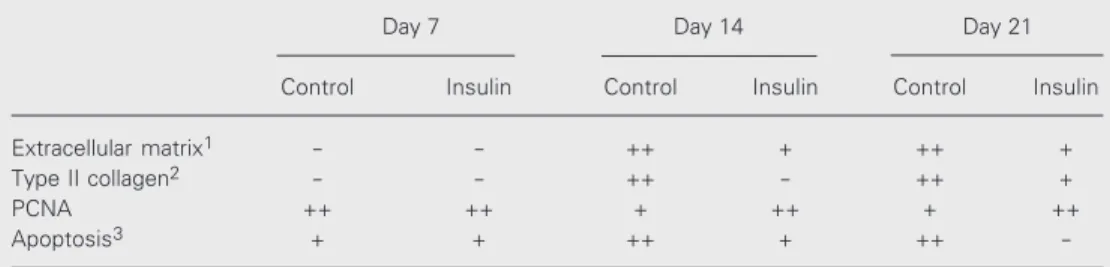
Table 1. Semiquantitative analysis of selected histological indexes demonstrating the deposition of extracel-lular matrix, expression of type II collagen, cell proliferation and apoptosis with time in culture.
Day 7 Day 14 Day 21
Control Insulin Control Insulin Control Insulin
Extracellular matrix1 - - ++ + ++ +
Type II collagen2 - - ++ - ++ +
PCNA ++ ++ + ++ + ++
Apoptosis3 + + ++ + ++
-PCNA, proliferating cell nuclear antigen. Semiquantitative parameters: (-) absent or weak, (+) moderate, (++) intense.
Control: 1% FCS treated cultures.
Insulin: 1% FCS + 60 ng/ml (0.01 µM) insulin.
1Amount of extracellular matrix positively stained by Alcian blue at pH 1.0.
2Type II collagen expression detected by immunohistochemistry.
label the 3'-end of fragmented nuclear DNA with the ApopTag plus peroxidase in situ
apoptosis detection kit (Intergen, Purchase, NY, USA) following the manufacturer’s rec-ommendations, including the proteinase K step for 4 min. Rodent mammary gland slides were used as a positive control.
Statistical analysis
All values were analyzed by the paired Student t-test, with the level of significance set at P < 0.05.
Results
Micromass culture morphology and extracellular matrix deposition
At day 2, phase contrast microscopy re-vealed distinct cartilaginous aggregates of whole mount cultures. On day 14, cartilage nodules stained positively for Alcian blue, demonstrating the presence of sulfated pro-teoglycans and thus confirming the cartilagi-nous origin or the matrix.
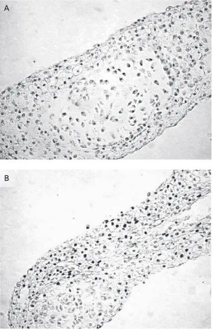
At day 21, light microscopy revealed the presence of round-shaped cells surrounded by ECM, resembling cartilage in vivo (Fig-ure 1A). The amount of ECM increased as a function of time in all cultures, though less conspicuously in insulin-treated cultures (Fig-ure 1B and Table 1).
Type II collagen was expressed in con-trol cultures by day 14 and persisted up to day 21. Under the influence of insulin, type II collagen was labeled with the polyclonal antibody only on day 21 (Table 1).
Figure 1. Micromass culture morphology and extracel-lular matrix deposition at day 21. A, Well-organized cartilage nodules resembling cartilage in vivo were formed in 1% FCS control cultures. B, In contrast, in insulin-treated cultures hypertrophic cells are scarce and the nodular organization is lacking, suggesting im-paired cell maturation. Hematoxylin-eosin. Final magni-fication: 10X (A) and 40X (B).
A AA AA
B BB BB
Cell size. Mean cell size (± SD) was determined by measuring cell diameter mi-croscopically (magnification 40X) at each culture time point (N ≥ 100 cells/culture).
Apoptosis
Cell proliferation
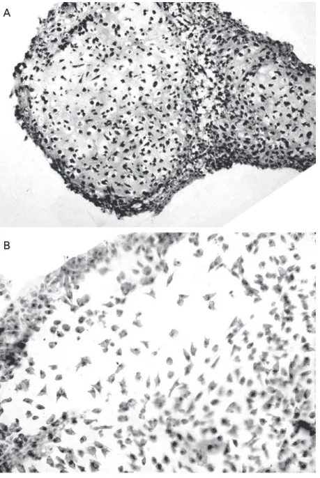
The results for PCNA immunostaining in all cultures and at different time points are presented in Table 1. At day 7 all cultures had a similar appearance with numerous cells positively stained for PCNA. Whereas the number of proliferating cells positively stained for PCNA gradually diminished in 1% FCS control cultures (Figure 2A), in insulin-treated cultures a high number of proliferating cells persisted up to day 21 (Figure 2B).
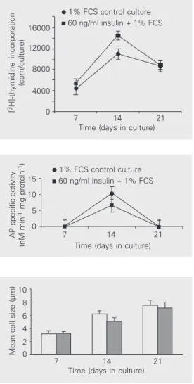
The rate of DNA synthesis, assessed by [3H]-thymidine uptake by the cultures, is shown in Figure 3. Under the influence of insulin, a statistically significant increase in DNA synthesis (P < 0.05) was observed at day 14.
Hypertrophy
At day 14, alkaline phosphatase activity was significantly lower (P < 0.05) (Figure 4) in insulin-treated cultures. Cell diameter was significantly smaller (P < 0.001 and P < 0.05 at days 14 and 21, respectively) (Figure 5) in insulin-treated cultures compared to 1% FCS control cultures.
Apoptosis
Internucleosomal fragmented DNA was detected by TUNEL after day 7 in all cul-tures. With time, apoptotic cells increased in 1% FCS control cultures (Table 1) and cor-responded to apoptosis identified by nuclear
Figure 2. Analysis of cell proliferation of chick limb mesenchyme. In later stages (day 21), when cell matu-ration prevails over cell prolifematu-ration, the number of proliferating cells positively stained for proliferating cell nuclear antigen (PCNA) gradually diminished in 1% FCS control cultures (A). In insulin-treated cultures a high number of proliferating cells (dark cells) persisted up to day 21 (B). PCNA immunohistochemistry coun-terstained with Mayer’s hematoxylin. Final magnifica-tion: 10X (A and B).
morphology in hematoxylin/eosin-stained sections. At day 21, the amount of apoptotic cells was distinctly smaller in insulin-treated cultures than in control cultures (Table 1).
Discussion
The present results demonstrate that in-sulin at the concentration of 60 ng/ml (0.01 µM) is mitogenic to chondrocytes and down-regulates chondrocyte maturation possibly through an antiapoptotic effect.
A A A A A
the cells not committed to the chondrogenic lineage and present in the original heteroge-neous population were possibly prevented from proceeding through the maturation pro-cess either because of their peripheral posi-tion in the aggregates or by a sorting out mechanism (22). An additional convenience of this culture system is to repeatedly follow an expected pattern of growth and differen-tiation and to achieve cartilage formation as a uniform three-dimensional tissue. In this micromass system, mesenchymal cells chon-drified, matured, and sustained hypertrophy as single cartilage nodules distributed ran-domly within the culture (12). Since this system closely reproduces the entire history of the chondrocyte, including cell morphol-ogy, matrix deposition and programmed cell death, it becomes a highly suitable model for the study of chondrocyte maturation under the influence of insulin.
The role of insulin as a growth-regulating hormone has been well established. Quarto et al. (23) demonstrated that insulin is a primary factor involved in the onset and progression of chondrogenesis. They also postulated that insulin acts directly on chon-drocyte maturation without utilizing the sec-ondary pathway of binding to IGF-I recep-tors. Indeed, in vivo (17) but not in vitro (24), the local production of IGF-I is apparently necessary for the growth-promoting role of insulin. Quarto et al. (25) also showed that FGF-2 induces chondrocyte proliferationif associated with insulin. Their findings dem-onstrated that FGF-2 does not induce cell proliferation by itself but must be associated with insulin, thus demonstrating a synergis-tic effect of the two factors. Insulin availabil-ity is also intimately linked to the progres-sion of normal and aberrant fetal growth. Hill and De Sousa (26) suggested that physi-ological concentrations of insulin might ex-ert direct growth-promoting actions on the epiphyseal growth plates of the fetal lamb.
The experiments reported here show that, under the influence of low concentration of
Mean cell size (µm)
10 8 6 4 2 0
7 14 21
Time (days in culture) Figure 5. Effect of insulin and
culture time on cell size. The cells treated with insulin were significantly smaller on days 14 (P < 0.001) and 21 (P < 0.05), respectively, compared to con-trol. Cell sizes were calculated based on the assumption that chondrocytes were spherical in shape. One hundred cells were measured at each time point in
each culture. Data are reported as mean ± SD for 10 independent experiments. Open columns, 1% FCS; closed columns, 60 ng/ml insulin.
In the model utilized in this study (12), the cells derived from mesenchymal embry-onic chick limb buds were prepared in micromass cultures. During the first 2 days of culture, aggregates were formed that ulti-mately differentiated into cartilage nodules consisting of round-shaped cells surrounded by ECM and resembling cartilage in vivo. In these high-density cultures, cell contact and the secretion of macromolecules into the microenvironment control cell growth and differentiation. Mesenchymal cells obtained from limb buds at stage 24 are phenotypi-cally committed to turning into cartilage and, indeed, in culture systems cartilage develops as the major phenotype. On the other hand,
[
3H]-thymidine incorporation
(cpm/culture) 16000 12000 8000 4000 0
7 14 21
Time (days in culture) 1% FCS control culture 60 ng/ml insulin + 1% FCS Figure 3. Effect of insulin and
1% FCS on DNA synthesis in chondrocyte micromass cul-tures. Data are reported as means ± SD for N = 6. Chondro-cytes responded to insulin by increasing DNA synthesis at day 14 (P < 0.05).
Figure 4. Effect of insulin and 1% FCS on alkaline phosphatase (AP) specific activity in micromass cul-tures of chick embryonic limb mesenchyme. Data are reported as means ± SD for N = 6. On day 14 the increase in AP specific activity was significantly greater (P < 0.05) in control cultures.
AP specific activity
(nM min
-1 mg protein -1)
15
10
5
0
7 14 21
insulin (60 ng/ml), chondrocytes proliferate but exhibit a significant impairment of matu-ration. This view is supported by the find-ings of lower alkaline phosphatase activity, lower mean cell diameter and reduced syn-thesis of type II collagen. Whereas in 1% FCS cultures the number of proliferating cells positively stained for PCNA was re-duced over time, in insulin-treated cultures the level of cell proliferation was sustained throughout the period of observation.
The rate of DNA synthesis, assessed by [3H]-thymidine uptake, was also significant-ly higher under the influence of insulin. Hill and De Sousa (26) showed that fetal tissues are more sensitive to the trophic effects of insulin and that, at physiological concentra-tions, insulin has a mitogenic action on iso-lated fetal lamb epiphyseal chondrocytes. Ballock and Reddi (3) also observed that insulin maintained chondrocyte viability in the absence of cell hypertrophy.
In vivo and in vitro studies have demon-strated that terminally differentiated chon-drocytes undergo programmed cell death (12,27). In vivo studies have shown that in the proximal tibial growth plate of young chicks, terminally differentiated chondro-cytes undergo programmed cell death (28,29). This process possibly provides a physiologi-cal mechanism for the rapid and controlled removal of terminally differentiated cells.
In the present study cell apoptosis was detected by the ApopTag Plus method which is based on the specific staining of
frag-mented DNA. The ability to observe the chondrocytes as a uniform three-dimensional tissue enabled us to visualize apoptosis at the single cell level. Also, we were able to rule out the possibility of DNA damage caused by artifacts generated by tissue digestion, cell isolation procedures, and activation of endogenous nuclease activity. The treatment with insulin was associated with an increased number of proliferating chondrocytes and a relatively low level of apoptosis.
Taken together, the data presented here suggest that insulin regulates chondrocyte maturation and hypertrophy through a pos-sible antiapoptotic effect. Bertrand et al.
(30,31) provided the first evidence of the antiapoptotic function of insulin. These in-vestigators postulate that this antiapoptotic action involves the activation by insulin of nuclear factor κB, a transcription factor play-ing a critical role in apoptosis inhibition. Yenush et al. (32) also raised the possibility that a phosphotyrosine-independent mech-anism promotes the antiapoptotic and growth actions of insulin. Additional studies will be necessary to further elucidate the precise role of insulin in the control of chondrocyte maturation for the induction of growth-pro-moting effects on the epiphyseal growth plate.
Acknowledgments
The authors thank A. Cohen and A. Balduíno for a critical reading of the manu-script.
References
1. Trippel SB, Wroblewski J, Makower A, Whelan MC, Schoenfeld D & Doctrow SR (1993). Regulation of growth-plate chondrocytes by insulin-like growth factor I and basic fibroblast growth factor.
Jour-nal of Bone and Joint Surgery, 2: 177-189.
2. Schmid TM & Linsenmayer TF (1985). Immunohistochemical local-ization of short-chain collagen (type X) in avian skeletal tissue.
Journal of Cell Biology,100: 598-605.
3. Ballock RT & Reddi AH (1994). Thyroxine is the serum factor that regulates morphogenesis of columnar cartilage from isolated chon-drocytes in chemically defined medium. Journal of Cell Biology, 126: 1311-1318.
4. Kato Y, Iwamoto M & Koike T (1998). Terminal differentiation and calcification in rabbit chondrocyte cultures grown in centrifuge tubes: regulation by transforming growth factor ß and serum fac-tors. Proceedings of the National Academy of Sciences, USA, 85: 9552-9556.
5. Alini M, Carey D, Hirata S, Grympas MD, Pidoux I & Poole AR (1994). Cellular and matrix changes before and at the time of calcification in the growth plate studied in vitro: arrest of type X collagen synthesis and net loss of collagen when calcification is initiated. Journal of
Bone and Mineral Research, 9: 1077-1087.
morphogenetic protein-2 act by distinct mechanisms to promote chick limb cartilage differentiation in vitro. Developmental Dynam-ics, 200: 103-116.
7. Holtzer H, Abbott J, Lash J & Holtzer S (1960). The loss of pheno-typic traits by differentiated cells in vitro. I. Differentiation of carti-lage cells. Proceedings ofthe National Academy of Sciences, USA, 46: 1533-1542.
8. Mayne R, Vail MS, Mayne P & Miller EJ (1976). Changes in the type of collagen synthesized as clones of chick chondrocytes grow and eventually lose division capacity. Proceedings of the National
Acade-my of Sciences, USA, 73: 1674-1678.
9. Ahrens PB, Solursh M & Reiters R (1977). Stage-related capacity for limb chondrogenesis in cell culture. Developmental Biology,60: 69-82.
10. San Antonio JD & Tuan RS (1986). Chondrogenesis of limb mesen-chyme in vitro: stimulation by cations. Developmental Biology, 115: 313-324.
11. Wong M & Tuan RS (1993). Nuserum, a synthetic serum replace-ment, supports chondrogenesis of embryonic chick limb bud mes-enchymal cells in micromass cultures. In Vitro Cellular and
Develop-mental Biology. Animal,29: 917-922.
12. Mello MA & Tuan RS (1999). High-density micromass cultures of embryonic limb bud mesenchymal cells: an in vitro model of endo-chondral skeletal development. In Vitro Cellular and Developmental
Biology. Animal, 35: 262-269.
13. Boskey AL, Stiner D, Doty SB, Binderman I & Leboy P (1992). Studies of mineralization in tissue culture: optimal conditions for cartilage calcification. Bone and Mineral,16: 11-36.
14. De Pablo F, Roth J, Hernandez E & Pruss RM (1982). Insulin is present in chick eggs and early chick embryos. Endocrinology, 111: 1909-1911.
15. Bassas L, De Pablo F, Lesniak MA & Roth J (1985). Ontogeny of receptors for insulin-like growth factor over insulin receptors in brain. Endocrinology, 117: 2321-2329.
16. Girbau M, Gomez JA, Lesniak MA & De Pablo F (1987). Insulin and insulin-like growth factor I both stimulate metabolism, growth and differentiation in the post neurula chick embryo. Endocrinology, 121: 1477-1483.
17. Alarid ET, Schlechter NL, Russell SM & Nicoll CS (1992). Evidence suggesting that insulin-like growth factor-I is necessary for the trophic effects of insulin on cartilage growth in vivo. Endocrinology, 130: 2305-2309.
18. Verhaeghe J & Bouillon R (1996). Principles of bone biology. In: Bilezikian JP, Raisz LG & Rodan GA (Editors), Effects of Diabetes
and Insulin on Bone Metabolism. Academic Press, San Diego, CA,
USA.
19. Hamburger V & Hamilton HL (1951). A series of normal stages in the development of the chick embryo. Journal of Morphology, 88: 49-92.
20. Cook HC (1996). Carbohydrates. In: Bancroft JD & Stevens A (Edi-tors), Theory and Practice of Histological Techniques. Churchill Liv-ingstone, New York.
21. Gavrieli Y, Sherman Y & Ben-Sasson SA (1992). Identification of programmed cell death in situ via specific labeling of nuclear DNA fragmentation. Journal of Cell Biology,119: 493-501.
22. Tacchetti C, Quarto R, Nitsch L, Hartmann DJ & Cancedda R (1987).
In vitro morphogenesis of chick embryo hypertrophic chondrocytes.
Journal of Cell Biology, 105: 999-1006.
23. Quarto R, Campanile G, Cancedda R & Dozin B (1992). Thyroid hormone, insulin and glucocorticoids are sufficient to support chon-drocyte differentiation to hypertrophy: a serum-free analysis.
Jour-nal of Cell Biology,119: 989-995.
24. Böhme K, Conscience-Egli M, Tshan T, Winterhalter KH & Bruckner P (1992). Induction of proliferation or hypertrophy of chondrocytes in serum-free culture: the role of insulin-like growth factor-I, insulin, or thyroxine. Journal of Cell Biology,116: 1035-1042.
25. Quarto R, Campanile G, Cancedda R & Dozin B (1997). Modulation of commitment, proliferation, and differentiation of chondrogenic cells in defined culture medium. Endocrinology, 138: 4966-4976. 26. Hill DJ & De Sousa D (1990). Insulin is a mitogen for isolated
epiphyseal growth plate chondrocytes from the fetal lamb.
Endocri-nology, 126: 2661-2670.
27. Lewinson D & Silbermann M (1992). Chondroclasts and endothelial cells collaborate in the process of cartilage resorption. Anatomical
Record,233: 504-514.
28. Hatori M, Klatte KJ, Teixeira CC & Shapiro IM (1995). End labeling studies of fragmented DNA in the avian growth plate: evidence of apoptosis in terminally differentiated chondrocytes. Journal of Bone
and Mineral Research, 10: 1960-1968.
29. Roach H (1997). New aspects of endochondral ossification in the chick: chondrocyte apoptosis, bone formation by former chondro-cytes, and acid phosphatase activity in the endochondral bone matrix. Journal of Bone and Mineral Research,12: 795-805. 30. Bertrand F, Atfi A, Cadoret A, L’Allemain G, Robin H, Lascols O,
Capeau J & Cherqui G (1998). A role for nuclear factor κB in the antiapoptotic function of insulin. Journal of Biological Chemistry, 273: 2931-2938.
31. Bertrand F, Desbois-Mouthon C, Cadoret A, Prunier C, Robin H, Capeau J, Atfi A & Cherqui G (1999). Insulin antiapoptotic signaling involves insulin activation of the nuclear factor κB-dependent sur-vival genes encoding tumor necrosis factor receptor-associated fac-tor 2 and manganese-superoxide dismutase. Journal of Biological
Chemistry, 274: 30596-30602.
32. Yenush L, Zanella G, Uchida T, Bernal D & White MF (1998). The pleckstrin homology and phosphotyrosine binding domains of insu-lin receptor substrate 1 mediate inhibition of apoptosis by insuinsu-lin.