Submitted19 October 2015 Accepted 11 March 2016 Published31 March 2016
Corresponding author Chris Beardsley,
Academic editor
Rodrigo Ramírez-Campillo
Additional Information and Declarations can be found on page 10
DOI10.7717/peerj.1881
Copyright 2016 Beardsley et al.
Distributed under
Creative Commons CC-BY 4.0
OPEN ACCESS
Test–re-test reliability and inter-rater
reliability of a digital pelvic inclinometer
in young, healthy males and females
Chris Beardsley1, Tim Egerton2and Brendon Skinner3
1Strength and Conditioning Research Limited, London, United Kingdom 2Sport Science Tutor, Congleton, United Kingdom
3Department of Sports Therapy, Staffordshire University, United Kingdom
ABSTRACT
Objective.The purpose of this study was to investigate the reliability of a digital pelvic
inclinometer (DPI) for measuring sagittal plane pelvic tilt in 18 young, healthy males and females.
Method.The inter-rater reliability and test–re-test reliabilities of the DPI for measuring
pelvic tilt in standing on both the right and left sides of the pelvis were measured by two raters carrying out two rating sessions of the same subjects, three weeks apart.
Results. For measuring pelvic tilt, inter-rater reliability was designated as good on
both sides (ICC=0.81–0.88), test–re-test reliability within a single rating session was designated as good on both sides (ICC=0.88–0.95), and test–re-test reliability between two rating sessions was designated as moderate on the left side (ICC=0.65) and good
on the right side (ICC=0.85).
Conclusion. Inter-rater reliability and test–re-test reliability within a single rating
session of the DPI in measuring pelvic tilt were both good, while test–re-test reliability between rating sessions was moderate-to-good. Caution is required regarding the interpretation of the test–re-test reliability within a single rating session, as the raters were not blinded. Further research is required to establish validity.
SubjectsKinesiology, Statistics Keywords Reliability, Pelvic tilt
INTRODUCTION
In addition, both in the literature and anecdotally, there are reports that greater anterior pelvic tilt may increase the risk of musculoskeletal injury during running (Schache et al., 1999;Schache, Blanch & Murphy, 2000;Schache et al., 2002). It has been suggested that such injuries could occur either through repetitive impingement of the vertebral facets (Schache et al., 1999;Schache et al., 2002) or by producing excessive lengthening of the hamstring, leading to strain injury (Schache et al., 1999;Schache et al., 2002). On this or another basis, some clinicians may decide to measure the extent of anterior pelvic tilt in their patients and clients, particularly those who undertake regular running activities.
Pelvic tilt can be measured either with a single measurement, at the center line, or with two measurements at either lateral border. Measurements taken in cadavers have shown that differences in bony anatomy lead to significant between-side differences in anterior pelvic tilt (Preece et al., 2008) and significant differences in pelvic tilt between sides have also been reported in live subjects (Herrington, 2011). The difference in pelvic tilt between sides has been taken as a measurement of pelvic torsion, which some investigations have associated with leg length discrepancy (Cummings, Scholz & Barnes, 1993;Young, Andrew & Cum-mings, 2000;Betsch et al., 2012;Wild et al., 2014). It has been variously suggested that pelvic torsion occurs as a natural adaptation to leg length discrepancy (Krawiec et al., 2003), that greater anterior pelvic tilt occurs on the side of the shorter leg compared to the contralateral leg (Knutson, 2005), and that this biomechanical feature may be common to both symptomatic and asymptomatic individuals alike (Herrington, 2011). Even so, the precise relationships between leg length discrepancy and pelvic torsion, as well as between leg length discrepancy and musculoskeletal injury risk, are contentious and remain poorly understood (Gurney, 2002;Juhl, Cremin & Russell, 2004;Knutson, 2005;Cooperstein & Lew, 2009).
Several methods are available for measuring pelvic tilt. Early studies often used radiography (Clayson et al., 1962;Flint, 1963) and this method continues to be used in relation to surgery affecting the hip and pelvis (Blondel et al., 2009;Lazennec et al., 2011) or when a standard is required against to validate other methods (Burdett, Brown & Fall, 1986;
Crowell et al., 1994;Petrone et al., 2003;Sprigle et al., 2003;Lazennec et al., 2011). Other methods include the Iowa Anatomical Position System (Day, Schmidt & Lehmann, 1984), the Metrecom Skeletal Analysis System (Barakatt et al., 1996), the antenna method (Moes, 1998), goniometers (Burdett, Brown & Fall, 1986;Sprigle et al., 2003), calipers (Sanders & Stavrakas, 1981;Gajdosik et al., 1985;Alviso, Dong & Lentell, 1988), inclinometers (Walker et al., 1987;Heino, Godges & Carter, 1990;Crowell et al., 1994;Youdas et al., 1996;Levine, Walker & Tillman, 1997;Hagins et al., 1998;Petrone et al., 2003;Preece et al., 2008;Gnat et al., 2009;Herrington, 2011), low-dose digital stereoradiography (Lazennec et al., 2011;
Guenoun et al., 2012), and magnetic resonance imaging (MRI) scans (Lalonde et al., 2006). Calliper-based inclinometers seem to be among the most common tools used by clinicians for measuring pelvic tilt for several reasons. They display good reliability for measuring iliac crest height differences (Walker et al., 1987; Hagins et al., 1998;
Petrone et al., 2003;Krawiec et al., 2003) and for measuring pelvic tilt (Heino, Godges & Carter, 1990;Crowell et al., 1994;Youdas et al., 1996;Gnat et al., 2009;Herrington, 2011;
have generally reported at least good (Walker et al., 1987;Heino, Godges & Carter, 1990;
Herrington, 2011) if not excellent reliability (Youdas et al., 1996; Hagins et al., 1998;
Krawiec et al., 2003;Gnat et al., 2009). Additionally, calliper-based inclinometers have also been found to display good convergent criterion reference validity by reference to radiography (Crowell et al., 1994;Petrone et al., 2003). Furthermore, these devices also have several practical advantages to the clinician, being quickly and easily utilized (Crowell et al., 1994), as well as being small, portable, relatively safe compared to radiography, and comparatively inexpensive in comparison with low-dose digital stereoradiography and MRI scanning devices. Calliper-based inclinometers also permit measurements to be taken on both sides of the pelvis, which may be important given the differences between sides that have previously been observed (Preece et al., 2008;Herrington, 2011).
Different models of calliper-based inclinometer have been investigated in the literature. The Palpation Meter (PALM, Performance Attainment Associates, St. Paul, MN, USA) is the calliper-based inclinometer that has been extensively explored (Hagins et al., 1998;Petrone et al., 2003;Krawiec et al., 2003;Gnat et al., 2009;Lee, Yoo & Gak, 2011;Herrington, 2011;
Fourchet et al., 2014). Other models that have been investigated include those developed and modified byWalker et al. (1987)andCrowell et al. (1994). The model used and developed by
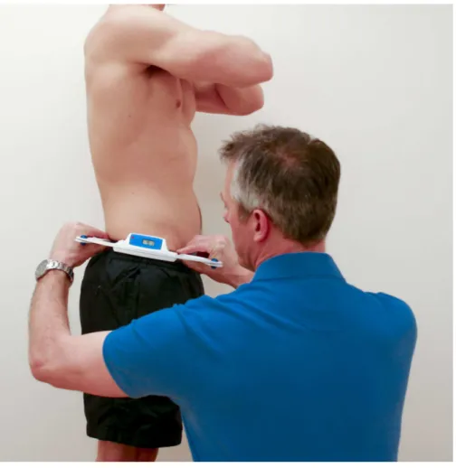
Crowell et al. (1994)included a spirit level to permit readings relative to the ground, finger-tip rings to allow superior palpation of the bony prominences, and a digital read-out for ease and speed of reading the output. The Digital Pelvic Inclinometer (DPI, Sub-4 Limited, UK) is a new, commercially-available, calliper-based inclinometer that is very similar to the model developed byCrowell et al. (1994)(Fig. 1). Like the model developed byCrowell et al. (1994), the DPI uses a digital display. This display allows the clinician to see the output of the device while performing the measurement procedure. In addition, the DPI also has recessed calliper ends, which allow simultaneous palpation of the bony prominences with the hands and the calliper arms. Finally, the DPI also contains a spirit level to facilitate measurements of pelvic angles relative to the ground as well as relative to the other side of the pelvis.
The purpose of this study was to investigate the inter-rater reliability and test–re-test reliability of the DPI in young, healthy males and females across two rating sessions with experienced, trained raters. The first hypothesis for this study was that inter-rater reliability for the DPI between two raters would be good. The second hypothesis was that test-rest reliability for the DPI would be good by reference to three separate measurements taken on a single rating session. The third hypothesis was that test–re-test reliability for the DPI would be good by reference to the mean of the measurements taken on each of two rating sessions on separate occasions.
METHOD
Experimental approach
Figure 1 Measuring pelvic tilt using the DPI.Standing position maintained by subject, while rater mea-sured pelvic tilt using the DPI.
Measurement procedures
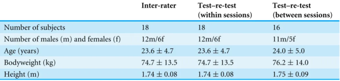
Table 1 Descriptive statistics for the subjects.
Inter-rater Test–re-test
(within sessions)
Test–re-test (between sessions)
Number of subjects 18 18 16
Number of males (m) and females (f) 12m/6f 12m/6f 11m/5f Age (years) 23.6±4.7 23.6±4.7 24.0±5.0 Bodyweight (kg) 74.7±13.5 74.7±13.5 76.2±14.0 Height (m) 1.74±0.08 1.74±0.08 1.75±0.09
The DPI comprises two precision arms, which are mounted upon a main body. The main body contains a tri-axial accelerometer, which records the angle of pelvic tilt across the two precision arms. The output from the tri-axial accelerometer is shown as an angle in degrees, in numerical form on a liquid crystal display. For each measurement of pelvic tilt, standard instructions were used per the manufacturer’s guidelines, as follows: ‘‘the practitioner places the index finger and thumb on each hand on each finger grip at the end of the DPI arms. With each index finger slightly prominent ready for concurrent palpation of the posterior superior iliac spine (PSIS) and anterior superior iliac spine (ASIS), the practitioner positions the DPI on the side of the innominate bone and takes a reading. The practitioner moves their index finger over the most prominent point of the iliac crests until the apex is established for the measuring. The practitioner then reads off the degree of inclination from the LCD.’’
Subjects and raters
Following a power analysis as described by Wolak, Fairbairn & Paulsen (2012), a convenience sample of 18 healthy subjects (12 males and 6 females) were recruited from a university physical therapy program. Of the 18 subjects, only 16 were included in the test–re-test reliability assessment between sessions (for subject characteristics relevant to each assessment, see Table 1).
Subjects qualified for the study if they met the following criteria: were≥18 years of age, were able to stand unsupported for the duration of the measurement process (<10 min), were free from existing low back injuries, had not experienced any low back injuries within the previous 3 months, and had no medical condition leading to clinically meaningful leg length inequality. In accordance with ethical requirements, the subjects received an explanation of the nature, purpose, and risks of the study and were given the opportunity to ask questions. All subjects signed an informed consent document prior to participating in the study. Written ethical approval for the study was granted by the Faculty of Health Sciences Ethics Panel, Staffordshire University.
Table 2 Descriptive statistics for pelvic tilt.
Right (degrees)
Left (degrees)
Difference (degrees)
Mean 10.6 10.5 0.1
Standard deviation 5.0 5.8 3.8
Statistics
Intra-class correlation coefficients (ICC) were used to assess the inter-rater, intra-rater (between sessions) and intra-rater (within sessions) reliability of pelvic tilt measured using the DPI for both right and left sides. ICCs are suitable for use in fully-crossed study designs assessing reliability of interval variables (Hallgren, 2012). Since the raters were not randomly selected for each subject but were the same for all subjects, a two-way Analysis of Variance (ANOVA) model was used (Shrout & Fleiss, 1979). Since absolute rather than ranked values of pelvic tilt are of interest, the ICC model type was set to require absolute agreement (McGraw & Wong, 1996). The unit of measurement used in the model differed between the statistics calculated. Since clinical practice commonly involves taking multiple measurements and recording the mean, the mean of the three ratings taken for each subject in a single session was used for hypothesis testing for inter-rater reliability and test–re-test reliability between sessions. Inter-rater reliability was assessed by combining the results of both testing sessions. Test–re-test reliability between sessions was assessed by combining the results of both raters. In contrast, for test–re-test reliability within single sessions, the reliability of the single, individual ratings was assessed, although again the results of both raters were combined together (Shrout & Fleiss, 1979). Before commencing the trial, it was decided that interpretation of the reported values for each ICC would be based upon the following criteria: <0.50=poor, 0.50–0.75 =moderate, and >0.75=good (Walmsley & Amell, 1996;Batterham & George, 2003;Portney & Watkins, 2008). To enhance clinical interpretation of the results, the standard error of measurement (SEM) and minimum difference to be considered real (MD) were estimated (Weir, 2005). Descriptive statistics were calculated as means with standard deviation. Statistical significance was set a priori atp<0.05. All statistical analysis was performed using R, using the irr (Gamer et al., 2007)
and ICC (Wolak, 2012) packages.
RESULTS
Descriptive statistics
Descriptive statistics (mean±standard deviation) for pelvic tilt on the right and left sides are presented inTable 2.
Reliability
The ICC, SEM, and MD reported when measuring inter-rater reliability, test–re-test reliability (within sessions) and test–re-test reliability (between sessions) are presented in Table 3.
Table 3 Inter-rater and test–re-test reliabilities of the DPI.Inter-rater and test–re-test reliabilities (be-tween sessions and within sessions) of the DPI for measuring pelvic tilt on the right and left sides, as as-sessed by intra-class correlation coefficient (ICC), standard error of measurement (SEM) and minimum difference (MD) to be considered real.
Inter-rater Test–re-test (between sessions) Test–re-test (within sessions)
Right Left Right Left Right Left
ICC 0.81* 0.88* 0.85* 0.65* 0.88* 0.95*
SEM 2.2 2.0 1.9 3.4 1.7 1.1
MD 6.0 5.5 5.4 9.4 4.8 2.9
Notes.
*=significant,p<0.05.
Table 4 Raw data showing pelvic tilt.Raw data showing pelvic tilt for each session (mean of three individual measurements), for each rater, for each subject.
Left side Right side
Rater 1 Rater 2 Rater 1 Rater 2
Subject Session 1 Session 2 Session 1 Session 2 Session 1 Session 2 Session 1 Session 2
1 14.07 15.33 10.17 12.83 13.50 10.50 9.83 8.60
2 20.00 14.83 14.83 16.27 20.15 17.50 16.83 16.73
3 8.70 5.67 14.67 3.53 10.15 11.00 6.33 7.57
4 10.27 6.67 17.17 8.97 12.35 11.33 8.33 11.40
5 11.13 No data 22.33 No data 14.20 No data 17.83 No data
6 13.70 14.17 16.67 11.37 11.95 16.33 13.50 10.00
7 11.60 9.67 9.17 10.87 10.15 10.17 9.83 9.73
8 12.13 7.00 12.33 7.80 8.80 10.33 3.50 12.37
9 10.17 5.67 12.17 7.17 9.10 10.33 7.67 8.67
10 14.93 11.83 14.67 7.13 16.75 10.17 11.83 11.33
11 1.67 −3.50 0.67 −10.47 2.80 0.17 −3.00 −3.33
12 11.03 1.67 10.00 −4.47 10.10 5.67 8.00 2.30
13 15.17 13.50 15.17 12.33 15.20 11.83 12.83 12.93
14 7.80 6.67 9.00 5.73 6.45 10.00 3.33 1.43
15 16.77 16.17 18.17 11.57 17.25 20.50 9.67 15.60
16 16.37 10.33 14.33 9.23 17.30 15.33 10.67 14.20
16 10.43 7.33 8.33 6.40 10.95 8.33 5.50 9.47
18 No data 18.00 No data 12.13 No data 16.00 No data 13.80
(between sessions), as only 16 subjects attended both sessions. Subject attendance in each session, along with the raw data for the mean pelvic tilt on left and right sides is shown in Table 4.
DISCUSSION
for the DPI would be good within a single rating session. The third hypothesis was that test–re-test reliability for the DPI would be good between two rating sessions.
By reference to pre-determined criteria for assessing reliability by reference to the magnitude of the ICC, the inter-rater reliability of the DPI for measuring pelvic tilt was designated as good on both sides (ICC=0.81–0.88), the test–re-test reliability of the DPI
for measuring pelvic tilt within a single rating session was designated as good on both sides (ICC =0.88–0.95), and the test–re-test reliability for the DPI for measuring pelvic tilt between two rating sessions was designated as moderate on the left side (ICC=0.65) and good on the right side (ICC=0.85).
For inter-rater and test-rest reliability, our findings (ICC=0.65–0.95; SEM=1.9–3.4
degrees; MD=2.9–9.4 degrees) are broadly in line with those of other investigations in similar devices measuring pelvic tilt. In their trial of a very similar type of caliper-based inclinometer to the DPI,Crowell et al. (1994)reported good intra-rater reliability (ICC=
0.92; SEM=0.93 degrees; MD=2.6 degrees) and good inter-rater reliability (ICC=0.95;
SEM =0.78 degrees; MD=2.2 degrees),Preece et al. (2008)reported good intra-rater reliability (albeit in cadavers) (ICC=0.98; SEM=1.1 degrees; MD=3.1 degrees),Gnat et al. (2009)reported good intra-rater reliability (ICC=0.99; SEM and MD not reported),
Herrington (2011)reported good intra-rater reliability (ICC=0.87; SEM=1.1 degrees; MD=2.5 degrees), and Fourchet et al. (2014)reported good inter-rater and intra-rater
reliability (coefficient of variation =15.8%). The reliability of the PALM in assessing linear differences in iliac crest height has also been found to be good (Hagins et al., 1998;
Petrone et al., 2003) but whether such findings can be considered as directly comparable with the measurement of pelvic tilt angle is unclear. The reliability of a three-dimensional (3D) camera-based motion capture system reported byLevine & Whittle (1996)was also found to be good but interestingly no better than the PALM (ICC =0.95; SEM=0.96 degrees; MD=2.7 degrees) and the caliper-based system used byGajdosik et al. (1985)
also displayed similar reliability (ICC=0.88; SEM=1.4 degrees; MD=4.0 degrees). Regarding pelvic tilt, our descriptive statistics (means of 10.5–10.6 degrees) are in line with the findings of other investigations, across various measurement devices. Using a PALM device,Herrington (2011)measured pelvic tilt in a population of 120 young, healthy subjects (65 males and 55 females, aged 23.8 years). It was reported that 85% of males and 75% of females displayed an anteriorly rotated pelvis, in the range of 6–7 degrees. Also using a PALM device,Lee, Yoo & Gak (2011)measured pelvic tilt in a population of 40 young, healthy subjects (23 males aged 23.8 years and 17 females aged 21.4 years) and found that anterior pelvic tilt was 7–8 degrees.Gajdosik et al. (1985)measured pelvic tilt in a population of 20 healthy males, aged 25.2 years, and reported a mean anterior pelvic tilt angle of 8.5±4.1 degrees. Using a 3D camera-based motion capture system,Levine
& Whittle (1996)measured pelvic tilt angle in a population of 20 healthy female subjects, aged 23.4 years, and reported a mean anterior pelvic tilt angle of 11.3±4.3 degrees. Using radiography,Vaz et al. (2002) measured pelvic tilt angle in 100 healthy students from medical professions, aged 27 years, and reported a mean anterior pelvic tilt angle of 12.3
±5.9 degrees. From this very brief review, it seems that calliper or calliper-inclinometer
slightly lower values of anterior pelvic tilt (6–8 degrees vs. 11–12 degrees) than those found using more sophisticated methods (Levine & Whittle, 1996;Vaz et al., 2002). It is interesting that the values reported here using the DPI (means of 10.5–10.6 degrees) are at the higher end of the spectrum reported in the literature and closer to those observed using more sophisticated methods. Whether this is a feature of the population measured, the presence of a spirit level in the DPI to standardize measurements relative to the ground, systematic bias in the DPI, or systematic bias in the raters is unclear.
Regarding differences between right and left sides, this investigation reported descriptive statistics (mean of 0.1 degrees greater anterior pelvic tilt on the right side) that are within the range of values observed by others. The literature is conflicting regarding whether the left or right sides tend to be more anteriorly rotated, or whether no difference is the norm. In respect of the prevailing direction of greater anterior tilt, some studies have reported very small differences that are likely within the bounds of measurement error (Gnat et al., 2009;Lee, Yoo & Gak, 2011). Other investigators have reported greater mean anterior tilt on the right side (Krawiec et al., 2003), which has been predicted based upon the apparent tendency for the right leg to be shorter in many populations (Knutson, 2005). However, greater mean anterior tilt on the left side has also been reported (Barakatt et al., 1996). In respect of the magnitude of difference between sides, as noted above, some studies have reported very small differences (Gnat et al., 2009;Lee, Yoo & Gak, 2011), while others have reported differences of around 2 degrees (Barakatt et al., 1996;Krawiec et al., 2003). It is noteworthy thatGnat et al. (2009)reported low mean values for the difference between sides in quiet standing (<0.5 degrees) but much greater values after exercise, particularly jumping (4.65±1.56 degrees).
LIMITATIONS
criteria prevented the inclusion of any subjects with medical conditions leading to clinically meaningful leg length discrepancies, our study was limited in that we did not perform any tests to assess whether any of the subjects had such leg length discrepancies, nor did we measure any other musculoskeletal parameters, such as hamstring and lumbopelvic flexibility using the sit-and-reach test, or actual hamstring muscle–tendon length. Such confounding factors might have affected our results.
In respect of the validity of the DPI, there are three substantial limitations of the present study. Firstly, criterion reference validity of the DPI for assessing anterior pelvic tilt on either side of the pelvis was not assessed. Future studies could explore this by correlating measurements taken using the DPI with measurements taken using gold standard methods (such as radiography) in the same group of subjects, as other investigators have done (Crowell et al., 1994;Petrone et al., 2003). Therefore, while the DPI displays good reliability between raters and between ratings taken in the same session, it may not produce valid measurements of pelvic tilt in comparison with values recorded using radiography or MRI. Secondly, the extent to which the measurements of anterior pelvic tilt on either side of the pelvis or the difference between these (pelvic torsion) might be predictive of increased injury risk or low back pain was not assessed. Thirdly, the extent to which measurements of anterior pelvic tilt on either side of the pelvis or the difference between these (pelvic torsion) might provide useful information about the extent of any existing leg length inequality was not explored.
CONCLUSIONS
The inter-rater reliability and test–re-test reliability of the DPI for measuring pelvic tilt angle on both right and left sides of the pelvis were assessed, in a convenience sample of young, healthy males and females. The inter-rater reliability of the DPI for measuring pelvic tilt was designated as good on both sides (ICC=0.81–0.88); the test–re-test reliability of the DPI for measuring pelvic tilt within a single rating session was designated as good on both sides (ICC=0.88–0.95); and the test–re-test reliability for the DPI for measuring pelvic tilt between two rating sessions was designated as moderate on the left side (ICC=
0.65) and good on the right side (ICC=0.85). Given that the raters were not blinded to the
measurements, our findings regarding the test–re-test reliability of the DPI for measuring pelvic tilt within a single rating session should be interpreted with caution. Nevertheless, these results indicate that the DPI produces acceptably reliable measurements, although further research is required to establish the validity of the DPI in measuring pelvic tilt.
ADDITIONAL INFORMATION AND DECLARATIONS
Funding
The authors received no funding for this work.
Competing Interests
Author Contributions
• Chris Beardsley conceived and designed the experiments, analyzed the data, wrote the paper, prepared figures and/or tables.
• Tim Egerton and Brendon Skinner performed the experiments, reviewed drafts of the paper.
Human Ethics
The following information was supplied relating to ethical approvals (i.e., approving body and any reference numbers):
The study was approved by the Faculty of Health Sciences Ethics Panel, Staffordshire University.
Data Availability
The following information was supplied regarding data availability: The raw data has been supplied asSupplemental Information.
Supplemental Information
Supplemental information for this article can be found online athttp://dx.doi.org/10.7717/ peerj.1881#supplemental-information.
REFERENCES
Alviso DJ, Dong GT, Lentell GL. 1988.Intertester reliability for measuring pelvic tilt in
standing.Physical Therapy 68(9):1347–1351.
Barakatt E, Smidt GL, Dawson JD, Wei SH, Heiss DG. 1996.Interinnominate motion
and symmetry: comparison between gymnasts and nongymnasts.Journal of Or-thopaedic & Sports Physical Therapy23(5):309–319DOI 10.2519/jospt.1996.23.5.309.
Batterham AM, George KP. 2003.Reliability in evidence-based clinical practice: a
primer for allied health professionals?Physical Therapy in Sport 4(3):122–128 DOI 10.1016/S1466-853X(03)00076-2.
Betsch M, Wild M, Große B, Rapp W, Horstmann T. 2012.The effect of simulating
leg length inequality on spinal posture and pelvic position: a dynamic rasterstereo-graphic analysis.European Spine Journal21(4):691–697
DOI 10.1007/s00586-011-1912-5.
Blondel B, Parratte S, Tropiano P, Pauly V, Aubaniac JM, Argenson JN. 2009.Pelvic tilt
measurement before and after total hip arthroplasty.Orthopaedics & Traumatology: Surgery & Research95(8):568–572.
Burdett RG, Brown KE, Fall MP. 1986.Reliability and validity of four instruments for
measuring lumbar spine and pelvic positions.Physical Therapy 66(5):677–684.
Chaléat-Valayer E, Mac-Thiong JM, Paquet J, Berthonnaud E, Siani F, Roussouly P.
2011.Sagittal spino-pelvic alignment in chronic low back pain.European Spine
Journal 20(5):634–640DOI 10.1007/s00586-011-1931-2.
Clayson SJ, Newman IM, Debevec DF, Anger RW, Skowlund HV, Kottke F. 1962.
Cooperstein R, Lew M. 2009.The relationship between pelvic torsion and anatomical leg length inequality: a review of the literature.Journal of Chiropractic Medicine
8(3):107–118DOI 10.1016/j.jcm.2009.06.001.
Crowell RD, Cummings GS, Walker JR, Tillman LJ. 1994.Intratester and intertester
reliability and validity of measures of innominate bone inclination.Journal of Orthopaedic & Sports Physical Therapy20(2):88–97DOI 10.2519/jospt.1994.20.2.88.
Cummings G, Scholz JP, Barnes K. 1993.The effect of imposed leg length difference on
pelvic bone symmetry.Spine18(3):368–373 DOI 10.1097/00007632-199303000-00012.
Day JW, Schmidt GL, Lehmann T. 1984.Effect of pelvic tilt on standing posture.Physical
Therapy64(4):510–516.
Flint MM. 1963.Lumbar posture: a study of roentgenographic measurement and the
influence of flexibility and strength.Research Quarterly. American Association for Health, Physical Education and Recreation34(1):15–20.
Fourchet F, Materne O, Rajeb A, Horobeanu C, Farooq A. 2014.Pelvic tilt: reliability of
measuring the standing position and range of motion in adolescent athletes.British Journal of Sports Medicine48(7):594–594.
Gajdosik R, Simpson R, Smith R, DonTigny RL. 1985.Pelvic tilt intratester
relia-bility of measuring the standing position and range of motion.Physical Therapy
65(2):169–174.
Gamer M, Lemon J, Fellows I, Singh P. 2007.IRR: various coefficients of interrater
reliability and agreement. R package v. 0.70.Available athttp:// www.r-project.org.
Gnat R, Saulicz E. 2008.Induced static asymmetry of the pelvis is associated with
functional asymmetry of the lumbo-pelvo-hip complex.Journal of Manipulative and Physiological Therapeutics31(3):204–211DOI 10.1016/j.jmpt.2008.02.012.
Gnat R, Saulicz E, Biały M, Kłaptocz P. 2009.Does pelvic asymmetry always mean
pathology? Analysis of mechanical factors leading to the asymmetry.Journal of Human Kinetics21:23–32.
Guenoun B, Zadegan F, Aim F, Hannouche D, Nizard R. 2012.Reliability of a new
method for lower-extremity measurements based on stereoradiographic three-dimensional reconstruction.Orthopaedics & Traumatology: Surgery & Research
98(5):506–513.
Gurney B. 2002.Leg length discrepancy.Gait & Posture15(2):195–206
DOI 10.1016/S0966-6362(01)00148-5.
Hagins M, Brown M, Cook C, Gstalder K, Kam M, Kominer G, Strimbeck K. 1998.
Intratester and intertester reliability of the palpation meter (PALM) in measur-ing pelvic position.Journal of Manual & Manipulative Therapy6(3):130–136 DOI 10.1179/jmt.1998.6.3.130.
Hallgren KA. 2012.Computing inter-rater reliability for observational data: an overview
and tutorial.Tutorials in Quantitative Methods for Psychology 8(1):23–34.
Heino JG, Godges JJ, Carter CL. 1990.Relationship between hip extension range of
motion and postural alignment.Journal of Orthopaedic & Sports Physical Therapy
Herrington L. 2011.Assessment of the degree of pelvic tilt within a normal asymp-tomatic population.Manual Therapy16(6):646–648
DOI 10.1016/j.math.2011.04.006.
Juhl JH, Cremin TMI, Russell G. 2004.Prevalence of frontal plane pelvic
pos-tural asymmetry—part 1.Journal of the American Osteopathic Association
104(10):411–421.
Knutson GA. 2005.Anatomic and functional leg-length inequality: a review and
recommendation for clinical decision-making. Part I, anatomic leg-length inequality: prevalence, magnitude, effects and clinical significance.Chiropractic & Manual Therapies13(1):1–10.
Krawiec CJ, Denegar CR, Hertel J, Salvaterra GF, Buckley WE. 2003.Static innominate
asymmetry and leg length discrepancy in asymptomatic collegiate athletes.Manual Therapy8(4):207–213DOI 10.1016/S1356-689X(03)00012-2.
Lalonde NM, Dansereau J, Pauget P, Cinquin P, Aissaoui R. 2006.Accessing the
influence of repositioning on the pelvis’ 3-D orientation in wheelchair users.IEEE Transactions on Neural Systems and Rehabilitation Engineering 14(1):76–82.
Lazennec JY, Rousseau MA, Rangel A, Gorin M, Belicourt C, Brusson A, Catonné Y.
2011.Pelvis and total hip arthroplasty acetabular component orientations in sitting
and standing positions: measurements reproductibility with EOS imaging system versus conventional radiographies.Orthopaedics & Traumatology, Surgery & Research
97(4):373–380DOI 10.1016/j.otsr.2011.02.006.
Lee JH, Yoo WG, Gak HB. 2011.The immediate effect of anterior pelvic tilt
tap-ing on pelvic inclination.Journal of Physical Therapy Science23(2):201–203 DOI 10.1589/jpts.23.201.
Levine D, Walker JR, Tillman LJ. 1997.The effect of abdominal muscle strengthening
on pelvic tilt and lumbar lordosis.Physiotherapy Theory and Practice13(3):217–226 DOI 10.3109/09593989709036465.
Levine D, Whittle MW. 1996.The effects of pelvic movement on lumbar lordosis in the
standing position.Journal of Orthopaedic & Sports Physical Therapy24(3):130–135 DOI 10.2519/jospt.1996.24.3.130.
Lim HS, Roh SY, Lee SM. 2013.The relationship between pelvic tilt angle and disability
associated with low back pain.Journal of Physical Therapy Science25(1):65–68 DOI 10.1589/jpts.25.65.
McGraw KO, Wong SP. 1996.Forming inferences about some intraclass correlation
coefficients.Psychological Methods1(1):30–46DOI 10.1037/1082-989X.1.1.30.
Moes CCM. 1998.Measuring the tilt of the pelvis.Ergonomics41(12):1821–1831
DOI 10.1080/001401398185992.
O’Sullivan P. 2012.It’s time for change with the management of non-specific
chronic low back pain.British Journal of Sports Medicine46(4):224–227 DOI 10.1136/bjsm.2010.081638.
Petrone MR, Guinn J, Reddin A, Sutlive TG, Flynn TW, Garber MP. 2003.The accuracy
length discrepancy.Journal of Orthopaedic & Sports Physical Therapy 33(6):319–325 DOI 10.2519/jospt.2003.33.6.319.
Portney LG, Watkins MP. 2008.Foundations of clinical research: applications to practice.
Upper Saddle River: Prentice Hall.
Preece SJ, Willan P, Nester CJ, Graham-Smith P, Herrington L, Bowker P. 2008.
Variation in pelvic morphology may prevent the identification of anterior pelvic tilt. Journal of Manual & Manipulative Therapy 16(2):113–117
DOI 10.1179/106698108790818459.
Sanders G, Stavrakas P. 1981.A technique for measuring pelvic tilt.Physical Therapy
61(1):49–50.
Schache AG, Bennell KL, Blanch PD, Wrigley TV. 1999.The coordinated movement of
the lumbo–pelvic–hip complex during running: a literature review.Gait & Posture
10(1):30–47DOI 10.1016/S0966-6362(99)00025-9.
Schache AG, Blanch PD, Murphy AT. 2000.Relation of anterior pelvic tilt during
running to clinical and kinematic measures of hip extension.British Journal of Sports Medicine34(4):279–283DOI 10.1136/bjsm.34.4.279.
Schache AG, Blanch P, Rath D, Wrigley T, Bennell K. 2002.Three-dimensional angular
kinematics of the lumbar spine and pelvis during running.Human Movement Science
21(2):273–293DOI 10.1016/S0167-9457(02)00080-5.
Shrout PE, Fleiss JL. 1979.Intraclass correlations: uses in assessing rater reliability. Psychological Bulletin86(2):420–428DOI 10.1037/0033-2909.86.2.420.
Sprigle S, Flinn N, Wootten M, McCorry S. 2003.Development and testing of a pelvic
goniometer designed to measure pelvic tilt and hip flexion.Clinical Biomechanics
18(5):462–465DOI 10.1016/S0268-0033(03)00049-4.
Vaz G, Roussouly P, Berthonnaud E, Dimnet J. 2002.Sagittal morphology and
equilib-rium of pelvis and spine.European Spine Journal 11(1):80–87 DOI 10.1007/s005860000224.
Walker ML, Rothstein JM, Finucane SD, Lamb RL. 1987.Relationships between
lumbar lordosis, pelvic tilt, and abdominal muscle performance.Physical Therapy
67(4):512–516.
Walmsley RP, Amell TK. 1996.The application and interpretation of intraclass
corre-lations in the assessment of reliability in isokinetic dynamometry.Isokinetics and Exercise Science6(2):117–124.
Weir JP. 2005.Quantifying test–re-test reliability using the intraclass correlation
coeffi-cient and the SEM.The Journal of Strength & Conditioning Research19(1):231–240.
Wild M, Kühlmann B, Stauffenberg A, Jungbluth P, Hakimi M, Rapp W, Betsch M.
2014.Does age affect the response of pelvis and spine to simulated leg length
discrep-ancies? A rasterstereographic pilot study.European Spine Journal23(7):1449–1456 DOI 10.1007/s00586-013-3152-3.
Wolak ME, Fairbairn DJ, Paulsen YR. 2012.Guidelines for estimating repeatability.
Wolak M. 2012.Functions facilitating the estimation of the intraclass correlation coefficient. R package v. 2.2.1.Available athttp:// www.r-project.org.
Youdas JW, Garrett TR, Egan KS, Therneau TM. 2000.Lumbar lordosis and pelvic
inclination in adults with chronic low back pain.Physical Therapy 80(3):261–275.
Youdas JW, Garrett TR, Harmsen S, Suman VJ, Carey JR. 1996.Lumbar lordosis and
pelvic inclination of asymptomatic adults.Physical Therapy 76(10):1066.
Young RS, Andrew PD, Cummings GS. 2000.Effect of simulating leg length