Vojnosanit Pregl 2012; 69(7): 581–588. VOJNOSANITETSKI PREGLED Strana 581
O R I G I N A L A R T I C L E UDC: 616.127-005.8-003.92/.93
DOI:10.2298/VSP110110008T
Histochemical and immunohistochemical analyses of the myocardial
scar fallowing acute myocardial infarction
Histohemijska i imunohistohemijska analiza ožiljka u miokardu posle akutnog
infarkta miokarda
Vujadin Tatiü*, Sašo Rafajlovski†‡, Vladimir Kanjuh§, Radoslav GajaninŒ, Dušan Sušþeviü¶, Bela Balint¶‡, Slobodan Obradoviü†‡
*Center for Pathology and Forensic Medicine, †Clinic of Internal Emergency Medicine,
¶
Institute of Transfusiology, Military Medical Academy, Belgrade, Serbia;
§
Serbian Academy of Science and Art, Belgrade, Serbia; ‡University of Defence, Faculty of Medicine of the Military Medical Academy, Belgrade, Serbia;
ŒFaculty of Medicine, Banjaluka, Bosnia and Herzegovina
Abstract
Background/Aim. The heart has traditionally been con-sidered as a static organ without capacity of regeneration after trauma. Currently, the more and more often asked question is whether the heart has any intrinsic capacities to regenerate myocytes after myocardial infarction. The aim of this study was to present the existence of the preserved muscle fibers in the myocardial scar following myocardial infarction as well as the presence of numerous cells of various size and form that differently reacted to the used immunohistochemical antibodies. Methods. Histological, histochemical and immunohistochemical analyses of myo-cardial sections taken from 177 patients who had died of acute myocardial infarction and had the myocardial scar following myocardial infarction, were carried out. More sections taken both from the site of acute infarction and scar were examined by the following methods: hematoxy-lin-eosin (HE), periodic acid schiff (PAS), PAS-diastasis, Masson trichrom, Malory, van Gieson, vimentin, desmin, myosin, myoglobin, alpha actin, smoth muscle actin (SMA), p53, leukocyte common antigen (LCA), prolifer-ating cell nuclear antigen (PCNA), Ki-67, actin HHF35, CD34, CD31, CD45, CD45Ro, CD8, CD20. Results. In all sections taken from the scar region, larger or smaller islets of the preserved muscle fibers with the signs of
hy-pertrophy were found. In the scar, a large number of cells of various size and form: spindle, oval, elongated with abundant cytoplasm, small with one nucleus and cells with scanty cytoplasm, were found. The present cells differently reacted to histochemical and immunohistochemical meth-ods. Large oval cells showed negative reaction to lympho-cytic and leukolympho-cytic markers, and positive to alpha actin, actin HHF35, Ki-67, myosin, myoglobin and desmin. Elongated cells were also positive to those markers. Small mononuclear cells showed positive reaction to lympho-cytic markers. Endothelial and smooth muscle cells in the blood vessel walls were positive to CD34 and CD31, and smooth muscle cells to SMA. Oval and elongated cells were positive to PCNA and Ki-67. The preserved muscle fibers in the scar were positive to myosin, myoglobin and desmin as well as elongated and oval cells. Other cells were negative to these markers. Conclusion. Our findings speak that myocardial regeneration is maybe possible and develops in human ischemic heart damages and that the myocardium is not a static organ without capacity of cell regeneration.
Key words:
myocardial infarction; cicatrix; myocardium; regeneration; myocytes, cardiac;
immunohistochemistry; histological techniques.
Apstrakt
Uvod/Cilj. Tradicionalno, smatrano je da je srce statiÿki organ i da je nesposobno da se regeneriše posle povrede. Danas, sve ÿešýe se postavlja pitanje da li srce ima unutraš-nju sposobnost da regeneriše miocite posle infarkta miokar-da. Cilj ove studije bio je da se prikaže postojanje oÿuvanih mišiýnih vlakana u ožiljku srÿanog mišiýa posle preležanog akutnog infarkta miokarda, kao i prisustvo mnogobrojnih
vimentin, dezmin, miozin, mioglobin, ơ-aktin, SMA, p53, LCA, PCNA, Ki-67, aktin HHF35, CD34, CD31, CD45, CD45Ro, CD8, CD20. Rezultati. U svim iseÿcima uzetim iz predela ožiljka naĀena su veýa ili manja ostrvca oÿuvanih mišiýnih vlakana srca sa znacima hipertrofije. U ožiljku je naĀen veliki broj ýelija razliÿite veliÿine i oblika: vretenaste, ovalnog oblika, izdužene sa dosta citoplazme, sitne sa jed-nim jedrom i ýelije sa oskudnom citoplazmom. Prisutne ý e-lije su razliÿito reagovale na primenu histohemijskih i imu-nohistohemijskih metoda. Velike, ovalne ýelije davale su ne-gativnu reakciju na limfocitne i leukocitne markere, a poziti-vne na alfa aktin, aktin HHF35, Ki-67, miozin, mioglobin i dezmin. Na ove markere bile su pozitivne i izdužene ýelije. Sitne monojedarne ýelije davale su pozitivnu reakciju na
lim-focitne markere. Endotelne ýelije i glatke mišiýne ýelije u zi-du krvnih sudova bile su pozitivne na CD34 i CD31, a glat-ke mišiýne ýelije i na SMA. Ovalne i izdužene ýelije bile su pozitivne i na PCNA i Ki-67. Oÿuvana mišiýna vlaka u oži-ljku bila su pozitivna na miozin, mioglobin i dezmin, kao i izdužene i ovalne ýelije. Ostale ýelije bile su negativne na ove markere. Zakljuÿak. Naš nalaz ide u prilog mišljenju da se miokardna regeneracija dešava u humanim ishemijskim povredama srca i da miokard nije statiÿki organ bez ýelijske obnove.
Kljuÿne reÿi:
infarkt miokarda; ožiljak; miokard; regeneracija; miocit srca; imunohistohemija; histološke tehnike.
Introduction
The heart has traditionally been considered as a static organ without capacity of regeneration after trauma 1, 2. Currently, the more and more often asked question is whether the heart has any endogenous capacities to regenerate myocytes after myocardial infarction because the need for regeneration of damaged myocar-dium has been imposed 1, 3, 4. There are numerous opinions that postnatal and adult hearts cannot regenerate and that the myocyte number present at birth dictates the lifetime heart function 1, 4–6. These opinions confirm that the postnatal hearts are composed of a fixed myocyte number and if they die, they are permanently lost, so that the myocardium has to perform its vital function with a reduced number of cells, which will result in their hypertrophy and finally to their death. Still, some authors 1, 3, 4, 7–10 believe that there is some possibility of myocyte division in the pathologic heart. In the course of the previous years many authors 5, 8, 11–14 have documented the presence of mytotic forms in the human cardiomyocytes in acute and chronic ischemic cardiomyopathy, idiopathic dilatational cardiomyopathy, chronic aortic stenosis and ventricular dysfunction.
The aim of this study was to show the presence of the preserved muscle fibers in the myocardial scar after experi-enced acute myocardial infarction as well as the presence of numerous cells of various size and form that differently re-acted to the used immunohistochemical antibodies.
Methods
Retrospective histological, histochemical and immu-nohistochemical analyses of sections from the myocardium of 177 deceased patients with acute myocardial infarction were carried out. Before death, all of them already had myocardial infarction and the cardiac muscle scar as its consequence.
From 1975 to 2000, at the Institute of Pathology and Forensic Medicine, Military Medical Academy, Belgrade, autopsies of 308 deceased patients due to acute myocardial infarction were performed. It was shown that 177 of them had already acute myocardial infarction. Among them there were 123 males and 54 females, aged 45–79 years, and most of them were aged 55–68 years.
The time period from the first infarction to death was 6 months to 2 years, and a period from death to autopsy was 7– 12 hours. On the basis of data from the disease history, 163 of the autopsies showed systemic hypertension higher than 160 mmHg and 14 of them diabetes mellitus. Neither pres-ence of some neoplasm nor chronic infection were found at any of 177 autopsies. Immediate cause of death in all pa-tients was acute myocardial reinfarction.
At all autopsies, the mass of the heart was measured, lo-calization, size of acute infarction and also the size of the scar as a consequence of previous infarction were precisely deter-mined and measured. Several sections from the acute infarc-tion site as well as from the scar and a borderline between the scar and the normal myocardial tissue were taken for analysis. Sections were fixed in 10% buffered neutral formalin, embed-ded into paraffin beds and cut by a microtome to 5–7 mi-crometers. The obtained sections were analysed by the fol-lowing histologic, histochemical and immunohistochemical methods: hematoxylin-eosin (HE), periodic acid schiff (PAS), PAS diastasis, Masson trichrom, Malory, van Gieson, vimentin, desmin, myosin, myoglobin, alpha actin, smooth muscle actin (SMA), p53, leukocyte common antigen (LCA), proliferating cell nuclear antigen (PCNA), Ki-67, actin HHF35, CD34, CD31, CD45, CD8, CD20 and CD45-Ro.
Results
Volumen 69, Broj 7 VOJNOSANITETSKI PREGLED Strana 583
The reaction of different cells found in myocardial scar tissue to used markers is summarized in Table 1. Histo-chemical staining of various tissue is shown in Table 2.
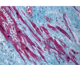
Microscopic analysis was carried out for all sections from the myocardial scar of the deceased patients who al-ready had bad myocardial infarction before the fatal one, re-vealed the preserved myocardial muscle fibers in the form of larger or smaller islets completely isolated from the sur-rounding preserved cardiac muscle, but surrounded by the connective tissue and without any contact with the normal muscle fibers (Figure 1).
Fig. 1 – Larger and smaller islets of the preserved muscle fibers in the myocardial scar (Malory, x200)
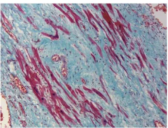
The preserved muscle fibers in the scar were hypertro-phic with large hypertrohypertro-phic and hyperchromatic nucleus and with the preserved transversal stria (Figure 2).
Fig. 2 – The preserved muscle fibers are hypertrophic with large hypertrophic and hyperchromatic nucleus (Masson
trichrom, x400)
Around the preserved muscle fibers there was the en-larged newly formed connective tissue, in some sites very dense and in the others loose. The newly formed connective tissue was well vascularized by the numerous newly formed blood vessels with thin walls, coated with the monolayer en-dothelium. The lumens of newly formed blood vessels were
Table 1 Reaction of cells present in the myocardial scar to used markers
Type of cells in the myocardial scar
Markers
Lympho-cytes
Leuko-cytes
Large oval cells abundant with
cytoplasm
Elongated cells
Endothelial cells
Smooth muscle cells in the blood
vessels wall
CD8 + – – – – –
CD20 + – – – – –
CD31 – – + + + –
CD34 + – – – + –
CD45 + + – – – –
CD45-Ro + – – – – –
Alpha ACTIN – – + + – +
Ki-67 – – + + – –
PCNA – – + + – –
ACTIN HHF35 – – + + – +
DESMIN – – + + – +
MYOGLOBIN – – + + – –
MYOSIN – – + + – –
LCA + + – – – –
SMA – – – – – +
PCNA – proliferating cell nuclear antigen; LCA – leukocyte common antigen; SMA – smooth muscle actin
Table 2 Tissue reactions to histochemical dyeing
Type of tissues Dyeing
Heart muscle fibers Collagen fibers Elastic fibers
PAS + – –
Masson trichrom + (red) + (green) + (green)
van Gieson + (yellow) + (red) + (red)
Malory + (red) + (green) + (green)
of various size and form and overfilled with blood. In the newly formed connective tissue (scar) there was a large number of both single cells and groups of cells of various sizes and forms (Figure 3).
Fig. 3 – A large number of cells of various sizes and forms can be seen in the newly formed connective tissue
(Masson trichrom, x200)
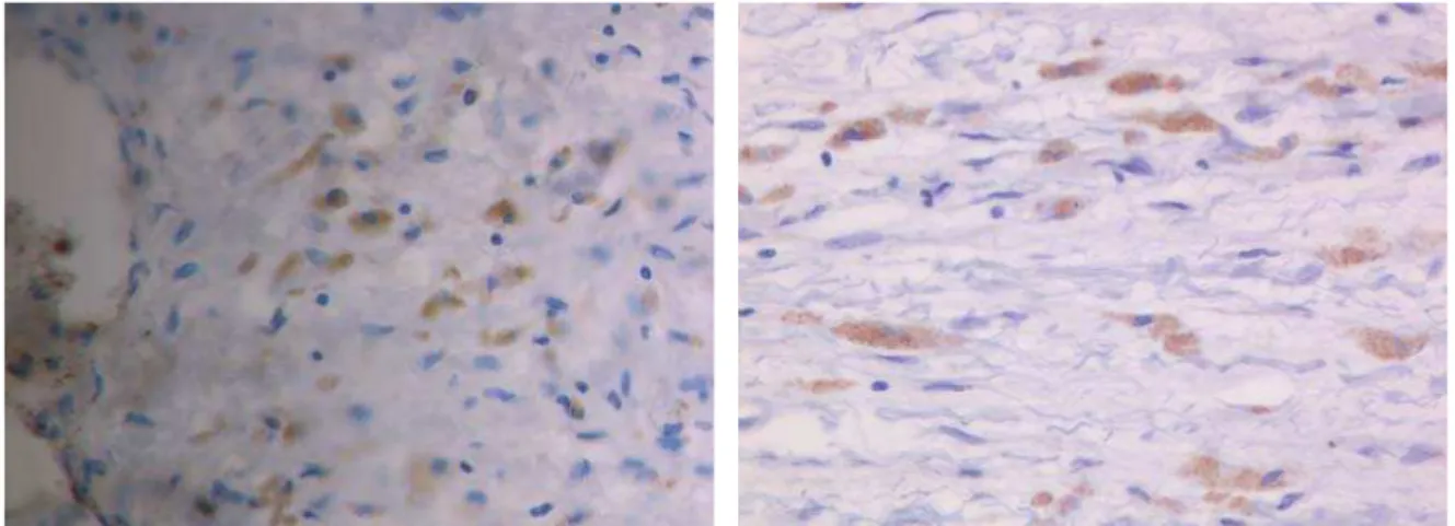
Some of the present cells were of the spindle form with a large hyperchromatic nucleus, the others were small with a little cytoplasm and one central nucleus. Some of these cells were oval with abundant cytoplasm and large circular nu-cleus. There were also elongated cells with abundant fine-grained cytoplasm and large circular nucleus. There were also cells with scanty cytoplasm so that only the nucleus was dominant. The present cells were mostly mononuclear and rarely with two or more nuclei. Only scattered blood vessels with a thick damaged wall could be seen in the scar. In more expressed vascularisation there was a larger number of cells. Myogenic, large oval cells with a large nucleus and abundant cytoplasm showed negative reaction to the lymphocytic and leukocytic markers CD34, CD45, CD20, CD8, CD45-Ro and LCA, but they were positive to alpha actin (Figure 4), actin
HHF35 (Figure 5), Ki-67 (Figure 6), myosin, myoglobin (Figure 7) and desmin. Elongated myogenic cells were also positive to these markers (which would speak for cardio-myocytes). Other cells found in the scar were negative to these markers. Small mononuclear cells that would corre-spond to the lymphocytes as well as leukocytes presented in the scar, showed reaction both to the lymphocytic and leuko-cytic markers (Figure 8).
Fig. 5 – Oval and elongated cells in the scar positive to actin HHF35 (x400)
Fig. 6 – Large oval cells in the scar positive to Ki-67; other cells are negative (x400)
Fig. 7 – Large oval and elongated cells in the scar positive to myoglobin (x400)
Volumen 69, Broj 7 VOJNOSANITETSKI PREGLED Strana 585
Fig. 8 – Lympocytes and leukocytes in the scar positive to LCA (x400)
CD34 and CD31 markers showed positive reactions to endothelial cells and smooth muscle cells in walls of the newly formed blood vessels in the scar. The usage of SMA markers resulted in positive reaction for the presence of the smooth muscle cells in walls of the newly formed blood ves-sels. The present oval cells abundant with cytoplasm and with the large nucleus as well as elongated mononuclear
cells showed positive reaction to PCNA markers (Figure 9), while spindle cells as well as other present cells were nega-tive to PCNA marker. Expression to Ki-67 nuclear antigen was positive in all nuclei of large, oval and elongated myo-genic cells (Figure 10).
Mitotic figures were noted in some nuclei of these cells suggesting cells division. These cells were also positive to PCNA marker for myocytic protein confirmation (Figures 9). The usage of myoglobulin, myosin and desmin markers showed positive reaction to the preserved muscle fibers in the scar (Figure 11), as well as to the elongated mononuclear cells and large, oval cells abundant with cytoplasm and with the large nucleus suggesting the presence of cardiac muscle fibers and most probably cardiomyocytes. Other cells were negative to these markers (Figures 1 and 2).
Discussion
Cardiac failure is the leading cause of both morbidity and mortality in developed countries 10. Hearts of adult per-sons are well supplied with blood and are capable to main-tain tissue integrity during physiologic cell function 1, 3, 4. Soon after birth the cardiac function is maintained by
in-Fig. 9 – Oval and elongated cells in the scar positive to PCNA; other cells were negative (x400)
Fig. 10 – Oval and elongated cells in the scar positive to Ki-67 (x400)
creasing both the number and volume of myocytes which to-gether contribute to development of the adult heart 2. The question is whether the heart possesses endogenous capacity to regenerate myocytes after myocardial infarction 1, 3, 4. Most of human bodily organs contain divisible and indivisi-ble cells. However, indivisiindivisi-ble cells can be in the Go phase and reenter the cellular cycle or become terminally differen-tiated without possible further division 3. But, these mecha-nisms of cell growth have not been accepted so far when the myocardium is concerned and it is believed that cardiomyo-cytes cannot be regenerated in cells by the division because in mammals their division was discontinued immediately after birth 1, 4–6, 15. On this basis human myocytes can live for the lifetime even if it were 100 years. It seems exceptionally extravagant that cardiomyocytes contracting 70 times a min-ute do that continually for 100 years. During that period they would contract 3.7 billion times 1, 3. If it is true, then cardio-myocytes are immortal cells 1, 3. It has been accepted that myocytes can enlarge their volume but not the number (the cardiac mass of a 42-weeks-old infant is 2.9 ± 6.2 g).
Nevertheless, some authors 3, 16 accept the possibility of myocyte division. In their studies they have measured the volume and number of human cardiac myocytes of patients died of decompensated cardiac hypertension and congestive cardiac failure. Mass of all the examined hearts was 500 g and more and it was characterized by noticeably increased myocyte number, more than cellular hypertrophy 3, 16. Some other authors 17 claim that methods used by these authors can only suggest, but not confirm the real increase in the cell number. It was believed that cardiac hypertrophy is a conse-quence of the present myocytes hypertrophy 4, 8, 18 and that the heart is a terminally differentiated postmitotic organ. After these studies had been published, generations of a great number of pathologists and cardiovascular experts accepted the fact that myocyte mitosis was not seen in the adult myo-cardium. However, in the course of the last 10–12 years a greater number of authors 1, 3–5, 7–11 have documented the presence of mitotic figures in human cardiomyocytes in cases of acute and chronic ischemic cardiomyopathy, idio-pathic dilatational cardiomyopathy, chronic aortic stenosis and ventricular dysfunction. Other authors 11, 19–21 emphasize that the formed parenchymatous cells are permanent in the normal myocardium, but that myocyte regeneration is also present in pathologic hearts, which they have microscopi-cally confirmed.
Recognition that stem cells are present in the myocar-dium raised a question of their role in the treatment of the af-fected human heart 9, 10, 17, 18, 22. Question related to the pres-ence and role of adult cardiac stem cells is based upon the fact that these stem cells regenerate in vivo in cases of the myocardial infarction 9. Urbanek et al. 22 emphasizes that the number of cardiac stem cells was increased in chronic myo-cardial infarction, and Quaini et al. 4 prove that the ventricu-lar myocytes are not terminally differentiated cells. Beltrami et al. 8 have analyzed sections taken from three patients died 4–12 days after myocardial infarction and compared findings of sections taken from the normal hearts of 9 control groups of patients. By using methods of immunohistochemistry in
sections taken from the deceased after myocardial infarction, the following was found: cardiomyocytes with elements characteristic for the cell division such as mitotic spindle and contractile ring formations, karyokinesis and cytokinesis. Similar findings were also found out in our own research. These findings confirm that there is a mitotic proliferation after myocardial infarction, and myocyte regeneration can contribute to increase in myocardial mass. This suggests that a prolonged cardiac failure can progressively influence upon mitotic activity. Regeneration initiation time depends on the span survival period after infarction and it starts already on the 10th day after experienced infarction. It reduces infarc-tion by 40%–50% after 20 days. Ten days after infarcinfarc-tion develops myocyte and blood vessels regeneration being im-proved in time 2, 8, 9.
The human heart, just like the brain, is mostly com-posed of terminally differentiated cells, but it is not a termi-nally differentiated organ because it contains stem cells that can develop its regeneration 2, 3, 13, 23–27. By the usage of Ki-67 nuclear antigen, Beltrami et al. 8 has found out positive reaction associated with cell division in all nuclei of the large oval and elongated cells. Similar results were found in our research. These cells were negative to lymphocyte, leu-kocyte and endothelial markers. Ki-67 is a nuclear antigen and in our research it was positive in myocytes of an infarc-ted heart suggesting that cardimyocytes are divisional. By using alpha actin Beltrami et al. 8 have found in these cells its accumulation in the contractile ring what was also confirmed in our study. These results show that in the adult heart after experienced infarction, there is a myocyte subpopulation not terminally differentiated and capable to divide soon after in-farcton. In our research cells that were positive to Ki-67 nu-clear antigen were also positive to the usage of: PCNA, alpha actin, actin HHF35, myoglobin, myosin and desmin, but showed negative reaction to lymphocyte, leukocyte and en-dothelial markers. Other authors' 1, 5, 8 finding are the same suggesting possible myocyte regeneration after myocardial infarction1, 2, 5, 8. It has been proved that heart stem cells are capable to differentiate into three types of heart cells: car-diomyocytes, smooth muscle cells in the blood vessel wall and endothelial cells 12. We have showed the presence of both smooth muscle and endothelial cells in the walls of the newly formed blood vessels in the scar after infarction. Stem cell factor can affect and activate all the three mentioned cells during myocardial ischemia and result in a significant increase in new myocyte formations 2, 12. Myocardium regen-eration requires myocyte and blood vessels formation be-cause myocytes cannot live and grow without blood vessels. However, blood vessels formation alone will not regenerate the dead myocardium and its contractile activity after infarc-tion7, 28, 29. The preserved muscle fibers that we have found isolated in the myocardial scar after infarction were hyper-trophic with the large nucleus suggesting their participation in synchronous contractions of heart muscle and possibly in prevention of the heart aneurysm development.
Volumen 69, Broj 7 VOJNOSANITETSKI PREGLED Strana 587
infarction6, 18, 30. Myocardium regeneration occurs in human ischemic and nonischemic heart damages. Heart stem cells localized within infarction or close to it can be divided and differentiated reconstructing consequently the myocardium. This response can increase the number of myocyte and blood vessels reducing in that way infarction size, improving its function and reducing mortality rate 6, 18, 30. The fact that myocardium is not a static organ and that cell reconstruction is not limited as well as both endothelial and vascular smooth muscle cells, requires reinterpretation of the heart biology and mechanism of its life.
Conclusion
The islets of the preserved cardiomyocytes (together with some other cells) surrounded by the connective tissue in the myocardial scar following myocardial infarction were found in our investigations. That it is about cardiomyocytes (and also about other cells) we have confirmed not only by
clasical histopathologic dyeing but also by immunohisto-chemical methods and by broad battery of tests to corre-sponding markers. On the other hand, the mentioned islets of the preserved cardiomyocytes were not found in sections taken from necrotic tissue in the repeated acute myocardial infarction resulting in the fatal outcome. The question is what, in fact, the found islets of the preserved cardiomyo-cytes in the scar of the preceded infarction represent? Are they the remains of the preserved myocardium in the infarc-ted region supplied by some arterial collateral? Is it corre-lated with the regenerated myocardium due to progenitory cells delivered by blood and differentiated into cardiomyo-cytetes, or with proliferated cardiac stem sells themselves? Also, what is the significance of these preserved islets of cardiomyocytes? Do they take part in myocardial contrac-tions? Are they the cause of cardiac arrhythmias in pathologic cases? The findings obtained and confirmed by this study are clear and therefore important and interesting, but further stud-ies are necessary to answer the above raised questions.
R E F E R E N C E S
1. Anversa P, Kajstura J, Leri A, Bolli R. Life and death of cardiac stem cells: a paradigm shift in cardiac biology. Circulation 2006; 113(11): 1451î63.
2. Leri A, Kajstura J, Anversa P. Cardiac stem cells and mecha-nisms of myocardial regeneration. Physiol Rev 2005; 85(4): 1373î416.
3. Anversa P, Leri A, Rota M, Hosoda T, Bearzi C, Urbanek K, et al. Concise review: stem cells, myocardial regeneration, and methodological artifacts. Stem Cells 2007; 25(3): 589î601. 4. Quaini F, Cigola E, Lagrasta C, Saccani G, Quaini E, Rossi C, et al.
End-stage cardiac failure in humans is coupled with the induc-tion of proliferating cell nuclear antigen and nuclear mitotic division in ventricular myocytes. Circ Res 1994; 75(6): 1050î63.
5. Laflamme MA, Murry CE. Regenerating the heart. Nat Biotech-nol 2005; 23(7): 845î56.
6. Limana F, Germani A, Zacheo A, Kajstura J, Di Carlo A, Borsellino G, et al. Exogenous high-mobility group box 1 protein induces myocardial regeneration after infarction via enhanced cardiac C-kit+ cell proliferation and differentiation. Circ Res 2005; 97(8): e73î83.
7. Beltrami AP, Urbanek K, Kajstura J, Yan SM, Finato N, Bussani R,
et al. Evidence that human cardiac myocytes divide after myo-cardial infarction. N Engl J Med 2001; 344(23): 1750î7. 8. Beltrami AP, Barlucchi L, Torella D, Baker M, Limana F, Chimenti
S, et al. Adult cardiac stem cells are multipotent and support myocardial regeneration. Cell 2003; 114(6): 763î76.
9. Pfister O, Mouquet F, Jain M, Summer R, Helmes M, Fine A, et al. CD31- but Not CD31+ cardiac side population cells exhibit functional cardiomyogenic differentiation. Circ Res 2005; 97(1): 52î61.
10.Kajstura J, Leri A, Finato N, Di Loreto C, Beltrami CA, Anversa P. Myocyte proliferation in end-stage cardiac failure in humans. Proc Natl Acad Sci U S A 1998; 95(15): 8801î5.
11.Pasumarthi KB, Nakajima H, Nakajima HO, Soonpaa MH, Field LJ. Targeted expression of cyclin D2 results in cardiomyocyte DNA synthesis and infarct regression in transgenic mice. Circ Res 2005; 96(1): 110î8.
12.Linke A, Müller P, Nurzynska D, Casarsa C, Torella D, Nascimbene A, et al. Stem cells in the dog heart are self-renewing, clono-genic, and multipotent and regenerate infarcted myocardium,
improving cardiac function. Proc Natl Acad Sci U S A 2005; 102(25): 8966î71.
13.Oh H, Bradfute SB, Gallardo TD, Nakamura T, Gaussin V, Mishina Y, et al. Cardiac progenitor cells from adult myocar-dium: homing, differentiation, and fusion after infarction. Proc Natl Acad Sci U S A 2003; 100(21): 12313î8.
14.Soonpaa MH, Field LJ. Survey of studies examining mam-malian cardiomyocyte DNA synthesis. Circ Res 1998; 83(1): 15î26.
15.Murry CE, Field LJ, Menasché P. Cell-based cardiac repair: re-flections at the 10-year point. Circulation 2005; 112(20): 3174î83.
16.Pasumarthi KB, Field LJ. Cardiomyocyte cell cycle regulation. Circ Res 2002; 90(10): 1044î54.
17.Schwartz RS, Curfman GD. Can the heart repair itself? N Engl J Med 2002; 346(1): 2î4.
18.Linzbach AJ. Heart failure from the point of view of quantita-tive anatomy. Am J Cardiol 1960; 5: 370î82.
19.Urbanek K, Quaini F, Tasca G, Torella D, Castaldo C, Nadal-Ginard B, et al. Intense myocyte formation from cardiac stem cells in human cardiac hypertrophy. Proc Natl Acad Sci U S A 2003; 100(18): 10440î5.
20.Beltrami AP, Urbanek K, Kajstura J, Yan SM, Finato N, Bussani R,
et al. Evidence that human cardiac myocytes divide after myo-cardial infarction. N Engl J Med 2001; 344(23): 1750î7. 21.Dawn B, Stein AB, Urbanek K, Rota M, Whang B, Rastaldo R, et
al. Cardiac stem cells delivered intravascularly traverse the ves-sel barrier, regenerate infarcted myocardium, and improve car-diac function. Proc Natl Acad Sci U S A 2005; 102(10): 3766î71.
22.Urbanek K, Rota M, Cascapera S, Bearzi C, Nascimbene A, De An-gelis A, et al. Cardiac stem cells possess growth factor-receptor systems that after activation regenerate the infarcted myocar-dium, improving ventricular function and long-term survival. Circ Res 2005; 97(7): 663î73.
23.Jackson KA, Majka SM, Wang H, Pocius J, Hartley CJ, Majesky MW, et al. Regeneration of ischemic cardiac muscle and vas-cular endothelium by adult stem cells. J Clin Invest 2001; 107(11): 1395î402.
multi-potent cardiac stem cells in ischemic heart failure. Proc Natl Acad Sci U S A 2005; 102(24): 8692î7.
25.Kawada H, Fujita J, Kinjo K, Matsuzaki Y, Tsuma M, Miyatake H, et al. Nonhematopoietic mesenchymal stem cells can be mobi-lized and differentiate into cardiomyocytes after myocardial in-farction. Blood 2004; 104(12): 3581î7.
26.Rubart M, Field LJ. Cardiac regeneration: repopulating the heart. Annu Rev Physiol 2006; 68: 29î49.
27.Scholzen T, Gerdes J. The Ki-67 protein: from the known and the unknown. J Cell Physiol 2000; 182(3): 311î22.
28.Nadal-Ginard B, Kajstura J, Leri A, Anversa P. Myocyte death, growth, and regeneration in cardiac hypertrophy and failure. Circ Res 2003; 92(2): 139î50.
29.Zak R. Development and proliferative capacity of cardiac muscle cells. Circ Res 1974; 35(2): suppl II: 17î26.
30. Marino TA, Haldar S, Williamson EC, Beaverson K, Walter RA, Marino DR, et al. Proliferating cell nuclear antigen in developing and adult rat cardiac muscle cells. Circ Res 1991; 69(5): 1353î60.