UNIVERSIDADE ESTADUAL PAULISTA “JÚLIO DE
MESQUITA FILHO”
FACULDADE DE MEDICINA
Mariana Miziara de Abreu Teodoro
Análise do padrão de resposta Th17 e de células T
regulatórias em pacientes com a forma digestiva da doença
de Chagas crônica
Dissertação apresentada à Faculdade de Medicina, Universidade Estadual Paulista “Júlio de Mesquita Filho”, Câmpus de Botucatu, para obtenção do título de Mestre em Doenças Tropicais.
Orientadora: Profa. Dra. Sueli Aparecida Calvi
Coorientadora: Profa. Dra. Ângela Maria Victoriano de Campos Soares
Coorientadora: Dra. Lucilene Delazari dos Santos
Mariana Miziara de Abreu Teodoro
Análise do padrão de resposta Th17 e de células T
regulatórias em pacientes com a forma digestiva da
doença de Chagas crônica
Dissertação apresentada à Faculdade de Medicina, Universidade Estadual Paulista “Júlio de Mesquita Filho”, Câmpus de Botucatu, para obtenção do título de Mestre em Doenças Tropicais.
Orientadora: Profa. Dra. Sueli Aparecida Calvi
Coorientadora: Profa. Dra. Ângela Maria Victoriano de Campos Soares
Coorientadora: Lucilene Delazari dos Santos
FICHA CATALOGRÁFICA ELABORADA PELA SEÇÃO TÉC. AQUIS. TRATAMENTO DA INFORM. DIVISÃO TÉCNICA DE BIBLIOTECA E DOCUMENTAÇÃO - CÂMPUS DE BOTUCATU - UNESP
BIBLIOTECÁRIA RESPONSÁVEL: ROSANGELA APARECIDA LOBO-CRB 8/7500
Teodoro, Mariana Miziara de Abreu.
Análise do padrão de resposta Th17 e de células T regulatórias em pacientes com a forma digestiva da doença de Chagas crônica / Mariana Miziara de Abreu Teodoro. - Botucatu, 2016
Dissertação (mestrado) - Universidade Estadual Paulista "Júlio de Mesquita Filho", Faculdade de Medicina de Botucatu Orientador: Sueli Aparecida Calvi
Coorientador: Ângela Maria Victoriano de Campos Soares Coorientador: Lucilene Delazari dos Santos
Capes: 40101096
1. Células T. 2. Chagas, Doença de. 3. Megaesôfago. 4. Megacólon. 5. Resposta imune.
Estudo
realizado
no
laboratório
do
Departamento de Doenças Tropicais e
Diagnóstico por Imagem da Faculdade de
Medicina de Botucatu – UNESP. Este
projeto contou com bolsa FAPESP
processo n
o2013/02223-7 e com auxílio à
pesquisa
FAPESP
processo
n
oE
“M
esmo que a vida pareça ruim, há sempre
algo que se pode fazer e obter sucesso.
Enquanto há esperança, há vida!
”
D
D
edico este estudo à minha querida orientadora Sueli.Obrigada pela oportunidade, incentivo, apoio,
dedicação, carinho, amizade e amor.
Sem você eu não teria chegado até aqui.
A
“N
enhum dever é mais importante que a gratidão.
”
(Cícero)
Agradeço à Deus, por toda força, paciência e aprendizado. Obrigada por me guiar e confortar com suas palavras e ensinamentos,
mas principalmente, por não me deixar perder o equilíbrio nos
momentos difíceis.
Meus agradecimentos especiais à minha família:
Agradeço aos meus pais, Oswaldo e Ana, por tudo que me ensinaram e
por me ajudarem a chegar até aqui; se hoje sou a pessoa que sou, é graças à todos os ensinamentos de vocês!
Vocês são meu orgulho, meu exemplo e minha vida! Amo vocês de todo meu coração!
Obrigada ao meu querido irmão, Bruno, por todo amor, por todas as risadas, pelo apoio e incentivo que sempre me deu. Obrigada por ser
tão especial!
força”, “Calma que vai dar tudo certo” e “Você consegue”. Obrigada por
estar comigo nos melhores e piores momentos da minha vida. Vocês são tudo pra mim! Amo vocês!
Agradeço ao meu querido marido, companheiro e eterno namorado, Leonardo, por toda paciência, apoio incondicional, confiança, suporte,
carinho e amor.
Você me faz ser uma pessoa melhor todos os dias!
Obrigada por estar sempre ao meu lado e me fazer enxergar o mundo de uma forma melhor e mais bonita!
Amo você!
Obrigada a meus sogros e sogras, cunhados e cunhadas por todo apoio sempre!
Agradeço também aos meus entes queridos que estão ou não mais presentes em minha vida, vocês contribuíram muito para que eu
chegasse até aqui, muito obrigada!
À minha querida orientadora Profa. Dra Sueli Aparecida Calvi, por se tornar parte da minha família. Obrigada pela oportunidade, por todos os
Os momentos que passei com você, carregarei para sempre! Obrigada
por mudar a minha vida! Você sempre será meu exemplo!
Meus sinceros agradecimentos:
À minha querida amiga Mariana Gatto, por toda ajuda, dedicação, cumplicidade, carinho e amizade. Obrigada por compartilhar comigo
todos os seus conhecimentos e por estar presente nos momentos bons e ruins que passamos. Você me ajudou em todas as partes deste trabalho sempre com a maior disposição. Te admiro muito e te levarei
pra sempre em meu coração.
À minha querida amiga Jéssica “Radinha”, por todo interesse em
aprender e por não medir esforços em me ajudar. Obrigada por todo companheirismo, carinho, atenção, amizade e principalmente, por fazer
com que nossas longas noites de experimentos, não parecessem tão longas assim. Obrigada por ser meu braço direito. Eu não teria
conseguido sem você!
À minha amiga e companheira de mestrado Laura, por toda ajuda, companheirismo e amizade. Nosso caminho durante o mestrado não foi
todas as risadas em nossas tardes de segunda-feira. Te desejo muito
sucesso!
Ao meu querido amigo Sebastião, pela amizade e pelas belas palavras de apoio nos momentos difíceis. Obrigada por sempre me ouvir,
aconselhar e dividir comigo os momentos de tristeza e alegria. Embora não tenha participado ativamente deste trabalho, você me ajudou muito.
Obrigada por sempre me lembrar que “não sou obrigada”. Ao colaborador deste trabalho, Dr. Rodrigo Mattos dos Santos, pela
grande ajuda na realização dos experimentos. Obrigada por me acalmar nos momentos de desespero, tentando me ajudar a encontrar soluções
para os problemas que apareceram ao longo do caminho.
Aos médicos Prof. Dr. Paulo Câmara Marques Pereira e a Dra. Érika Alessandra Pellison Nunes da Costa, por toda contribuição na triagem
dos pacientes para a realização deste trabalho.
À equipe do Laboratório de Citometria de Fluxo do Hemocentro de Botucatu, Aline, Léia e Carol, muito obrigada pela ajuda. Em especial
agradeço à Dra. Marjorie de Assis Golim, pelas análises e auxílio durante o desenvolvimento deste trabalho. Vocês foram essenciais para
Às minhas amigas de laboratório Drika, Thaty e Vanessa, obrigada pelos
bons momentos que vivemos juntas, por todos almoços e conversas. Esses momentos foram extremamente importantes para deixar mais
leve nossas vidas.
À Profa. Dra. Ângela Maria Victoriano de Campos Soares, por me acolher num momento tão difícil e me orientar com tanta excelência e
paciência neste último ano.
À banca do Exame Geral de Qualificação, em especial à Profa. Dra. Lenice do Rosário de Souza e Profa. Dra. Luciane Alarcão Dias-Melício,
pela cooperação e disposição em analisar esse trabalho. Obrigada pelas valiosas observações.
À todos os professores e funcionários do programa de Pós-graduação em Doenças Tropicais, do departamento de Doenças Tropicais e Diagnóstico por Imagem, da UNIPEX, da FMB-UNESP e do Hospital das
Clínicas de Botucatu.
Aos meus queridos amigos de faculdade, Fernanda, Marina, Ana Laura, Rodrigo, Priscila e Sarah, por sempre acreditarem e torcerem por mim.
Aos amigos de Bauru, Botucatu e de toda uma vida, obrigada por sempre tentarem estar por perto, mesmo com a distância e com o pouco
tempo disponível.
Aos amigos da comissão organizadora do “VIII Encontro da Pós-Graduação” pela companhia, conversas e trocas de experiências.
À Fundação de Amparo à Pesquisa do Estado de São Paulo (FAPESP) pela bolsa de estudo e auxílio à pesquisa concedidos.
Aos doadores de sangue e principalmente aos pacientes, que
voluntariamente e sem nenhum tipo de bonificação aceitaram participar deste estudo e dividiram comigo suas experiências de vida, me
ensinando grandes lições.
E, finalmente, à todos que de alguma forma contribuíram para que este trabalho fosse realizado e não foram citados.
Muito obrigada!
S
S
umário
umário
RESUMO
ABSTRACT
REVISÃO BIBLIOGRÁFICA ... 1
REFERÊNCIAS BIBLIOGRÁFICAS ... 8
CAPÍTULO 1 (artigo enviado para publicação) ... 13
CONCLUSÃO ... 33
R
R
esumo
esumo
A Doença de Chagas (DC), cujo agente etiológico é o protozoário Trypanosoma
cruzi (T. cruzi), tornou-se nos últimos anos um grave problema de saúde pública,
mesmo em países não-endêmicos. Um importante desafio para os pesquisadores da
doença de Chagas é estabelecer os eventos que levam alguns pacientes a
desenvolver a doença crônica assintomática, ou forma indeterminada, enquanto
outros evoluem para uma doença severa, com lesões em tecidos cardíacos ou
gastrointestinais. Alguns estudos têm sugerido que a ausência de sintomas clínicos
em indivíduos com a forma crônica indeterminada está associada a um controle
eficaz da resposta imune efetora contra o parasita, que evita uma resposta
inflamatória excessiva, o que resultaria em lesão tecidual. Não existem estudos com
o objetivo de confirmar este mecanismo em pacientes com a forma digestiva da DC,
especialmente em relação ao controle modulador de mecanismos efetores
envolvendo células Th17, por células Treg. No presente estudo, nós avaliamos em
pacientes com a forma digestiva da DC e em pacientes com a forma indeterminada
ou assintomática a taxa de ativação Th17/Treg, através da avaliação da freqüência
de células Th17 e Treg por citometria de fluxo; da expressão do mRNA para Rorγt e
Foxp3, fatores de transcrição envolvidos na diferenciação dessas células,
respectivamente; bem como os níveis de IL-17a e IL-10 em sobrenadantes de
cultura de células mononucleares de sangue periférico (PBMC’s). Foi demonstrado
no presente estudo, que em pacientes com a forma digestiva da DC há um
desequilíbrio na taxa de ativação de células Th17/Treg, favorecendo o perfil Th17,
o perfil Treg. Os nossos resultados apoiam a hipótese de que o desequilíbrio na taxa
de ativação Th17/Treg observado no grupo de pacientes digestivos, favorecendo
Th17 em detrimento de Treg, geraria um ambiente não regulador, que resulta em
uma resposta inflamatória exacerbada, responsável pelas lesões dos tecidos
A
A
bstract
bstract
Chagas disease (CD) whose etiological agent is the protozoan Trypanosoma
cruzi (T. cruzi) in recent years became a serious problem of public health even
in nonendemic countries. One important challenge for the researchers of Chagas
disease is to establish the events that lead some patients to develop
asymptomatic or indeterminate chronic disease, while others undergo severe
disease with lesions in cardiac or gastrointestinal tissues. Some studies have
suggested that absence of clinical symptoms in individuals with the
indeterminate chronic form is associated to a fine control of effector immune
responses against the parasite that avoid a perpetuated inflammatory process
which results in tissue injury. There are no studies aiming to confirm this
mechanism in patients with digestive form of the disease, particularly in relation
to the modulatory control by Treg cells of effector mechanisms involving Th17.
Here, we studied in patients with the digestive form of CD and in those with the
indeterminate or asymptomatic form of the disease, the ratio Th17/Treg
activation by evaluating the frequency of Th17 and Tregs cells by flow
cytometry, mRNA expression for Rorγt and Foxp3, the transcription factors
involved in the differentiation of these cells respectively as well as the levels of
IL-17a and IL-10 in peripheral blood mononuclear cells culture supernatants.
imbalance in the ratio Th17/Treg cells activation in favor of Th17, while in
patients with the indeterminate form, this imbalance favour Treg activation. Our
findings support the hypothesis that in contrast with indeterminate patients a
non regulatory environment observed in patients with digestive CD, which
results in an exacerbated inflammatory response exerted by Th17, due to lower
R
R
evisão
evisão
B
A doença de Chagas (DC), também conhecida como tripanosomíase
americana é causada pelo protozoário Trypanosoma cruzi (T. cruzi). É uma doença
negligenciada e considerada um grave problema de saúde pública com sérias
deficiências no tratamento e a falta de uma vacina eficaz, até o momento. É
endêmica na América Latina, embora, nas últimas décadas sua detecção tenha
aumentado nos EUA, Canadá e em vários países da Europa, devido, principalmente,
a migração de latino-americanos. Atualmente, existem aproximadamente 10 milhões
de pessoas infectadas no mundo todo e mais de 25 milhões vivendo em áreas de
risco. 1
A principal forma de transmissão da DC é a transmissão vetorial, mas
também pode ser transmitida por transfusão sanguínea, transplante de órgãos,
acidentes laboratoriais e transmissão vertical (da mãe para o feto). 2 Mais
recentemente, uma importante forma de transmissão foi evidenciada no Brasil, a via
oral, através de alimentos contaminados com as fezes do inseto vetor. 3
A DC evolui em duas fases, a aguda, que dura de 2 a 4 meses e é
caracterizada pela alta parasitemia. Na maioria das vezes não apresenta sinais e
sintomas específicos, o que dificulta sua detecção, porém se detectada e tratada a
tempo pode ser curada; e a fase crônica caracterizada pela baixa parasitemia, e que
persiste por toda a vida. 4
Na fase crônica, os pacientes podem apresentar 4 diferentes formas clínicas:
indeterminada, cardíaca, digestiva e mista; sendo que nesta última, temo
manifestações cardíacas e digestivas ao mesmo tempo. A grande maioria dos
pacientes que evoluem para a fase crônica permanece com sorologia positiva,
pacientes com a forma indeterminada da DC, de 30 a 40% podem evoluir para forma
sintomática, sendo que destes, aproximadamente, 10 % evoluem para a forma
digestiva. 5 Porém, não existe, até o momento, um marcador que determine se o
indivíduo irá evoluir ou não para as formas clínicas da doença.
A cardiomiopatia chagásica é resultado de um intenso processo inflamatório
com a presença de um grande número de células no miocárdio. Esta infiltração
celular pode ser uma resposta para o tropismo cardíaco do parasita e ou falhas nos
mecanismos de regulação do processo inflamatório. Esses mecanismos ainda não
estão totalmente entendidos, mas, provavelmente, depende de fatores genéticos do
hospedeiro, pois algumas pessoas, apesar da infecção, nunca desenvolvem doença
cardíaca. 6
As manifestações da forma digestiva da DC são atribuídas à destruição de
nervos do plexo mioentérico, o que leva a movimentos peristálticos descoordenados,
hipertrofia muscular e dilatação. 7 O megaesôfago e o megacólon são as maiores
causas de morbidade na forma clínica digestiva da DC crônica, sendo que para o
desenvolvimento do megaesôfago é necessária uma redução de aproximadamente
85% do número de neurônios, e no megacólon, uma perda de pelo menos 50% do
número de neurônios. 8 Patologicamente, ambos os órgãos, esôfago e cólon, exibem
um grande alargamento luminal e hipertrofia muscular. Microscopicamente,
infiltrados inflamatórios e fibroses encontradas foram associadas com lesões de
células musculares e do sistema nervoso intramural. 8,9 Os infiltrados inflamatórios
são compostos principalmente por linfócitos T CD3+CD4+, linfócitos B CD20+, células
NK CD57+ e macrófagos CD68+. 10,11 A persistência do kDNA do T. cruzi na lesão
crônica sugere um papel do parasita na manutenção da ativação celular e do
A causa da destruição neural na DC tem sido debatida na literatura. Na fase
aguda, quando o T. cruzi está presente em números elevados no tecido, o parasita
pode ser responsável pelas lesões neurais. Em contraste, a carga parasitária é
muito baixa nas lesões chagásicas durante a fase crônica. Além disso, a frequente
ocorrência de ganglionite e periganglionite em pacientes desenvolvendo
megaesôfago e/ou megacólon aponta para a participação das células do sistema
imune nesse processo. 15 Além disso, foi demonstrada, uma forte associação entre a
taxa de desenervação e a presença de células com potencial citotóxico. 16 Apesar da
importância desta forma da doença em áreas endêmicas, os mecanismos
imunológicos envolvidos no dano tecidual não têm recebido atenção adequada nos
últimos anos. 17
Muitas das manifestações clínicas observadas na DC são consequências da
resposta imune do hospedeiro contra o parasita. 18 Em cada fase da infecção existe
uma resposta específica, com repertório de células, citocinas e outras substâncias
que reduzem a carga parasitária, auxiliando na defesa do organismo, mas que
também estão envolvidas na patogenia da doença.
Assim, após a infecção, a imunidade celular tenta isolar o parasita a fim de
evitar sua disseminação e a resposta humoral, simultânea, produz, inicialmente,
anticorpos da classe IgM e, após 2 a 3 semanas, da classe IgG. No entanto, a
ineficiência dos mecanismos imunes para eliminar o T. cruzi garante sua
persistência no organismo e desencadeia resposta inflamatória, resultando em
danos aos tecidos durante a fase crônica da doença. 19
Linfócitos de pacientes chagásicos e animais infectados com T. cruzi são
autoimune. 20 Alguns autores acreditam que persistência do parasita e
autoimunidade coexistem na doença de Chagas e que estratégias de invasão do
parasita e defeitos na homeostasia imune do hospedeiro são importantes para o
desenvolvimento das formas clínicas da doença. 21 Estudos demonstraram que uma
forte resposta inflamatória pode ser encontrada em estágios avançados da doença,
mesmo na ausência do parasita. 22-24
O repertório de células do sistema imune, com alto nível de ativação,
observado durante a fase aguda da infecção chagásica, compreende macrófagos,
células NK, T CD4+ e T CD8+. 25-28 O T. cruzi pode induzir a síntese de IL-12 e de
outras citocinas por macrófagos e células dendríticas.29-31 A resposta Th1 que se
segue caracteriza-se pela produção de IFN-γ por linfócitos T, com consequente
aumento na ativação das células fagocíticas para destruir parasitas internalizados,
aumentando H2O2, o óxido nítrico e a produção de TNF-α. 32-35 As células NK
desempenham também um papel importante na resistência à infecção aguda,
principalmente devido à sua produção precoce de IFN-γ. 36,37 Animais tratados com
anti-IFN-γ apresentam-se mais susceptíveis à infecção pelo parasita, enquanto
aqueles que recebem IFN-γ tornam-se mais resistentes. 38-40 Michailowsky et al. 41
demonstraram ação sinérgica do IFN-γ com o tratamento antiparasitário na fase
aguda da infecção. No entanto, esta citocina também tem sido relacionada ao
desenvolvimento de cardiopatia na fase crônica da infecção, o que sugere que tenha
um duplo papel na DC. 42
O TNF-α é outra citocina pró-inflamatória com papel destacado na modulação
da resposta imune protetora na infecção pelo T. cruzi. 43,44 Essa substância controla
Assim como a IL-12 participa da diferenciação em resposta Th1, a IL-4
promove a resposta Th2, indutora de imunidade humoral, na ausência de IL-12 e
IFN-γ. 46 Além disso, o aumento de secreção de IL-10, durante a fase crônica, pode
estar associado à proteção do hospedeiro contra resposta inflamatória intensa,
induzida pelo perfil Th1. Essa citocina, também do perfil Th2, é produzida pelos
macrófagos e regula a expressão ou função de IL-12 e INF-γ.47,48 Dutra et al. 49
detectaram níveis mais altos de IL-5, IL-10, IL-13, e IFN-γ em pacientes chagásicos
crônicos em relação a indivíduos não infectados, sugerindo que o equilíbrio entre
essas citocinas pode ser o ponto principal do controle da morbidade, durante a fase
crônica da infecção.
A resistência de um organismo às infecções requer a geração de uma
resposta imune que, não só, controle o patógeno invasor, mas, também, que limite
os danos colaterais aos tecidos do próprio hospedeiro, resultantes dessa resposta. 50
Assim, ao mesmo tempo em que os elementos efetores agem no sentido de eliminar
o parasita, mecanismos reguladores são ativados, minimizando a lesão tecidual
provocada pela própria resposta imune. 50,51 As células T usam diferentes
mecanismos para regular a resposta imune durante a doença de Chagas, e as
interações parasita-hospedeiro podem ser influenciadas pela relação células T
reguladoras/efetoras. 52
As células T regulatórias (Tregs) foram descritas como uma população de
células T CD4+CD25+Foxp3+, em que sua principal característica é o fator de
transcrição Forkhead Box P3, (Foxp3) que regulam as respostas imunes inata e
adaptativa. 52 As Tregs modulam as respostas imunes, não só para antígenos
próprios e tumorais, mas também a antígenos exógenos e agentes infecciosos. 53-60
CD4+ e CD8+, regular negativamente a ativação e capacidade citolítica de células T
CD8+, e modular as respostas imunes humorais. 61-63 Desta forma, as células Treg
servem para limitar o dano tecidual, mas também podem impedir a eliminação da
infecção por supressão de respostas imunes cruciais. 64
As Tregs através da expressão de IL-10 e TGF-β atuam beneficiando os
pacientes com a forma indeterminada da DC, mantendo o equilíbrio entre células
efetoras que matam os parasitas e evitando o desenvolvimento da imunopatologia
tecidual. 52
Embora não haja evidência para a verdadeira função desta população de
células na doença de Chagas, já foi demonstrado que, no sangue periférico de
pacientes indeterminados há um aumento na frequência de circulação de células
Tregs CD4+CD25+Foxp3+ e esses pacientes também apresentam um aumento
significativo na concentração de células Foxp3+ no tecido do coração, sugerindo que
estas células podem estar envolvidas no controle da morbidade da doença de
Chagas. 65-67
Nos últimos anos, a explicação de resistência e suscetibilidade a infecções
parasitárias baseada na dicotomia Th1 e Th2 e na atuação das células Tregs têm
sofrido uma análise mais cuidadosa, a partir de estudos mostrando que durante o
processo de diferenciação para células efetoras as células CD4+ podem seguir
destinos alternativos que não envolvem Th1, Th2 ou Treg. Nesse sentido, as
células CD4+ cultivadas na presença de TGF-β, IL-6, IL-1β, IL-23 e IL-21 passam a
expressar o fator de transcrição RORγt (“retinoid-related orphan receptor gamma”) e
transformam-se na subpopulação Th17 que desempenha um papel atribuído
IL-17A, IL-17F, IL-21, IL-22 e TNF-α, promovem um processo inflamatório intenso
mediado por neutrófilos e estão associadas a doenças autoimunes e à resistência
contra várias infecções bacterianas e parasíticas, mas também podem
correlacionar-se com imunopatologia em diversas outras infecções. 69 Dentre as citocinas já
citadas, importantes para a diferenciação de Th17 68, destacamos a IL-23, produzida
principalmente por APCs ativadas, em particular células de Langerhans, macrófagos
e células dendríticas. O receptor de IL-23 está presente em células de memória T,
células NK T, macrófagos, células dendríticas e células T naive. É a citocina chave
que impulsiona a diferenciação de células T naive para células Th17 uma vez que na
sua ausência o perfil de resposta CD4 resulta na diferenciação de células Treg.
Estudos demonstram que a IL-17 é produzida durante a fase aguda da DC em
camundongos e controla a inflamação cardíaca modulando a resposta Th1. 70
Miyazaki et al 71 relataram que a IL-17 produzida durante infecção com T. cruzi
resulta na ativação eficiente da resposta imune contra o parasita, uma vez que
camundongos deficientes dessa citocina apresentaram níveis mais baixos de IFN-γ,
prolongada parasitemia e mortalidade quando comparados aos controles. Outros
estudos associam a IL-17 à destruição de neurônios em modelos experimentais de
EAE (experimental autoimmune encephalomyelitis). 72 Poucos estudos relacionam
as células Th17 e suas citocinas a doenças digestivas, particularmente no que se
refere a lesões da DC
Os estudos acima apontam para a necessidade de se estabelecer os
desequilíbrios imunológicos envolvidos na patogênese das lesões digestivas na
REFERÊNCIAS BIBLIOGRÁFICAS
1- OMS [Internet]. OMS [Atualizada em: junho/2010; acesso em: 10/06/2012]. Chagas disease (American trypanosomiasis). Disponível em: www.who.int/mediacentre/factsheets/fs340/en/.
2- Lana M, Tafuri WL. Trypanosoma cruzi e Doença de Chagas. In: Neves DP, Melo AL, Linardi PM, Vitor RWA. Parasitologia humana. Edição (1). São Paulo: Atheneu; 2005. 85-108.
3- Drauzio Varella.com.br [Internet]. São Paulo: Estação saúde – educação e cultura LTDA. [Acesso em: 10/07/2012]. Doença de Chagas. Disponível em:
www.drauziovarella.com.br/doencas-e-sintomas/doenca-de-chagas/.
4- Dutra WO, Menezes CAS, Villani FNA, Costa GC, Silveira ABM, d’Avilla Reis D, et al. Cellular and genetic mechanisms involved in the generation of protective and pathogenic immune responses in human Chagas disease. Mem Inst Oswaldo Cruz. 2009; 104(1): 208-18.
5- Umezawa ES, Stolf AM, Corbett CE, Shikanai-Yasuda MA. Chagas’ disease. Lancet. 2001; 357: 797– 99.
6- Gutierrez FRS, Guedes PMM, Gazzinelli RT, Silva JS. The role of parasite persistence in pathogenesis of Chagas heart disease. Paras Immunol. 2009; 31:673-85.
7- Tostes Jr S, Lopes ER, Pereira FE, Chapadeiro E. Miocardiopatia chagásica crônica humana: estudo quantitativo de linfócitos CD4+ e CD8+ nos exsudatos inflamatórios. Rev Soc Brasil Med Trop. 1994; 27: 127-34.
8- Köberle F. Chagas’ disease and Chagas’ syndromes: the pathology of American trypanosomiasis. Adv Parasitol. 1968; 6: 63-116.
9- Adad SJ, Cançado CG, Etchebehere RM, Teixeira VP, Gomes UA, Chapadeiro E, Lopes ER. Neuron count reevaluation in the myoenteric plexus of chagasic megacolon after morphometric neuron analysis. Virchows Arch. 2001; 438: 254-58.
10- Corbett CE, Ribeiro U Jr, Prianti MG, Habr-Gama A, Okumura M, Gama-Rodrigues J. Cell-mediated immune response in megacolon from patients with chronic Chagas’ disease. Dis Colon Rectum. 2001; 44: 993-98.
11- d’Avila Reis D, Lemos EM, Silva GC, Adad SJ, McCurley T, Correa-Oliveira R, Machado CR. Phenotypic characterization of the inflammatory cells in chagasic megaoesophagus. Trans R Soc Trop Med Hyg. 2001; 95: 177-78.
12- Jones EM, Colley DG, Tostes S, Lopes ER, Vnencak-Jones CL, McCurley TL. Amplification of a T. cruzi DNA sequence from inflammatory lesions in human chagasic cardiomyopathy. Am J Trop Med Hyg. 1993; 48: 348-57.
13- Vago AR, Macedo AM, Adad SJ, Reis DD, Corrêa-Oliveira R. PCR detection of Trypanosoma cruzi DNA in oesophageal tissues of patients with chronic digestive Chagas’ disease. Lancet. 1996; 348: 891-92.
14- Vago AR, Andrade LO, Leite AA, d’Avila Reis D, Macedo AM, Adad SJ, Tostes S Jr, Moreira MC, Filho GB, Pena SD. Genetic characterization of Trypanosoma cruzi directly from tissues of patients with chronic Chagas disease: differential distribution of genetic types into diverse organs. Am J Pathol. 2000; 156: 1805-1809.
16- da Silveira AB, Adad SJ, Correa-Oliveira R, Furness JB, D’Avila Reis D. Morphometric study of eosinophils, mast cells, macrophages and fibrosis in the colon of chronic chagasic patients with and without megacolon. Parasitology. 2007; 134: 789-96.
17- Oliveira EC, Fujisawa MM, Hallal Longo DEM, Farias AS, Moraes JC, Guariento ME, et al. Neuropathy of Gastrointestinal Chagas’ Disease: Immune Response to Myelin Antigens. Neuroimmunomodulat. 2009; 16: 54–62.
18- Tarleton RL, Zhang L. Chagas disease etiology: autoimmunity or parasite persistence? Parasitol Today. 1999; 15: 94–9.
19- Tarleton RI. Parasite persistence in the actiology of Chagas disease. Int J Parasitol 2001; 31:550-4.
20- Cunha-Neto E, Duranti M, Gruber A, Zingales B, De Messias I, Stolf N, et al. Autoimmunity in Chagas disease cardiopathy: biological relevance of a cardiac myosin-specific epitope crossreactive to an immunodominant Trypanosoma cruzi antigen. Proc Natl Acad Sci. 1995;92: 3541–45.
21- Tarleton RL, Zhang L. Chagas disease etiology: autoimmunity or parasite persistence? Parasitol Today. 1999; 15: 94–9.
22- Acosta AM, Santos-Buch CA. Autoimmune myocarditis induced by Trypanosoma cruzi. Circulation 1985; 71: 1255– 61.
23-
24- Leon JS, Daniels MD, Toriello KM, Wang K & Engman DM. A cardiac myosin-specific autoimmune response is induced by immunization with Trypanosoma cruzi proteins. Infect Immun 2004; 72: 3410– 17.
25- Cardillo F, Voltarelli JC, Reed SG, Silva JS. Regulation of Trypanosoma cruzi infection in mice by gamma interferon and interleukin-10: the role of NK cells. Infect Immun. 1996; 64:128-34.
26- Minoprio P, Eisen H, Joskowicz M, Pereira P, Coutinho A. supression of polyclonal antibody production in murine Trypanosoma cruzi infected mice by treatment with anti-L3T4 antibodies. JImmunol. 1987; 139: 545-50.
27- Tarleton RL. Depletion of CD8+ T cells increases susceptibility and reverses vaccine-induce immunity in mice infected with Trypanosoma cruzi. J Immunol. 1990; 144: 717-24.
28- Tarleton RL, Koller BH, Latour A, Postan M. Susceptibility of β2-microglobulin-deficient mice of Trypanosoma cruzi infection. Nature. 1992; 356: 338-40.
29- Aliberti JC, Cardoso MA, Martins GA, Gazzinelli RT, Vieira LQ, Silva JS. Interleukin-12 mediates resistance to Trypanosoma cruzi in mice and is produced macrophages in response to live
trypomastigotes. Infect Immun. 1996; 64:1961-7.
30- Camargo MM, Almeida IC, Pereira MES, Ferguson MAJ, Travassos LR, Gazzinelli RT. GPI anchored mucin-like glycoproteins isolated from Trypanosoma cruzi trypomastigotes initiate the synthesis of pro-inflammatory cytokines by macrophages. J Immunol. 1997; 158: 5890-1.
31- Hunter CA, Slifer T, Araújo F. Interleukin-12-mediated resistance to Trypanosoma cruzi is dependent on tumor necrosis factor alpha and gamma interferon. Infect Immun. 1996; 64: 2381-6.
32- Nogueira N, Ellis J, Chaplan S, Cohn Z. Trypanosoma cruzi: in vivo and in vitro correlation between T-cell activation and susceptibility in inbred strains of mice. Exp Parasitol. 1981; 51(3) 325– 34.
oxide-mediated mechanism inhibitable by interleukin-10 and transforming growth factor-beta. Eur J Immunol. 1992; 22 (10) 2501–6.
34- Vespa GNR, Cunha FQ, Silva JS. Nitric oxide is involved in control of Trypanosoma cruzi-induced parasitemia and directly kills the parasite in vitro. Infect Immun. 1994; 62 (11) 5177–82.
35- Silva JS, Vespa GNR, Cardoso MAG, Aliberti JCS, Cunha FQ. Tumor necrosis factor alpha mediates resistance to Trypanosoma cruzi infection in mice by inducing nitric oxide production in infected gamma interferon-activated macrophages. Infect Immun. 1995; 63 (12) 4862–67.
36- Rottenberg ME, Cardoni RL, Andersson R, Segura EL, Örn A. Resistance to Trypanosoma cruzi requires T helper/inducer cells as well as natural killer cells. Scand J. Immunol. 1988; 28: 573–82.
37- Cardillo F, Voltarelli JC, Reed SG, Silva JS. Regulation of Trypanosoma cruzi infection in mice by gamma interferon and interleukin 10: role of NK cells. Infect Immun. 1996; 64 (1) 128–34.
38- Silva JS, Morrissey PJ, Grabstein KH, Mohler MK, Andreson D, Reed SG. Interleukin-10 and interferon-γ regulation of experimental Trypanosoma cruzi infection. J Exp Med 1992; 175: 169- 74.
39- Ortiz-Ortiz L, Ortega T, Capin R, Martinez T. Enhanced mononuclear phagocytic activity during Trypanosoma cruzi infection in mice. Int Arch Allergy Appl Immunol 1976; 50: 232-42.
40- Torrico F, Heremans H, Rivera MT, Van Marck E,Billiau A, Carlier Y. Endogenous IFN-γ is required for resistance to acute Trypanosoma cruzi infection in mice. J Immunol 1991; 146: 3626-32.
41- Michailowsky V, Murta SMF, Carvalho-Oliveira L, Pereira MES, Ferreira LRP, Brener Z, et al. Interleukin-12 enhances in vivo parasiticidal effect of benzidazole during acute experimental infection with a naturally drug-resistant strain of Trypanosoma cruzi. Antimicrob. Agents Chemother 1998; 42(10): 2549-56.
42- Gomes JA, Bahia-Oliveira LM, Rocha MO, Martins-Filho OA, Gazzinelli G, Corrêa-Oliveira R. Evidence that development of severe cardiomyopathy in human Chagas’ disease is due to a Th1-specif immune response. Infect Immun 2003; 71: 1185-93.
43- Abrahamsohn IA, Coffman R. Cytokine and Nitric Oxide regulation of the immunosuppresion in Trypanosoma cruzi infection. J Immunol. 1995; 155: 3955-63.
44- Aliberti JC, Souto JT, Marino AP, Lannes-Vieira J, Teixeira MM, Farber J, et al. Modulation of chemokine production and inflammatory responses in interferon-gamma and tumor necrosis factor-R1-deficient mice during Trypanosoma cruzi infection. Am J Pathol 2001; 158: 1433-40.
45- Abrahamsohn IA. Cytokines in innate and acquires immunity to Trypanosoma cruzi. Bras J Medical and Biological Research 1998; 31: 117-21.
46- Seder RA, Paul WE. Acquisition of lymphokine-producing phenotype by CD4+ T cells. Annu Rev Immunol 1994; 12: 635-73.
47- Brener Z, Gazzinelli RT. Immunological control of Trypanosoma cruzi infection and pathogenesis of Chagas’ disease. Int Arch Allergy Immunol 1997; 114:103-10.
48- Gazzinelli RT, Oswald IP, Hieny S, James S, Sher A. The microbicidal activity of interferon-γ
treated macrophages against Trypanosoma cruzi involves an L-arginine-dependent, nitrogen oxide-mediated mechanism inhibitable by interleukin-10 and transforming growth factor-β. Eur J Immunol 1992; 22: 2501-06.
49- Dutra WO, Gollob KJ, Pinto-Dias JC, Gazzinelli G, Correa-Oliveira R, Coffman RL, et al.Cytokine mRNA profile of peripheral blood mononuclear cells isolated from individuals with Trypanosoma cruzi chronic infection. Scand J Immunol 1997; 44: 74-80.
51- Kotner J, Tarlenton R. Endogenous CD4+CD25+ regulatory T cells have a limited in the control of Trypanosoma cruzi infection in mice. Infect Immun. 2007; 75:861-9.
52- de Araújo FF, Vitelli-Avelar DM, Teixeira-Carvalho A, Renato Zuquim Antas P, Assis Silva Gomes J, et al. Regulatory T Cells Phenotype in Different Clinical Forms of Chagas’ Disease. PLoS Negl Trop Dis. 2011: 5(5): e992.
53- Onizuka S, Tawara I, Shimizu J, Sakaguchi S, Fujita T, E. Nakayama E. Tumor rejection by in vivo administration of anti-CD25 (interleukin-2 receptor alpha) monoclonal antibody. Cancer Res. 1999; 59:3128–33.
54- Sakaguchi S. Naturally arising CD4+ regulatory T cells for immunologic self-tolerance and negative control of immune responses. Annu Rev Immunol. 2004; 22:531–62.
55- Sakaguchi S. Regulatory T cells: key controllers of immunologic self-tolerance. Cell. 2000; 101:455–458.
56- Kretschmer K, Apostolou I, Hawiger D, Khazaie K, Nussenzweig MC, von Boehmer H. Inducing and expanding regulatory T cell populations by foreign antigen. Nat Immunol. 2005; 6:1219–1227.
57- Belkaid Y. The role of CD4+CD25+ regulatory T cells in Leishmania infection. Expert Opin Biol Ther. 2003; 3:875–885.
58- Hisaeda H, Maekawa Y, Iwakawa D, Okada H, Himeno K, Kishihara K, et al. Escape of malaria parasites from host immunity requires CD4_ CD25_ regulatory T cells. Nat. Med. 2004; 10:29–30.
59- Mendez S, Reckling SK, Piccirillo CA, Sacks D, Belkaid Y. Role for CD4+ CD25+ regulatory T cells in reactivation of persistent leishmaniasis and control of concomitant immunity. J Exp Med. 2004; 200: 201–210.
60- Suvas S, Kumaraguru U, Pack CD, Lee S, Rouse BT. CD4+CD25+ T cells regulate virus-specific primary and memory CD8+ T cell responses. J Exp Med. 2003; 198:889–901.
61- Thornton AM, Piccirillo CA, Shevach EM. Activation requirements for the induction of CD4+CD25+ T cell suppressor function. Eur J Immunol. 2004; 34:366–76.
62- Piccirillo CA, Shevach EM. Cutting edge: control of CD8+ T cell activation by CD4+CD25+ immunoregulatory cells. J Immunol. 2001; 167: 1137–40.
63- Zhao DM, Thornton AM, Dipaolo RJ, Shevach EM. Activated CD4+CD25+ T cells selectively kill B lymphocytes. Blood 2006; 107: 3925–3932.
64- Belkaid Y, Rouse BT. Natural regulatory T cells in infectious disease. Nat Immunol. 2005; 6: 353– 60.
65- de Araújo FF, Vitelli-Avelar DM, Teixeira-Carvalho A, Antas PRZ, Gomes JAS, Sathler-Avelar R, et al. Regulatory T cells phenotype in different clinical forms of Chagas disease. PLoS Negl. Trop. Dis. 2011; 5: 992–9.
66- de Araújo FF, da Silveira AB, Correa-Oliveira R, Chaves AT, Adad SJ, Fiuza JA. Characterization of the presence of Foxp3(+) T cells from patients with different clinical forms of Chagas’ disease. Hum Pathol. 2011; 42: 299–301.
67- Araujo FF, Gomes JAS, Rocha MOC, Williams-Blangero S, Pinheiro VM, Morato MJ, et al.
Potential role of CD4+CD25High regulatory T cells in morbidity in Chagas disease. Front Biosci. 2007; 12: 2797–2806.
69- Stockinger B, Veldhoen M, Martin B. Th17 cells: linking innate and adaptative immunity. Semin Immunol 2007; 19: 353-61.
70- da Matta Guedes PM, Gutierrez FR, Maia FL, Milanezi CM, Silva GK, Pavanelli WR, et al. IL-17 Produced during Trypanosoma cruzi Infection Plays a Central Role in Regulating Parasite-Induced Myocarditis. PLoS Negl Trop Dis 2010; 4(2): e604.
71 - Miyazaki Y, Hamano S, Wang S, Shimanoe Y, Iwakura Y, Hiroki Yoshida H. IL-17 is necessary for host protection against acute-phase Trypanosoma cruzi infection. Journ Immunol. 2010; 185:1150-57.
C
________________________________________________________________
Artigo enviado para publicação na PLOS One
Th17 and Treg cells evaluation in patients with the digestive form of
chronic Chagas disease
Mariana Miziara de Abreu1; Mariana Gatto1; Jéssica Cristina Bilizário Noguerol
Andrade1; Laura Denise Mendes da Silva1; Marjorie de Assis Golim2; Érika
Alessandra Pellison Nunes da Costa1; Rodrigo Mattos dos Santos1; Paulo
Câmara Marques Pereira1; Ângela Maria Victoriano de Campos Soares3; Sueli
Aparecida Calvi1
1
Tropical Diseases Department, Botucatu School of Medicine – UNESP, Botucatu, São
Paulo, Brazil
2
Flow Cytometry Laboratory, Hemocenter, Botucatu School of Medicine – UNESP,
Botucatu, São Paulo, Brazil
3
Microbiology and Immunology Department, Biosciences Institute – UNESP, Botucatu, São
Paulo, Brazil
Corresponding author: [email protected]
ABSTRACT
Chagas disease (CD) whose etiological agent is the protozoan Trypanosoma
cruzi (T. cruzi) in recent years became a serious problem of public health even
in nonendemic countries. One important challenge for the researchers of Chagas
disease is to establish the events that lead some patients to develop
asymptomatic or indeterminate chronic disease, while others undergo severe
disease with lesions in cardiac or gastrointestinal tissues. Some studies have
suggested that absence of clinical symptoms in individuals with the
indeterminate chronic form is associated to a fine control of effector immune
responses against the parasite that avoid a perpetuated inflammatory process
which results in tissue injury. There are no studies aiming to confirm this
mechanism in patients with digestive form of the disease, particularly in relation
to the modulatory control by Treg cells of effector mechanisms involving Th17.
Here, we studied in patients with the digestive form of CD and in those with the
indeterminate or asymptomatic form of the disease, the ratio Th17/Treg
activation by evaluating the frequency of Th17 and Tregs cells by flow
cytometry, mRNA expression for Rorγt and Foxp3, the transcription factors
involved in the differentiation of these cells respectively as well as the levels of
IL-17a and IL-10 in peripheral blood mononuclear cells culture supernatants.
We showed that in patients with digestive form of the disease there is an
patients with the indeterminate form, this imbalance favour Treg activation. Our
findings support the hypothesis that in contrast with indeterminate patients a
non regulatory environment observed in patients with digestive CD, which
results in an exacerbated inflammatory response exerted by Th17, due to lower
Treg activation, may be responsible for tissue lesions in these patients.
INTRODUCTION
Chagas disease (CD) whose etiological agent is the protozoan Trypanosoma
cruzi (T. cruzi) is a neglected disease, however is a serious problem of public
health. It is endemic in Latin America, but in recent decades its detection has
increased in the USA, Canada and several European countries, where most of
the Latin immigrants are now living. Currently, there are about 10 million
people infected worldwide and more than 25 million living in risk areas. The
clinical course of chronic disease is variable and ranges from the absence of
symptoms to severe disease. Most chronically infected individuals present the
indeterminate or asymptomatic chronic disease characterized by positive
serology and no clinical manifestations. However, about 30 to 40% of them
develop symptomatic CD with cardiovascular (cardiac form) or gastrointestinal
(digestive form) involvements [1].
The clinical manifestations of the digestive CD form are attributed to the
destruction of myenteric plexus nerves which leads to uncoordinated peristaltic
megacolon are the major causes of high morbidity in patients with this clinical
form. Pathologically, both, esophagus and colon, exhibit a large luminal
enlargement and muscle hypertrophy. Histologically, inflammatory infiltrate and
fibrosis were found associated withs lesions in muscle cells and in intramural
nervous system [3-4]. Inflammatory infiltrates composed primarily by T
lymphocytes CD3+CD4+, B cells CD20+, NK cells CD57+ and macrophages
CD68+ have been attributed to constant cell activation due to parasite persistence
since T. cruzi kDNA is frequently detected in chronic lesions [5-9].
Host resistance to infection requires the generation of immune response effector
mechanisms able to effectively control the invading pathogen. However, at same
time, these mechanisms, if exacerbated, can be closely related to tissue damage.
Thus, while the effector elements act to eliminate the parasite, regulatory
mechanisms must be activated in order to minimizing tissue damage caused by
them [10-11].
Studies have shown that the main host effector mechanisms against T. cruzi
involve Th1 response with production ofIFN-γ and TNF-α which activate cells
to eliminate the parasite [12-15]. However, there are few studies evaluating the
role of Th17, another important proinflammatory subpopulation of CD4 cells.
Naïve CD4 + cells cultured in the presence of TGF-β, IL-6, IL-1β, 23 and
IL-21 express the transcription factor RORγt ("retinoid-related orphan receptor
IL-17A, IL-17F, IL-21, IL-22 and TNF-α and promote an intense inflammatory
process mediated by neutrophils may be associated with resistance against
various bacterial and parasitic infections, but also with immunopathology in
others [16]. These findings lead us to suppose that protection induced by Th17
occurs in situations where a concomitant modulatory response is induced.
Regulatory T cells (Treg cells) are probably the major CD4+ cells subset
involved in this control.
Treg cells have been described as a CD4+CD25+ T cells that expresses the
transcription factor Foxp3. By producing antinflammatory cytokines such as
IL-10 and TGF-β these cells regulate innate and adaptive immune responses [17].
They have the ability to suppress the proliferation of CD4+ T cells and IFN-γ
production, downregulate activation and cytolytic capacity of CD8+ T cells as
well as modulate humoral immune responses [18-20] Thus, Treg cells can
prevent the elimination of infection by removal of crucial effector immune
responses, but at same time they can induce a protective response by limiting
tissue damage, caused by exacerbated responses [21].
Based on these findings, here, we hypothesized that in contrast with
asymptomatic or indeterminated disease, disbalance between Th17 and Treg
activation in favor of Th17 profile may be associated to symptomatic disease.
Confirming this, our results showed that Th17 cells are preferentially induced in
activated in indeterminate patients, which allow us to conclude that the
imunoregulatory environment observed in these patients may be responsible for
the maintenance of the asymptomatic state.
MATERIALS AND METHODS
Patients
All patients were recruited from the General Service of Infeccious diseases of
the Clinical Hospital of Botucatu Medical School, from July 2013 to March
2014. They were conducted to this service by the Blood center from the same
hospital when, at the time of blood donation, they had presented at least one
positive serological test (ELISA, IF, IHA) for Chagas disease. When they began
to be monitored, they underwent more 3 serological tests of different principles
(ELISA/Chemiluminescence, IHA and IF), and those who were positive for at
least two of these tests were considered as chronic patients.
The groups were divided into:
Digestive (DIG): 23 chronic CD patients with megacolon and/or megaesophagus
that presented EKG and chest x-ray without alterations; Radiological
examination of esophagus, stomach duodenum or colon with alterations.
Indeterminated (IND): 19 chronic CD patients that did do not present changes in
the electrocardiogram, chest x-ray and radiological examination of esophagus,
Healthy Subjects (HS): 20 donors individuals from the Blood Center of the
Clinical Hospital of the Botucatu School of Medicine, with more than 70 kg,
with negative serology for DC, of both sexes, older than 18 years, and no
epidemiological history of CD.
Pregnant women and other patients presenting other simultaneously infectious
diseases were excluded from the study. The Research Ethics of the Botucatu
Medical School approved this study and all patients and healthy subjects signed
a free and informed consent form.
From the 42 patients with CD included in the study, 20 were males and 22
females; 10 males and 13 females with digestive form and 10 men and 9 women
with indeterminated form. The average age of DIG patients is 55.9 years and the
IND is 54.2 years, and the age of the patients ranged between 23 and 72 years.
From the 23 patients with digestive CD, 11 had megaesophagus, 1 megagastria,
one megaesophagus and concomitant megacolon and 10 had megacolon.
Of the 23 patients included in DIG, 15 had a family history of CD. Already in
IND, only 3 patients did not have an immediate family member with CD, and 5
of all the patients had a mother with CD.
Blood Sample Collection
Blood samples (25 ml) were taken from a forearm vein once in all the groups.
Blood was collected in heparinized tubes and then centrifuged at 450 g for 10
(PBMCs) to RNA extraction, evaluated Th17 and Treg cells by flow citometry
and to measure cytokines in their culture supernatants.
Peripheral blood mononuclear cells (PBMCs) isolation
PBMCs were obtained from peripheral blood by separation on Histopaque®
gradient m (Sigma-Aldrich, St. Louis, MO, USA). The halo rich in lymphocytes
and monocytes was removed and washed by centrifugation with the culture
medium RPMI 1640 (Sigma-Aldrich) at 400g for 15 minutes and then
centrifuged at 450 g for 15 minutes. The cells were then resuspended in complete
medium, consisting of RPMI-1640 supplemented with 2 mM L-glutamine, 40
µg/ml gentamicin and 10% fetal bovine serum (Nutricell, Campinas, São Paulo,
Brazil). The identification and viability of the cells were performed using Trypan
Blue staining and cell concentration was adjusted to 1 x 10⁷ and 1x10⁶ cells/ml to
perform the protocols.
Foxp3 and Roryt mRNA expression
Total RNA was extracted from PBMCs at 1×10⁷ cells/ml by the TRIzol method
(Invitrogen, São Paulo, Brazil). The RNA concentration was determined by
absorbance at 260 nm; all samples showed an absorbance value of
approximately 2.0. One microgram of RNA was used for the synthesis of 20 µL
of complementary DNA (cDNA) by ImProm-II™ Reverse Transcription System
(Promega, Brazil). Foxp3 and Roryt mRNA levels were determined by real-time
PCR. Primer sequences are shown in Table 1. The β-actin gene was used as an
internal control. Relative quantification of each target mRNA was performed
using a standard curve-based method for relative real-time PCR data processing
Power SYBR Green PCR Master Mix [22]. The real-time PCR cycler conditions
were 95°C for 10 minutes, followed by 40 annealing cycles at 95°C for 15
seconds and extension at 60°C for 1 minute. Fluorescence signals were collected
during the annealing and extension cycle of amplification (60°C for 1 minute).
Amplification of specific transcripts was confirmed based on the melting curve
profiles generated at the end of each run. The control samples' mean expression
was assigned a relative value of 1.0, and concentrations in all other samples
were normalized proportionately.
Table 1. Primers for Foxp3, RORγt and β- actina.
Gene Forward Sequence Reverse Sequence GenBank Produc
t Lenght RORγ t 5’CCACAGATTTTGCAAGGGATC A 3’
5’TGAGAAGGACAGGGAGCCAA 3’ NM_001001523. 1
85
Foxp3 5’CCACAGATGAAGCCTTGGTC 3’ 5’ACAGTCTCTGGAGCAGCAGC 3’ XM_011543919.
1 101 β -actina 5’CTGGAACGGTGAAGGTGACA 3’ 5’AAGGGACTTCCTGTAACAATGC A 3’ XM_006715764. 1 140
Frequency of Treg and Th17 cells
To evaluate the frequency of Treg cells, 100uL of 1x10⁷ PBMCs/mL were
placed in Falcon tubes for flow cytometry (Becton and Dickinson, BD
Company) and centrifuged at 1800 rpm for 5 minutes. After centrifugation, the
supernatant was discarded, and the cells resuspended in 100ul of Stain Buffer
(FBS) (BD Pharmingen ™ (BD-Becton and Dickinson Company), and
incubated with anti-CD4 and anti-CD25 monoclonal antibodies surface
(BD-Becton Dickinson and Company) for 20 minutes at room temperature in the
dark. After the cells, were washed, fixed and permeabilized and then incubated
in the dark at room temperature, washed, fixed with 1% formaldehyde and
stored at 4°C until reading.
On the other hand, to evaluate the frequency of Th17 cells, 1 ml of 1x10⁶
PBMCs/ml were placed in Falcon tubes for flow cytometer (Becton and
Dickinson, BD Company) (1mL / tube) and centrifuged at 1800 rpm for 5
minutes. After centrifugation, the supernatant was discarded, and the cells were
resuspended in 50uLof FBS, followed by incubation for 10 minutes at room
temperature with 10uL of human AB serum for blocking non-specific Fc
bindings. After, the cells were incubated with anti-CD3 and anti-CD4
monoclonal antibodies (BD-Becton Dickinson and Company) for 30 minutes at
4°C in the dark. Cells were washed, fixed and permeabilized and then incubated
with anti-IL-17a antibody for 30 minutes in the dark at 4°C, washed, fixed with
1% formaldehyde and stored at 4°C until reading.
For each test, there was a control tube in which cells were incubated with
isotype control antibodies conjugated to the same fluorochromes used in the test.
Analysis and cell acquisition were performed by flow cytometry (FACSCalibur
TM
, Becton, Dickinson and Company, Franklin Lakes, NJ, USA) using the Cell
Quest software (Becton, Dickinson and Company, Franklin Lakes, NJ, USA).
Regarding the gating strategies to distinguish between cells populations
analyzed by flow cytometry, FSC-SSC profile was used to distinguish total
and the expression of CD3, CD4 and CD25. Acquisition was standardized to
50,000 events per sample.
Cytokine production
PBMCs (1 x 10⁶ cells/ml in complete medium) were plated in 24-well culture
plates and incubated at 37°C and 5% CO2 for 24 hours. After incubation, the
supernatants were aspirated and evaluated for the concentrations of the
cytokines IL-17a and IL-10 by CBA (Cytometric Beads Array) technique. In
this analysis flow cytometry FACSCaliburTM (Becton, Dickinson and
Company), and the Cell Quest and FCAP ArrayTM Softwares (Becton,
Dickinson and Company, Franklin Lakes, NJ, USA) were used.
Statistical Analysis
Statistical analysis was performed using the GraphPad Prism Version 5.01 for
Windows, GraphPad Software, Inc., (San Diego, California - USA).Significant
differences for the results referring frequencies of Treg and Th17 cells were
calculated by the ANOVA test for independent samples followed by means
comparison by Multiple Tukey-Kramer test. For cytokines production and
Foxp3 e Rorγt expression Kruskall-Wallis test followed by medians comparison
by Multiple comparisons of Dunns were performed. The significance level for
RESULTS
Frequency of Th17 and Treg cells
In a first set of experiments, we investigated the ratio Th17/Treg activation in
the two groups of patients, by evaluating by flow cytometry the frequency of
CD4 cells positive for intracellular IL-17a (Th17) and for surface CD25 and
intracellular Foxp3 (Treg).
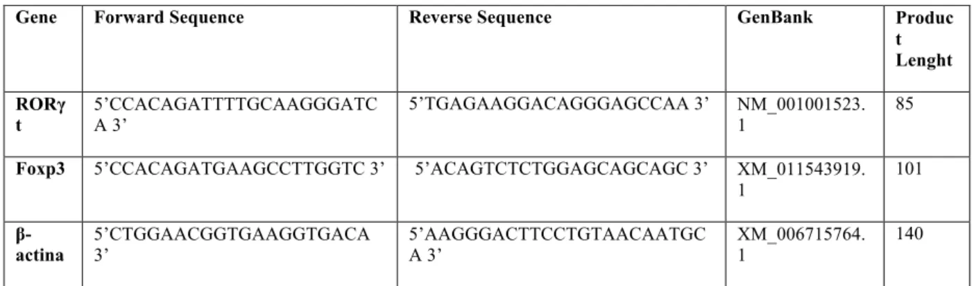
Patients with digestive CD exhibited a significantly higher frequency of Th17
cells compared with indeterminate patients and HS (p<0,05) (Figure 1A). In
contrast they presented lower percentage of Treg cells in comparison to HS
(p<0,05) (Figure 1B) and mainly in relation to IND (p<0,05).
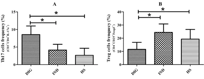
Rorγt and Foxp3 gene expression
On other approach to investigate the ratio Th17/Treg activation was to evaluate
in the 3 groups, the gene mRNA expression for the two transcription factors
involved in the differentiation of these cells, Rorγtand Foxp3 for Th17 and Treg
cells respectively.
We observed that DIG expressed significant higher levels of mRNA for Rorγt
compared to IND (p<0,05) (Figure 2A). An inverse ratio was detected for IND,
that expressed higher amount of Foxp3 (p<0,05) (Figure 2B).
Figure 2. Gene Expression of Rorγt and Foxp3: mRNA expression of Th17 and Treg cells transcription factors, Rorγt (A) and Foxp3 (B) respectively, in patients with digestive and indeterminated form of CD and healthy subjects. Each dot represents a different patient and each bar represents the median.. *p<0.05.
Cytokine production
We also evaluated the production of IL-17 and IL-10 the two cytokines
predominantly released for Th17 and Treg respectively. Our data showed that
and IL-10 (p<0,05) (Figure 3B) compared to controls . However, despite there
was no significative differences between the two groups of patients, those with
indeterminated form trended to release lower levels of IL-17 in relation to
digestive ones. In contrast, indeterminate patients trended to release higher
levels of IL-10.
Figure 3. Production of IL-17a and IL-10 in PBMC’s culture: Levels of IL-17a (A) and IL-10 (B) in supernatants were analyzed after 24 hours of PBMCs culture (1x10⁶ cells/ml), obtained from patients with digestive and indeterminated form of CD and healthy subjects. Each dot represents a different patient and each bar represents the median. *p<0.05.
DISCUSSION
One of the most important challenges for the researchers of Chagas disease is to
establish the factors that lead some patients to develop asymptomatic chronic
disease, while others undergo severe disease. This issue is probably
multifactorial, but one factor in particular has been supported by some studies
from an environment, where there is a fine regulation of effector mechanisms of
the immune response against the parasite, which avoid tissue lesion [23].
However, to our knowledge there are no studies investigating this question in
relation to the possible control of effector mechanisms involving Th17, one
proinflammatory CD4 subset. In addition, there are no studies involving
patients with digestive form of Chagas disease. We hypothesized that in contrast
with asymptomatic or indeterminated disease, disbalance between Th17 and
Treg cells activation in favor of Th17 may be associated to symptomatic
digestive disease. Confirming our hypothesis the results showed that patients
with digestive CD showed a significantly higher percentage of Th17 cells
compared with indeterminate patients, which was confirmed by the results
relative to Rorγt transcription factor expression. These results are contrary to
found in the literature for cardiac patients.
Guedes et al. [24] observed that Chagas patients with no or mild
cardiomyopathy had higher frequency of CD4+IL-17+ compared to patients with
moderate/severe cardiomyopathy and controls. Magalhães et al. [25] showed
that no chagasic patients have a higher frequency of CD4+ IL-17+ compared to
patients with heart CD.
Some studies have shown that these cells, despite participating in the induction
of a protective inflammatory response during infection with T. cruzi culminating
Th17 cells inhibit the inflammatory response in the myocardium induced by Th1
cells through its capacity of atracting and activating neutrophils producers of
IL-10 [27]. However, as there are no studies evaluating patients with digestive CD
and Th17 cells, our results are promising in showing that unlike the studies to
date with cardiac patients, patients with the digestive form show an increased
frequency of Th17 cells, that instead regulatory, play a predominat
proinflammatory role, which may be involved in the lesions of these patients.
Our hypothesis was also confirmed by the results showing that patients with
gastrointestinal CD showed a significantly lower frequency of Treg cells as well
as lower expression of the transcription factor Foxp3 in comparison with
patients with indetermined CD. These results are supported by the literature
since Silveira et al. [28] have observed higher frequency of Foxp3+ cells in
colon biopsies of patients without megacolon compared to those with this
lesion. Araújo et al. [29] reported that patients with indeterminate CD had higher
frequency of Foxp3+CD4+CD25high cells compared to patients with cardiac CD.
Still, to confirm our results, IL-17 and IL-10 levels were compared in the two
group of patients. Patients DIG showed only a modest tendency to release
higher IL-17 levels in relation to IND. However, there was a clear trend of these
patients to present lower levels of IL-10. This finding confirms our hypothesis
and is supported by studies detecting increased IL-10 levels in patients with
information are in contrast with the results found by Pisseti et al. [31], which
reported an increase in IL-10 levels in patients with gastrointestinal CD
compared with indeterminated form suggesting that this cytokine might be in
favor of a Th2 shift, inhibiting a Th1 effector response.
Taken together our findings support the argument that in contrast with
indeterminated patients a non regulatory environment observed in patients with
digestive CD, which results in an exacerbated inflammatory response exerted by
Th17 may be responsible for tissue lesions in these patients.
CONCLUSIONS
We showed that in patients with digestive form of CD there is an imbalance in
the ratio Th17/Treg cells activation in favor of Th17, while in patients with the
indeterminated form, this imbalance favour Treg activation. Our findings
support the hypothesis that in contrast with indeterminated patients, a non
regulatory environment observed in patients with digestive CD, which results in
an exacerbated inflammatory response exerted by Th17, due to lower Treg
activation, may be responsible for tissue lesions in these patients.
ACKNOWLEDGMENTS
The authors gratefully acknowledge all patients and the healthy volunteers
for their willingness to participate in this study. We also thank the Infectious
REFERENCES
[1] WHO. Chagas disease (American trypanosomiasis). Avaiable in
www.who.int/mediacentre/factsheets/fs340/en/. Accessed 10 june 2012.
[2] Tostes Jr S, Lopes ER, Pereira FE, Chapadeiro E. (1994) Miocardiopatia chagásica crônica humana: estudo quantitativo de linfócitos CD4+ e CD8+ nos exsudatos inflamatórios. Rev Soc Brasil Med Trop. 27: 127-34.
[3] Köberle F. (1968) Chagas’ disease and Chagas’ syndromes: the pathology of American trypanosomiasis. Adv Parasitol. 6: 63-116.
[4] Adad SJ, Cançado CG, Etchebehere RM, Teixeira VP, Gomes UA, Chapadeiro E, Lopes ER. (2001) Neuron count reevaluation in the myoenteric plexus of chagasic megacolon after morphometric neuron analysis. Virchows Arch. 438: 254-58.
[5] Corbett CE, Ribeiro U Jr, Prianti MG, Habr-Gama A, Okumura M, Gama-Rodrigues J. (2001) Cell-mediated immune response in megacolon from patients with chronic Chagas’ disease. Dis Colon Rectum. 44: 993-98.
[6] d’Avila Reis D, Lemos EM, Silva GC, Adad SJ, McCurley T, Correa-Oliveira R, Machado CR. (2001) Phenotypic characterization of the inflammatory cells in chagasic megaoesophagus. Trans R Soc Trop Med Hyg. 95: 177-78.
[7] Jones EM, Colley DG, Tostes S, Lopes ER, Vnencak-Jones CL, McCurley TL. (1993) Amplification of a T. cruzi DNA sequence from inflammatory lesions in human chagasic cardiomyopathy. Am J Trop Med Hyg. 48: 348-57.
[8] Vago AR, Macedo AM, Adad SJ, Reis DD, Corrêa-Oliveira R. (1996) PCR detection of
Trypanosoma cruzi DNA in oesophageal tissues of patients with chronic digestive Chagas’ disease. Lancet. 348: 891-92.
[9] Vago AR, Andrade LO, Leite AA, d’Avila Reis D, Macedo AM, Adad SJ, Tostes S Jr, Moreira MC, Filho GB, Pena SD. (2000) Genetic characterization of Trypanosoma cruzi
directly from tissues of patients with chronic Chagas disease: differential distribution of genetic types into diverse organs. Am J Pathol. 156: 1805-1809.
[10] Belkaid Y. (2008) Role of Foxp3-positive regulatory T cells during infection. Eur J Immunol. 38:918-21.
[11] Kotner J, Tarlenton R. (2007) Endogenous CD4+CD25+ regulatory T cells have a limited in the control of Trypanosoma cruzi infection in mice. Infect Immun. 75:861-9.