J of Evolution of Med and Dent Sci/ eISSN- 2278-4802, pISSN- 2278-4748/ Vol. 3/ Issue 13/Mar 31, 2014 Page 3476
AN ATTEMPT TO UNRAVEL FEATURES OF PNEUMATOSIS CYSTOIDES
INTESTINALIS
Sunil Kumar B1, Suneeta Padhy2, Inbasekar Poovizhi3, Ramaswamy Anikode Subramanian4 HOW TO CITE THIS ARTICLE:
Sunil Kumar B, Suneeta Padhy, Inbasekar Poovizhi, Ramaswamy Anikode Subramanian. An Attempt to Unravel Features of Pneumatosis Cystoides Intestinalis . Journal of Evolution of Medical and Dental Sciences 2014; Vol. 3, Issue 13, March 31; Page: 3476-3483, DOI: 10.14260/jemds/2014/2308
ABSTRACT: Pneumatosis cystoides intestinalis (PCI) is a rare disease characterized by presence of multiple gas filled cysts in subserosal or submucosal wall of large intestine or small intestine. PCI are most commonly due to an underlying disease or can be idiopathic. Understanding of etiology and pathogenesis is necessary in each individual case for appropriate management. Thirty eight enteral resected specimens were studied from January 2008 to September 2011 in PESIMSR. Clinical and morphological characteristics of the 3 cases with histological diagnosis of PCI found, were studied and compared with other studies.
KEYWORDS: Pneumatosis cystoides intestinalis.
INTRODUCTION: Pneumatosis cystoides intestinalis (PCI) is a rare disorder, characterized histologically by the presence of multiple gas filled cysts in the subserosal or submucosal wall of the large or small intestine.1 PCI is most commonly due to an underlying disease or can be idiopathic. Understanding of the etiology and pathogenesis is necessary in each case for appropriate management.
MATERIALS AND METHODS: This is a retrospective study conducted on all resected enteral specimens, received in department of pathology, PESIMSR, Kuppam from January 2008 to September 2011. Clinical details and histopathological slides were retrieved from the archives in the department of pathology.
AIM OF STUDY: To study the frequency, clinical features, and histomorphological characteristics of histologically diagnosed PCI.
RESULTS: During the prescribed study period, a total no of 38 enteric specimens were analyzed. The most common indication found for surgical resection was perforation. Most of these resections occurred in the age group of >40 years (table1) with a male preponderance (M-24: F-14). Small bowel resections were common among all the resections (table 2). Three cases with histological features of PCI were observed and all were found in small bowel resections.
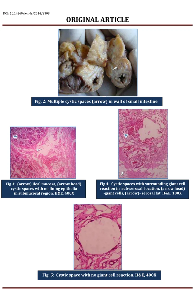
J of Evolution of Med and Dent Sci/ eISSN- 2278-4802, pISSN- 2278-4748/ Vol. 3/ Issue 13/Mar 31, 2014 Page 3477 Case 2: 55 year old male, presented with pain abdomen and fever. Segment of small intestine was received in our department which showed edematous mucosa. Microscopically, these cystic spaces had no lining epithelium and was surrounded by foreign body giant cells.
Case 3: 16 year old male, presented with acute pain abdomen. Small intestine perforation was found intraoperatively. Grossly perforation was confirmed. Microscopically, these cystic spaces had no lining epithelium; however no foreign body giant cells were observed (fig. 5, 6).
Abdominal ultrasonagraphic studies were done in all 3 cases, which did not detect presence of PCI. All the patients were symptom free after six month follow up.
DISCUSSION: PCI was first described by Du Vernoy in 1730, in autopsy specimens. Mayer named these entities as PCI in 1825.2, 3 PCI diagnosis in surviving patients was first established by Hahn in 18993. A PCI diagnosis via preoperative radiological findings was first described by Baumann-Schender in 1939.3 PCI are classified into primary and secondary. Secondary PCI term was coined by Koss in 1952, who analyzed 213 pathological specimens and attributed 85% of the cases to a secondary disease.3 The incidence of PCI is unknown, because it is usually asymptomatic.3
PCI is found to be more common in males, compared to females (3-3.5:1)4, 5 Knechtle et al6 had equal incidence among males and females3,6 .Most of the studies have stated, colon to be the most common site of PCI compared to small intestine.2 In our study all three cases showed PCI in the small intestine. There are many symptoms of PCI, including abdominal pain, abdominal distention, diarrhea, mucous stool, bloody stool and constipation.2, 7
Imaging studies which are useful in diagnosing PCI are plain abdominal X-rays, Opaque enema, Computerized tomography, Ultrasonography, MRI, and colonoscopy. Among these abdominal X-rays are the most reliable examination.8 But some subtle cases are missed on radiography and diagnosis is possible only on pathological examination9.Though radiological examination detects most of these cases, pathological examination is a must in few cases where a endoscopy biopsy is warranted. Though pathological examinations are useful, only few studies have described the microscopic features. A larger study on microscopy will probably reveal features of prognostic importance.
Only 4 cases of PCI were found in literature where histological features were described. Saber A et al10 will be designated as case 4, Sakurai Y et al1 will be designated as case 5, Mutha S et al11 will be designated as case 6 and Chun-Hsiung Liu et al 7 as case 7. Age of presentation in literature (case 4, 5, 6, and 7) was 45 to 79 years. Our cases 1 & 2 had similar age group, but case 3 was in younger age group (16 years). Range of age group in Arikanoglu Z et.al study was 29-74 years and in Wu LL et al study was 2-81 years.2, 3
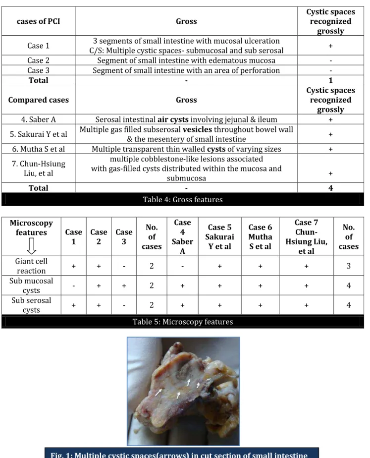
All the cases in literature had a common presenting symptom as pain abdomen, which correlated with our clinical symptoms. Pain abdomen was the most common symptom seen. Abdominal distension was seen in 3 literature cases (case 5, 6 & 7). None of our cases showed distention of abdomen (table 3); however emesis was seen in one case (case 1). Grossly our cases showed mucosal ulceration, edema and perforation. In addition, case1 showed multiple cystic spaces in wall.
J of Evolution of Med and Dent Sci/ eISSN- 2278-4802, pISSN- 2278-4748/ Vol. 3/ Issue 13/Mar 31, 2014 Page 3478 All cases had giant cell reaction except case 3 and 4 (table 5). Subserosal location of cysts was common finding, but an exclusive submucosal cyst was seen in case 3 which presented with acute abdomen and perforation. Subserosal cysts are known to be associated with secondary forms of PCI.3 Etio-Pathogenesis is bacterial, mechanical or pulmonary for PCI12 (fig. 7).
In case 1, no pulmonary cause was detected but, intraoperative finding showed an entero-vesical fistula. Microscopy showed no underlying cause and only chronic inflammatory process with PCI was noted. Case 2 showed volvulus intestinal obstruction.13 Case 3 showed intestinal perforation. So in case 1, 2 and 3 a mechanical etiological factor had a role. But in case 3, whether perforation led to PCI or PCI led to perforation remained an enigma. In case 4 and 5 bacterial, mechanical and pulmonary factors had a role in pathogenesis.
In case 7pathogenesis was related to pulmonary causes. Mechanical factors are the most common cause of PCI. Complications of PCI include intestinal obstruction, pneumoperitoneum, intussusception, volvulus, hemorrhage and intestinal perforation.10 Volvulus was a complication seen in 51 year old male in literature, which correlated with our case 2 showing similar age group and location13. No therapy is required for asymptomatic cases.10 For primary PCI, Metronidazole and hyperbaric oxygen are routinely used.14 For Secondary PCI with or without complication, surgery is indicated.10 In all our cases, the therapy was surgical because they presented with intestinal obstruction.
CONCLUSION: Pathogenesis in PCI is unclear and many factors play a role.10 Therapy of PCI may be conservative or surgical depending on the etiology or complication.14 Perforation may lead to PCI and one of the complications of PCI is perforation, so in a case with PCI with perforation it would be difficult to determine which was the initiating factor.10 PCI is an under recognized feature, often mistaken for artifact especially in subtle cases. Awareness of this rare entity and high index of suspicion with knowledge of etiopathogenesis can avoid major bowel surgery. Understanding of the salient microscopic features are essential, as small endoscopic biopsies will be diagnostic in subtle cases.
REFERENCES:
1. Sakurai Y, Hikichi M, Isogaki J, Furuta S, Sunagawa R, Inaba K et al. Pneumatosis cystoides intestinalis associated with massive free air mimicking perforated diffuse peritonitis . World J Gastroenterol. 2008; 14: 6733 – 56.
2. Wu LL, Yang YS, Dou Y, Liu QS. A systematic analysis of pneumatosis cystoids intestinalis. World J Gastroenterol 2013;19: 4973-8
3. Arikanoglu Z, Aygen E, Camci C, Akbulut S, Basbug M, Dogru O, Cetinkaya Z, Kirkil C. Pneumatosis cystoides intestinalis: A single center experience. World J Gastroenterol 2012; 18: 453-7
4. Koss LG. Abdominal gas cysts (pneumatosis cystoides intestinorum hominis); an analysis with a report of a case and a critical review of the literature. AMA Arch Pathol 1952; 53:523-49
5. Jamart J. Pneumatosis cystoides intestinalis: A statistical study of 919 cases. Acta Hepatogastroenterol. 1979; 26:419-22
J of Evolution of Med and Dent Sci/ eISSN- 2278-4802, pISSN- 2278-4748/ Vol. 3/ Issue 13/Mar 31, 2014 Page 3479 7. Chun-Hsiung Liu, MD, Hong Haw Chen, Wan-Ting Huang. Primary Pneumotosis Cystoides
Intestinalis. Chang. Gung Med J. 2003; 26:144-7
8. Rivera Vaquerizo PA, Martins CA, Lorente García MA, Colmenarejo MB, Flores RP. Pneumatosis cystoides intestinalis. Rev Esp Enf Erm Dig .2006; 98: 959-61
9. H, Khan M, Hudgins J, Lee K, Du L, Amorosa L. Gastrointestinal sarcoidosis associated with pneumatosis cystoides intestinalis. World J Gastroenterol. 2013; 19: 1135-9
10.Saber A. Pneumatosis intestinalis with complete remission: a case report .Cases journal .2009; 2: 7079 – 82.
11.Mutha S, Kumbhalkar DT, Bobhak SK. Pneumatosis cystoides intestinalis – a case report. Indian J Pathol Microbiol. 1999; 43:157 -8.
12.Hughes DTD, Gordon KCD, Swann JC, Bolt GL. Pneumatosis cystoides intestinalis. Gut. 1966; 7: 553–7.
13.Hicham El B, Abdelmalek, Benjelloun EB, Majdoub KI, Khalid M, Taleb KA. Ileal cystic pneumatosis revealed a volvulus of the small intestine. Pan Afr Med J. 2010; 6: 1-4
14.Braumann C, Menenkes C, Jacobi CA. Pneumatosis intestinalis – a pitfall for surgeon? Scand J of Surg .2005; 94: 47 – 50.
Age group (years) No. of cases
0-20 7
21-40 9
41-60 14
>60 8
Table 1: Total no of enteral resections-38
Resections No. of cases Small intestine 27 Large intestine 7
Both 4
Table 2
Case 1
Case 2
Case 3
Total (our cases)
Case4 Case5 Case6 Case7
Total (literature
cases) Abdomen
pain + + + 3 + + + + 4
Fever - + - 1 - - - - -
Abdomen
distention - - - + + + 3
Vomiting + - - 1 - - - - -
J of Evolution of Med and Dent Sci/ eISSN- 2278-4802, pISSN- 2278-4748/ Vol. 3/ Issue 13/Mar 31, 2014 Page 3480
cases of PCI Gross
Cystic spaces recognized
grossly
Case 1 3 segments of small intestine with mucosal ulceration
C/S: Multiple cystic spaces- submucosal and sub serosal + Case 2 Segment of small intestine with edematous mucosa - Case 3 Segment of small intestine with an area of perforation -
Total - 1
Compared cases Gross
Cystic spaces recognized
grossly 4. Saber A Serosal intestinal air cysts involving jejunal & ileum + 5. Sakurai Y et al Multiple gas filled subserosal vesicles throughout bowel wall
& the mesentery of small intestine + 6. Mutha S et al Multiple transparent thin walled cysts of varying sizes + 7. Chun-Hsiung
Liu, et al
multiple cobblestone-like lesions associated with gas-filled cysts distributed within the mucosa and
submucosa +
Total - 4
Table 4: Gross features
Microscopy
features Case 1 Case 2 Case 3 No. of cases Case 4 Saber A Case 5 Sakurai
Y et al
Case 6 Mutha S et al
Case 7 Chun-Hsiung Liu, et al No. of cases Giant cell
reaction + + - 2 - + + + 3
Sub mucosal
cysts - + + 2 + + + + 4
Sub serosal
cysts + + - 2 + + + + 4
Table 5: Microscopy features
J of Evolution of Med and Dent Sci/ eISSN- 2278-4802, pISSN- 2278-4748/ Vol. 3/ Issue 13/Mar 31, 2014 Page 3481
Fig. 2: Multiple cystic spaces (arrow) in wall of small intestine
Fig 3: (arrow) Ileal mucosa, (arrow head) cystic spaces with no lining epithelia
in submucosal region. H&E, 400X
Fig 4: Cystic spaces with surrounding giant cell reaction in sub-serosal location. (arrow head)
-giant cells, (arrow)- serosal fat. H&E, 100X
J of Evolution of Med and Dent Sci/ eISSN- 2278-4802, pISSN- 2278-4748/ Vol. 3/ Issue 13/Mar 31, 2014 Page 3482
Fig. 6a: Subgross picture of small intestine- cystic spaces in the wall. (arrow-mucosa,
arrow head-cystic space)
Fig. 6b: Cystic space with no giant cell reaction subserosal in location H&E, 100X.
(arrow-mucosa, arrow head-cystic space)
J of Evolution of Med and Dent Sci/ eISSN- 2278-4802, pISSN- 2278-4748/ Vol. 3/ Issue 13/Mar 31, 2014 Page 3483
AUTHORS:
1. Sunil Kumar B. 2. Suneeta Padhy 3. Inbasekar Poovizhi
4. Ramaswamy Anikode Subramanian
PARTICULARS OF CONTRIBUTORS:
1. Associate Professor, Department of Pathology, Akash Institute of Medical Science and Research, Bangalore.
2. Resident Specialist, Rajiv Gandhi institute of medical sciences, Srikakulum, Andhra Pradesh, India
3. Post Graduate, Department of Pathology, PESIMSR, Kuppam, Andhra Pradesh, India. 4. Professor, Department of Pathology, PESIMSR,
Kuppam, Andhra Pradesh, India.
NAME ADDRESS EMAIL ID OF THE CORRESPONDING AUTHOR:
Dr. Sunil Kumar B, No. 498, 10th Cross,
8th Main, Sadashivanagar,
Bangalore- 560080, Karnataka, India.
E-mail: drsunil2011@yahoo.com


