The Immunopathogenic Potential of
Arcobacter butzleri
–
Lessons from a
Meta-Analysis of Murine Infection Studies
Greta Gölz1*, Thomas Alter1, Stefan Bereswill2, Markus M. Heimesaat2
1Institute of Food Hygiene, Freie Universität Berlin, Berlin, Germany,2Department of Microbiology and Hygiene, Charité - University Medicine Berlin, Berlin, Germany
Abstract
Background
Only limited information is available about the immunopathogenic properties ofArcobacter infectionin vivo. Therefore, we performed a meta-analysis of published data in murine infection models to compare the pathogenic potential ofArcobacter butzleriwithCampylobacter jejuni and commensalEscherichia colias pathogenic and harmless reference bacteria, respectively.
Methodology / Principal Findings
Gnotobiotic IL-10-/-mice generated by broad-spectrum antibiotic compounds were perorally infected withA.butzleri(strains CCUG 30485 or C1),C.jejuni(strain 81-176) or a commensal intestinalE.colistrain. Either strain stably colonized the murine intestines upon infection. At day 6 postinfection (p.i.),C.jejuniinfected mice only displayed severe clinical sequelae such as wasting bloody diarrhea. Gross disease was accompanied by increased numbers of colonic apoptotic cells and distinct immune cell populations including macrophages and monocytes, T and B cells as well as regulatory T cells upon pathogenic infection. WhereasA. butzleriandE.coliinfected mice were clinically unaffected, respective colonic immune cell numbers increased in the former, but not in the latter, and more distinctly uponA.butzleri strain CCUG 30485 as compared to C1 strain infection. Both,A.butzleriandC.jejuniinduced increased secretion of pro-inflammatory cytokines such as IFN-γ, TNF, IL-6 and MCP-1 in
large, but also small intestines. Remarkably, even though viable bacteria did not translocate from the intestines to extra-intestinal compartments, systemic immune responses were induced inC.jejuni, but alsoA.butzleriinfected mice as indicated by increased respective pro-inflammatory cytokine concentrations in serum samples at day 6 p.i.
Conclusion / Significance
A.butzleriinduce less distinct pro-inflammatory sequelae as compared toC.jejuni, but more pronounced local and systemic immune responses than commensalE.coliin a strain-dependent manner. Hence, data point towards thatA.butzleriis more than a commensal in vertebrate hosts.
a11111
OPEN ACCESS
Citation:Gölz G, Alter T, Bereswill S, Heimesaat MM (2016) The Immunopathogenic Potential of Arcobacter butzleri–Lessons from a Meta-Analysis
of Murine Infection Studies. PLoS ONE 11(7): e0159685. doi:10.1371/journal.pone.0159685
Editor:Sergei Grivennikov, Fox Chase Cancer Center, UNITED STATES
Received:March 10, 2016
Accepted:July 5, 2016
Published:July 20, 2016
Copyright:© 2016 Gölz et al. This is an open access article distributed under the terms of the
Creative Commons Attribution License, which permits
unrestricted use, distribution, and reproduction in any medium, provided the original author and source are credited.
Data Availability Statement:All relevant data are within the paper.
Funding:This work was supported by grants from the German Research Foundation (DFG) to SB and MMH (SFB633, TP A7 and B6, respectively), and from the German Federal Ministery of Education and Research (BMBF) to SB (TP1.1).
Introduction
The gram-negativeArcobacterspecies belong to theCampylobacteraceaefamily and can be found in a plethora of habitats. In animals,Arcobacterspp. are mostly regarded as gastrointesti-nal commensals [1]. In humans, however,Arcobacterspp. have been shown to induce single diarrheal cases, but also disease outbreaks have been reported [2,3]. Patients become infected by contaminated food or water and present with symptoms of acute gastroenteritis such as abdominal pain, acute or even prolonged diarrhea for up to several weeks [4,5]. Since identifi-cation ofArcobacterspp. may fail in routine diagnostic laboratories, robust epidemiological data onArcobacterassociated human disease are lacking. In a prospective German study, for instance, noArcobacterat all could be isolated in hospitalized patients suffering from commu-nity acquired acute gastroenteritis [6]. Van den Abeele and colleagues, however, reported in a large survey anArcobacterprevalence of 1.3% in stool samples derived from more than 6700 Belgian enteritis patients [5]. In studies from New Zealand, Thailand and Mexico,Arcobacter
spp. such asA.butzleriandA.cryaerophiluscould be detected in 0.9–8.0% of fecal samples obtained from diarrheal patients [7–9]. Isolation rates, however, were highly depending on the respective cultivation methods applied in the respective microbiology laboratories [5]. It is therefore highly likely that the prevalence rates reported so far have been rather underesti-mated. In line with this, a very recent Canadian study revealedA.butzleriisolation rates of 59.6% and 0.8% from stool samples determined by PCR-based and culture-dependent meth-ods, respectively [10]. Remarkably, neither differences could be found in fecalA.butzleri preva-lences between diarrheal and non-diarrheal patients, nor did patient age, sex or place of habitation correlate withA.butzleripositive results in fecal samples derived by quantitative real-time PCR [10]. Thus, it is still an open and unanswered question whetherArcobacterspp. need to be regarded as ordinary commensals or rather pathogenic species. Nevertheless, based upon retrospective studiesArcobacteris estimated the fourth most commonCampylobacterales
genus isolated from diarrheal patients [4,5,11]. Furthermore, the International Commission on Microbiological Specifications for Foods have ratedA.butzleriandA.cryaerophilusas seri-ous hazards for human health among the 21 so far describedArcobacterspecies [12]. Until now information regarding the underlying mechanisms ofArcobacterinfection and bacteria-host interactions are scarce due to lack of suitablein vivoinfection models. Very recently our group showed in gnotobiotic (i.e. secondary abiotic) IL-10-/-mice, a well-established murine model ofC.jejuniinfection, thatA.butzleriinduced intestinal and systemic immune responses [13,14]. These immune reponses were highly dependent on Toll-like-receptor (TLR) -4 consti-tuting the main receptor for lipooligosaccharide (LOS) and lipopolysaccharide (LPS) from gram-negative bacteria [15,16]. In the present study we assessed the immunopathological potential ofA.butzeriby comparing our published, but also so far unpublished data from gno-tobiotic IL-10-/-mice infected with the pathogenC.jejunior a commensal intestinalE.coli
strain [17,18]. In this meta-analysis we aimed to unravel whetherA.butzleriexhibited immu-nopathological features of a pathogen or a commensal.
Materials and Methods
Ethics statement
Study design
Data were pooled from separate published [13,14,17,18] as well as so far unpublished animal trials.
Generation of gnotobiotic IL-10
-/-mice
IL-10-/-mice (in C57BL/10 background, B10) were bred and kept in the facilities of the “For-schungseinrichtungen für Experimentelle Medizin”(FEM, Charité - Universitätsmedizin, Ber-lin, Germany) under specific pathogen-free (SPF) housing conditions. Gnotobiotic IL-10 -/-mice were generated by broad-spectrum antibiotic treatment as described earlier [19]. In brief, mice were kept in sterile cages and and hadad libitumaccess to an antibiotic cocktail consisting of ampicillin/sulbactam (1 g/L; Pfizer, Berlin, Germany), vancomycin (500 mg/L; Hexal, Holz-kirchen, Germany), ciprofloxacin (200 mg/L; Hexal, HolzHolz-kirchen, Germany), imipenem (250 mg/L; Fresenius Kabi, Graz, Austria), and metronidazole (1 g/L; Braun, Melsungen, Germany) in drinking water starting at 3 weeks of age immediately after weaning and continued for 3–4 months before the infection experiment [20]. Three days prior infection, the antibiotic cocktail was replaced by sterile tap water (ad libitum). These so generated gnotobiotic (i.e. secondary abiotic) mice were continuously kept in a sterile environment (with autoclaved food and drink-ing water), handeled under strict aseptic conditions and displayed a virtually depleted gastroin-testinal microbiota.
Bacterial strains
A commensalE.colistrain was isolated from our naive and conventionally colonized C57BL/6 wildtype mice as described earlier [19]. No known virulence factors of pathogenicE.colisuch asstx-1 and -2,catA,hlyA,cspA,katPandastAcould be detected by PCR analysis in a reference laboratory [18]. TheA.butzlerireference strain CCUG 30485 was derived from a fecal sample of a diarrheal patient [21], whereas the C1 strain was isolated from fresh chicken meat [22]. As a pathogenic reference strain,C.jejunistrain 81-176 was chosen.
Infection of mice
Gnotobiotic IL-10-/-mice were infected with 109colony forming units (CFU) ofC.jejunistrain 81-176,A.butzlerireference strain CCUG 30485 or strain C1, or a commensalE.colistrain by gavage in a total volume of 0.3 mL PBS on two consecutive days (day 0 and day 1) as described earlier in more detail [13,14,17,18].
C.jejuniand bothA.butzleristrains were grown on Karmali-Agar and Columbia Agar
sup-plemented wit 5% sheep blood (both from Oxoid, Wesel, Germany) for two days at 37°C under microaerobic conditions using CampyGen gas packs (Oxoid), whereas the commensalE.coli
was cultivated on MacConkey agar (Oxoid) for one day at 37°C in aerobic atmosphere.
Clinical Score
Sampling procedures
Mice were sacrificed by isofluran treatment (Abbott, Greifswald, Germany). Then, cardiac blood was taken and tissue samples removed from spleen, liver, mesenteric lymph nodes (MLN), ileum, and colon, all under sterile conditions. Ileal and colonicex vivobiopsies were collected from each mouse in parallel, for microbiological, immunohistochemical and immu-nological analyses. Immunohistopathological changes were determined in colonic samples that had been immediately fixed in 5% formalin and embedded in paraffin. Sections (5μm) were
stained with respective antibodies forin situimmunohistochemistry as described earlier [13,
20].
Immunohistochemistry
In situimmunohistochemical analysis of colonic paraffin sections was performed as described
previously [13,17,18,23,24]. In brief, primary antibodies against cleaved caspase-3 (Asp175, Cell Signaling, USA, 1:200), CD3 (#N1580, Dako, Denmark, dilution 1:10), FOXP3 (FJK-16s, eBioscience, 1:100), B220 (eBioscience, 1:200), and F4/80 (# 14–4801, clone BM8, eBioscience, San Diego, CA, USA, 1:50) were used [13]. For each animal, the average number of positively stained cells within at least six high power fields (HPF, 400 x magnification) were determined microscopically by a double-blinded investigator [13].
Quantitative analysis of
bacterial
colonization and translocation
ViableA.butzleri,C.jejuniand commensalE.coliwere detected in feces or at time of necropsy (day 6 p.i.) in luminal samples taken from the ileum or colon and dissolved in sterile PBS. Serial dilutions were cultured on Karmali- and Columbia-Agar supplemented with 5% sheep blood (Oxoid) for two days at 37°C under microaerobic conditions using CampyGen gas packs (Oxoid) forA.butzleriandC.jejunidetection, whereasE.coliwas cultivated on MacConkey and Columbia-Agar supplemented with 5% sheep blood (Oxoid) for 48 hours in aerobic atmo-sphere. In order to quantitatively assess bacterial translocation, MLN, spleen, and liverex vivo
biopsies were homogenized in 1 mL sterile PBS, whereas cardiac blood (200μL) was directly
streaked onto respective solid media and cultivated accordingly. The respective weights of fecal or tissue samples were determined by the difference of the sample weights before and after asservation. The detection limit of viable pathogens by direct plating was 100 CFU per gram.
Cytokine detection
Ileal and colonicex vivobiopsies were cut longitudinally, washed in PBS, and strips of approxi-mately 1 cm2intestinal tissue placed in 24-flat-bottom well culture plates (Nunc, Wiesbaden, Germany) containing 500μL serum-free RPMI 1640 medium (Gibco, life technologies, Paisley,
UK) supplemented with penicillin (100 U/ mL) and streptomycin (100μg/ mL; PAA
Laborato-ries). After overnight incubation at 37°C, culture supernatants and serum samples were tested for IFN-γ, TNF, IL-6, and MCP-1 by the Mouse Inflammation Cytometric Bead Assay (CBA;
BD Biosciences) on a BD FACSCanto II flow cytometer (BD Biosciences).
Statistical analysis
Results
Intestinal colonization efficacies of
E.
coli,
A.
butzleri
and
C.
jejuni
and
infection-induced clinical sequelae of gnotobiotic IL-10
-/-mice
In the present study we aimed to compare the immunopathological potential ofA.butzeriwith the gram-negative intestinal pathogenC.jejuniand a commensalE.colistrain isolated from the intestinal microbiota of a conventional mouse. To address this, we applied the gnotobiotic IL-10-/-mouse model generated by broad-spectrum antibiotic treatment. Following peroral infection with comparable bacterial loads of approximately 109CFU on two consecutive days (namely days 0 and 1) by gavage,E.coli,A.butzleristrains CCUG 30485 and C1 as well asC.
jejuniwere stably colonizing the intestinal tract of gnotobiotic IL-10-/-mice, as indicated by
high median bacterial loads of between 108and 109CFU per g feces (Fig 1). At day of necropsy (i.e. day 6 p.i.), small and large intestinalA.butzleriloads were between one and three orders of magnitude lower in colonic and ileal luminal contents, respectively, as compared toE.coliand
C.jejuniinfected mice (p<0.05–0.001;Fig 2). Analysis of the clinical outcome of infection
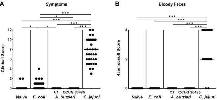
revealed thatC.jejuniinfected mice were severely compromized at day 6 p.i., as indicated by increased clinical scores (Fig 3A), and presented with wasting ulcerative enterocolitis including bloody diarrhea (Fig 3B). Infection with the commensalE.colior with eitherA.butzleristrain,
Fig 1. Kinetic survey of fecal bacterial shedding in perorally infected gnotobiotic IL-10-/-mice.Gnotobiotic IL-10-/-mice were
generated by antibiotic treatment and perorally colonized either with (A) a commensalE.colistrain,(B)A.butzleristrain C1, (C)A. butzleristrain CCUG 30485 or(D)C.jejunistrain 81-176 at day 0 and day 1 by gavage. Bacterial loads were determined in fecal samples (CFU / g, colony forming units per gram) over six days (d) post infection (p.i.) by culture. Medians (black bars) are indicated.
however, induced, if any, only rather minor symptoms (Fig 3A), and neither gross nor occult blood could be detected in fecal samples at day 6 p.i. at all (Fig 3B). Taken together, following stable infection,C.jejuni, but neitherA.butzleristrain nor commensalE.coliinduced macro-scopic disease.
Fig 2. Bacterial colonization alongside the intestinal tract of perorally infected gnotobiotic IL-10-/-mice.Gnotobiotic IL-10
-/-mice were generated by antibiotic treatment and perorally colonized either with a commensalE.colistrain,A.butzleristrain C1,A.butzleri strain CCUG 30485 orC.jejunistrain 81-176 at day 0 and day 1 by gavage. Colonization densities were determined in luminal samples derived from the (A) ileum and(B)colon (CFU / g, colony forming units per gram) at day 6 post infection by culture. Medians (black bars) and levels of significance (*p<0.05;**p<0.01;***p<0.001) determined by Mann-Whitney U test are indicated.
doi:10.1371/journal.pone.0159685.g002
Fig 3. Macroscopic sequelae (disease activity) in perorally infected gnotobiotic IL-10-/-mice.Gnotobiotic IL-10
-/-mice were generated by antibiotic treatment and perorally colonized either with a commensalE.colistrain,A.butzleristrain C1,A.butzleristrain CCUG 30485 orC.jejunistrain 81-176 at day 0 and day 1 by gavage. Naive mice served as uninfected controls.(A)Disease activity (symptoms) and(B)occurrence of blood in fecal samples (bloody diarrhea) were quantitatively assessed at day 6 postinfection applying respective standardized scores. Medians (black bars) and levels of significance (*p<0.05;***p<0.001) determined by Mann-Whitney U test are indicated.
Induction of apoptosis in the colon of infected gnotobiotic IL-10
-/-mice
We next raised the question whether despite absence of macroscopic diseaseA.butzlerihad the potential to induce more distinct microscopic sequelae of infection than a gram-negative com-mensal. Given that apoptosis is a commonly used diagnostic marker in the histopathological evaluation and grading of intestinal disease [23] and a hallmark ofC.jejuniinduced enterocoli-tis in gnotobiotic IL-10-/-mice [17], we quantitatively assessed numbers of caspase-3+ cells within the colonic epithelium of infected mice. Whereas apoptotic cell numbers in the colonic mucosa increased multi-fold until day 6 followingC.jejuniinfection (p<0.001;Fig 4), there was a trend towards higher abundance of colonic apoptotic cells inA.butzleristrain CCUG 30485 as compared toE.coliinfected and naive mice (n.s. after pooling of data sets;Fig 4). Hence, our data indicate that the potential ofA.butzlerito induce macroscopic or microscopic intestinal disease does not exceed that of a commensal bacterial strain.
Large intestinal immune cell responses in infected gnotobiotic IL-10
-/-mice
Given that recruitment of pro-inflammatory immune cells to sites of inflammation is a key fea-ture of infectious enteric diseases including campylobacteriosis [23], we next quantitatively assessed effector as well as innate and adaptive immune cells within the large intestinal mucosa and lamina propria byin situimmunohistochemical staining of colonic paraffin sections. At day 6 p.i., naive andE.coliinfected gnotobiotic IL-10-/-mice displayed comparable colonic numbers of CD3+ T lymphocytes, FOXP3+ regulatory T cells (Tregs), B220+ B lymphoctyes and F4/80+ macrophages and monocytes (Fig 5). UponA.butzleriinfection with either strain
orC.jejuni, however, colonic T cell numbers increased (p<0.001;Fig 5A), and reached highest
counts inC.jejuniinfected mice. Whereas large intestinal numbers of Tregs and B lymphocytes
inE.coliandA.butzleriC1 strain infected mice did not differ from naive controls, respective
cell numbers increased followingA.butzleristrain CCUG 30485 orC.jejuniwith highest Treg and B cell counts at day 6 followingC.jejuniinfection (p<0.001;Fig 5B, 5C and 5D). Interest-ingly, numbers of macrophages and monocytes increased uponC.jejuniandA.butzleri infec-tion (p<0.001;Fig 5D), but notably, less distinctly inC.jejuniinfected mice (p<0.05–0.001; Fig 5D). Hence,A.butzleri(and more distinctly strain CCUG 30485 than strain C1) as well as
C.jejuni, but not commensalE.coliinfection, resulted in recruitment of pro-inflammatory
immune cells into the colonic mucosa and lamina propria at day 6 p.i.
Large intestinal pro-inflammatory cytokine secretion following
A.
butzleri,
E.
coli
or
C.
jejuni
infection of gnotobiotic IL-10
-/-mice
We next compared colonic secretion of pro-inflammatory cytokines upon infection with either
A.butzleri,E.coliorC.jejuni. Colonic IFN-γ, TNF, IL-6, and MCP-1 concentrations increased
until day 6 followingC.jejuniinfection (p<0.05—p<0.001;Fig 6), whereas respective cyto-kines were also higher in the colon ofA.butzleristrain CCUG 30485 (p<0.05–0.001;Fig 6) and for MCP-1 also in C1 strain infected as compared to naive mice (p<0.05;Fig 6D). Remarkably, colonic TNF, IL-6 and MCP-1 levels did not differ between strain CCUG 30485
andC.jejuniinfected mice at day 6 p.i. (Fig 6B, 6C and 6D). Unexpectedly, increased large
intestinal IFN-γand TNF concentrations could also be observed in commensalE.colias
not differ fromC.jejuniinfected mice, hence supporting a strain-dependent pro-inflammatory potential ofA.butzleriin the colon upon peroral infection.
Small intestinal pro-inflammatory cytokine secretion following
A.
butzleri,
E.
coli
or
C.
jejuni
infection of gnotobiotic IL-10
-/-mice
Even though gnotobiotic IL-10-/-mice are considered a suitable model for severeC.jejuni
induced colonic disease [17,18], we addressed whether peroral infection with the respective
Fig 4. Apoptotic cells in colonic epithelium of perorally infected gnotobiotic IL-10-/-mice.Gnotobiotic IL-10-/-mice were
generated by antibiotic treatment and perorally colonized either with a commensalE.colistrain,A.butzleristrain C1,A.butzleri strain CCUG 30485 orC.jejunistrain 81-176 at day 0 and day 1 by gavage. Naive mice served as uninfected controls. The average numbers of apoptotic cells (positive for caspase-3, Casp3) from at least six high power fields (HPF, 400x magnification) per animal were determined microscopically in immunohistochemically stained colonic paraffin sections at day 6 postinfection. Medians (black bars) and levels of significance (*p<0.05;***p<0.001) determined by Mann-Whitney U test are indicated.
bacterial strains might also affect pro-inflammatory cytokine secretion in the small intestinal tract. In fact, 6 days followingC.jejuniinfection, increased ileal IFN-γ, TNF and IL-6, but not
MCP-1 concentrations could be measured (p<0.05–0.001;Fig 7). Remarkably, elevated IFN-γ,
IL-6 and MCP-1 levels could be determined at day 6 post CCUG 30485 strain infection (p<0.05–0.001;Fig 7A, 7C and 7D), that did, however, not differ from ileal secretion inC.
jejuniinfected mice. Moreover, both, IFN-γand IL-6 increased uponA.butzleriC1 strain
Fig 5. Colonic immune cell responses in perorally infected gnotobiotic IL-10-/-mice.Gnotobiotic IL-10
-/-mice were generated by antibiotic treatment and perorally colonized either with a commensalE.colistrain,A.butzleristrain C1,A.butzleristrain CCUG 30485 orC.jejunistrain 81-176 at day 0 and day 1 by gavage. Naive mice served as uninfected controls. The average number of cells positive for(A)CD3 (T lymphocytes),(B)FOXP3 (regulatory T cells, Tregs),(C)B220 (B lymphocytes) and(D)F4/80 (macrophages and monocytes) from at least six high power fields (HPF, 400x magnification) per animal were determined microscopically in immunohistochemically stained colonic paraffin sections at day 6 postinfection. Medians (black bars) and levels of significance (*p<0.05;**p<0.01;***p<0.001) determined by Mann-Whitney U test are indicated.
infection (p<0.05 and p<0.001, respectively;Fig 7A and 7C), but less distinctly for the former as compared to CCUG 30485 strain infection (p<0.05;Fig 7A). Taken together,A.butzleri strain C1 and more distinctly strain CCUG 30485 as well asC.jejuni, but notE.coliinfection is accompanied with increased inflammatory cytokines in the ileum pointing towards a pro-nounced, but strain-dependent pro-inflammatory potential ofArcobacteralso in the small intestinal tract.
Fig 6. Colonic pro-inflammatory cytokine secretion in perorally infected gnotobiotic IL-10-/-mice.Gnotobiotic IL-10-/-mice were
generated by antibiotic treatment and perorally colonized either with a commensalE.colistrain,A.butzleristrain C1,A.butzleristrain CCUG 30485 orC.jejunistrain 81-176 at day 0 and day 1 by gavage. Naive mice served as uninfected controls. Concentrations of(A) IFN-γ,(B)TNF,(C)IL-6, and(D)MCP-1 were determined in supernatants of colonicex vivobiopsies at day 6 postinfection by cytometric bead assay. Medians (black bars) and levels of significance (*p<0.05;**p<0.01;***p<0.001) determined by Mann-Whitney U test are indicated.
Bacterial translocation to extra-intestinal and systemic compartments
following
A.
butzleri,
E.
coli
or
C.
jejuni
infection of gnotobiotic IL-10
-/-mice
We next addressed whether respective bacterial infections were accompanied by translocation of viable bacteria from the intestines to extra-intestinal compartments including the systemic circulation. In more than 75% of diseasedC.jejuniinfected mice suffering from severe entero-colitis and more than half of uncompromizedE.coliinfected animals, respective strains could be isolated from MLN at day 6 p.i, whereasA.butzleriwas virtually undetectable (Fig 8A).
Fig 7. Ileal pro-inflammatory cytokine secretion in perorally infected gnotobiotic IL-10-/-mice.Gnotobiotic IL-10-/-mice were
generated by antibiotic treatment and perorally colonized either with a commensalE.colistrain,A.butzleristrain C1,A.butzleristrain CCUG 30485 orC.jejunistrain 81-176 at day 0 and day 1 by gavage. Naive mice served as uninfected controls. Concentrations of(A) IFN-γ,(B)TNF,(C)IL-6, and(D)MCP-1 were determined in supernatants of ilealex vivobiopsies at day 6 postinfection by cytometric bead assay. Medians (black bars) and levels of significance (*p<0.05;**p<0.01;***p<0.001) determined by Mann-Whitney U test are indicated.
Furthermore, 5.9% ofE.coliinfected, 33.3% ofA.butzleristrain C1 infected, 20.0% ofA.
but-zleristrain CCUG 30485, and 9.5% ofC.jejuniinfected mice harbored viable bacteria in their
livers (Fig 8C). In systemic compartments such as the spleen and cardiac blood, however, via-ble bacteria were virtually undetectavia-ble (Fig 8B and 8D). Hence, in contrast to commensalE.
coliand pathogenicC.jejuni,A.butzlericould not be cultured from MLN, whereas neither strain translocated further to extra-intestinal including systemic compartments.
Systemic pro-inflammatory immune responses following
A.
butzleri,
E.
coli
or
C.
jejuni
infection of gnotobiotic IL-10
-/-mice
We next addressed whether despite lack of translocating bacteria to extra-intestinal tissue sites, systemic immune responses were induced upon peroral infection. In fact, at day 6 followingC.
Fig 8. Bacterial translocation in perorally infected gnotobiotic IL-10-/-mice.Gnotobiotic IL-10-/-mice were generated by antibiotic
treatment and perorally colonized either with a commensalE.colistrain,A.butzleristrain C1,A.butzleristrain CCUG 30485 orC. jejunistrain 81-176 at day 0 and day 1 by gavage. Naive mice served as uninfected controls. Bacterial translocation was quantitatively assessed in homogenates of(A)mesenteric lymph nodes (MLN)(B)spleen,(C)liver, and(D)cardiac blood at day 6 postinfection by culture (direct plating). Medians (black bars), numbers of mice harboring the respective bacterial species out of the total number of analyzed animals (in parenthesis), and levels of significance (*p<0.05;**p<0.01;***p<0.001) determined by Mann-Whitney U test are indicated.
jejuniinfection, increased IFN-γ, TNF, IL-6 and MCP-1 serum levels could be measured
(p<0.001;Fig 9), whereasE.coliinfection resulted in elevated IFN-γand TNF serum concen-trations (Fig 9A and 9B). Moreover, MCP-1 serum levels increased until day 6 followingA.
butzleriinfection with either strain (p<0.001;Fig 9D) and were comparable to those obtained
fromC.jejuniinfected mice, whereas serum IFN-γwas higher in CCUG 30485, but not C1
strain infected mice as compared to naive animals (p<0.01;Fig 9A). Hence, despite absence of viable bacteria from the circulation increased levels of pro-inflammatory cytokines in sera could be observed at day 6 p.i. with highest concentrations inC.jejuniinfected mice, whereas
Fig 9. Systemic pro-inflammatory cytokine secretion in perorally infected gnotobiotic IL-10-/-mice.Gnotobiotic IL-10-/-mice were
generated by antibiotic treatment and perorally colonized either with a commensalE.colistrain,A.butzleristrain C1,A.butzleristrain CCUG 30485 orC.jejunistrain 81-176 at day 0 and day 1 by gavage. Naive mice served as uninfected controls. Concentrations of(A) IFN-γ,(B)TNF,(C)IL-6, and(D)MCP-1 were determined in serum samples taken at day 6 postinfection. Medians (black bars) and levels of significance (*p<0.05;**p<0.01;***p<0.001) determined by Mann-Whitney U test are indicated.
increased MCP-1 serum levels were comparable inC.jejuni,A.butzleristrains CCUG 30485 and C1 infected mice.
Taken together,A.butzleriis able to induce pro-inflammatory responses in perorally infected gnotobiotic IL-10-/-mice in a strain-dependent manner. Overall, however, the pro-inflammatory potential ofA.butzleriis far less pronounced than forC.jejuni, but more distinct as compared to a commensalE.colistrain. Particularly in the small intestines, increased cyto-kine levels could be observed that did not differ betweenA.butzleriandC.jejuniinfected mice.
Discussion
In the present study we aimed to shed more light onto the controversy whetherArcobacter
should be regarded as an ordinary commensal species (such asE.coli)or rather a serious intes-tinal pathogen (such asCampylobacter) in vivo. To address this, we performed a comparative survey on intestinal, extra-intestinal and systemic sequelae upon infection of gnotobiotic IL-10-/-mice with a commensalE.colistrain, the intestinal pathogenC.jejuniand two differentA.
butzleristrains. TheC.jejuniinfected gnotobiotic IL-10-/-mice developed wasting,
non-self-limiting acute enterocolitis within one week [17,18,20,24], whereas mice infected with a com-mensalE.colistrain did not exhibit any macroscopic or microscopic sequelae [17,18]. As upon
E.colichallenge,A.butzleriinfected mice were clinically virtually uncompromized. This is rather surprising given thatin vitrostudies revealed adhesive, invasive and also cytotoxic prop-erties ofA.butzleri[22,25–31]. Furthermore,A.butzleriinfection of a human colon cell line resulted in a compromized epithelial barrier pointing towards a potential mechanism by which diarrhea is induced inArcobacterinfected humans [32].
Notably, before our previous reports onA.butzleriinfected gnotobiotic IL-10-/-mice [13–
16], only one singlein vivostudy in mice had been published showing that the adherent prop-erties of initially low-adherentA.butzleristrains were enhanced upon serial intraperitoneal passages [33]. Our murineA.butzleriinfection studies, however, clearly revealed that despite absence of overt gross disease, distinct infection-induced intestinal, extra-intestinal and even systemic sequelae could be observed in anA.butzleristrain dependent manner [13–16]. These results indicate that gnotobiotic IL-10-/-mice might serve as infection model to investigate
Arcobacter-host interactions to some extent. One could argue, however, that differences in
phe-notypes observed inC.jejuniandA.butzleriinfected mice might have been due to differences in bacterial colonization status of mice, given thatA.butzleriloads in the large and small intes-tines were between 2 and 3 orders of magnitude lower as compared toC.jejuni(but alsoE.
coli). Considering the high bacterial burdens of 108–109CFU viable bacteria per gram luminal colon sample and 103–106CFU per gram luminal ileum sample however, it is questionable whether the observed differences might have such an biological impact explaining the discrep-ancies in disease outcome.
Despite the lack of clinical and histopathological sequelae, however,A.butzleriinduced a marked influx of effector cells as well as of innate and adaptive immune cells into the colonic mucosa and lamina propria of infected gnotobiotic IL-10-/-mice, again in a strain-dependent fashion. Increases in Tregs, T and B lymphocytes were more pronounced followingC.jejunias compared toA.butzleriinfection, but interestingly, the other way round was true for macro-phages and monocytes. These results are well in line with leukocytic infiltrates that were observed in the intestinal lamina propria ofA.butzleriinfected albino rats [34]. It is tempting to speculate that these innate immune cells eradicate the bacteria and limit the systemic out-come of arcobacteriosis. This assumption is further supported by the fact that viableA.butzleri
Furthermore, increased colonic abundances of immune cells were accompanied by elevated concentrations of pro-inflammatory cytokines, not only in the large, but also small intestines followingC.jejunias well asA.butzleriinfection. These findings are in line with results from
anin vitrostudy demonstrating thatA.butzleriinfection of THP-derived macrophages
resulted in an increased expression of pro-inflammatory cytokines including TNF and IL-6 [35]. Remarkably, despite the devastating and non-self-limiting phenotype followingC.jejuni, but notA.butzleriinfection, levels of distinct pro-inflammatory cytokines were comparable in intestinal and even in systemic compartments ofC.jejuniorA.butzleriinfected mice as indi-cated by comparable IL-6 and MCP-1 concentrations in ileum and colon, and the latter addi-tionally in serum samples. Hence,A.butzleriinduce not only intestinal, but also systemic immune responses, and this exceeds the pathogenic properties of a“merely”bacterial commensal.
Overall, the observedA.butzleriinduced immune responses were more pronounced upon strain CCUG 30485 (initially isolated from a diarrheal patient) as compared to strain C1 (derived from fresh chicken meat) as indicated by higher abundances of apoptotic and immune cell populations in the colonic mucosa and higher pro-inflammatory cytokine levels such as TNF (in colon) and IFN-γ(in colon, ileum and serum) in strain CCUG 30485 versus strain C1
infected mice. It is even highly likely that differentA.butzleristrains induce distinct host-dependent immune responses given that in humans some strains induce overt disease, whereas in chickens other strains behave like commensals [36]. This is supported byin vitroresults revealing that differentA.butzleristrains exerted different adhesive and invasive properties [22,25,31,37], even though no direct correlation between respective phenotypes and corre-sponding gene patterns or functional adhesion and invasion associated gene domains could be found [22,25]. Nevertheless, bothA.butzleristrains applied in our study exerted similar viru-lence gene patterns and comparable capabilities of adhesion and invasionin vitro[22,31].
Previousin vivostudies revealed that the virulence potential ofArcobacterwas not only strain-dependent, but also correlated with host factors such as animal species and breed. For instance, certain turkey strains such as Beltsville white turkeys could be colonized byA.butzleri
with variable loads and displayed mortality rates in a strain dependent manner, whereasA.
but-zleriwas unable to readily colonize turkey poults and conventional chicken [38].
Recent investigations revealed thatA.butzleriinduced small and large intestinal as well as extra-intestinal and systemic immune responses were TLR-4 dependent [15,16]. The fact that
Arcobacterstrains express variable LPS or LOS structures might further determine whether a
specific strain rather acts as a pathogen or a commensal in a susceptible or resistant host. To date, however, neitherA.butzleriLOS nor LPS have been isolated. In halophilicA.halophilus, however, the carbohydrate backbone of LOS has been characterized in detail [39].
In conclusion,A.butzleriinduce less distinct pro-inflammatory sequelae as compared toC.
jejuni, but more pronounced local (i.e intestinal) and systemic immune responses than
com-mensalE.coliin a strain-dependent manner. Overall, these results are in line with the relatively low pathogenic potential ofA.butzleriobserved in humans, but do, in fact, point towards a immunopathogenic potential ofA.butzleriin vertebrate hosts in general.
Acknowledgments
Author Contributions
Conceived and designed the experiments: GG TA SB MMH. Performed the experiments: GG MMH. Analyzed the data: GG MMH. Contributed reagents/materials/analysis tools: TA. Wrote the paper: GG SB TA MMH.
References
1. Ho HT, Lipman LJ, Gaastra W.Arcobacter, what is known and unknown about a potential foodborne zoonotic agent! Vet Microbiol. 2006; 115(1–3):1–13. PMID:16621345
2. Collado L, Figueras MJ. Taxonomy, epidemiology, and clinical relevance of the genusArcobacter. Clin Microbiol Rev. 2011; 24(1):174–92. doi:10.1128/CMR.00034-10PMID:21233511
3. Lappi V, Archer JR, Cebelinski E, Leano F, Besser JM, Klos RF, et al. An outbreak of foodborne illness among attendees of a wedding reception in Wisconsin likely caused byArcobacter butzleri. Foodborne Pathog Dis. 2013; 10(3):250–5. doi:10.1089/fpd.2012.1307PMID:23379282
4. Vandenberg O, Dediste A, Houf K, Ibekwem S, Souayah H, Cadranel S, et al.Arcobacterspecies in humans. Emerg Infect Dis. 2004; 10(10):1863–7. PMID:15504280
5. Van den Abeele AM, Vogelaers D, Van Hende J, Houf K. Prevalence ofArcobacterspecies among humans, Belgium, 2008–2013. Emerg Infect Dis. 2014; 20(10):1731–4. doi:10.3201/eid2010.140433 PMID:25271569
6. Jansen A, Stark K, Kunkel J, Schreier E, Ignatius R, Liesenfeld O, et al. Aetiology of community-acquired, acute gastroenteritis in hospitalised adults: a prospective cohort study. BMC Infecti Dis. 2008; 8:143.
7. Taylor DN, Kiehlbauch JA, Tee W, Pitarangsi C, Echeverria P. Isolation of group 2 aerotolerant Cam-pylobacterspecies from Thai children with diarrhea. J Infect Dis. 1991; 163(5):1062–7. PMID:2019754 8. Jiang ZD, Dupont HL, Brown EL, Nandy RK, Ramamurthy T, Sinha A, et al. Microbial etiology of
travel-ers' diarrhea in Mexico, Guatemala, and India: importance of enterotoxigenicBacteroides fragilisand Arcobacterspecies. J Clin Microbiol. 2010; 48(4):1417–9. doi:10.1128/JCM.01709-09PMID: 20107088
9. Mandisodza O, Burrows E, Nulsen M.Arcobacterspecies in diarrhoeal faeces from humans in New Zealand. N Z Med J. 2012; 125(1353):40–6. PMID:22522270
10. Webb AL, Boras VF, Kruczkiewicz P, Selinger LB, Taboada EN, Inglis GD. Comparative detection and quantification ofArcobacter butzleriin stools from diarrheic and non-diarrheic human beings in south-western Alberta, Canada. J Clin Microbiol. 2016. doi:10.1128/JCM.03202-15
11. Prouzet-Mauleon V, Labadi L, Bouges N, Menard A, Megraud F.Arcobacter butzleri: underestimated enteropathogen. Emerg Infect Dis. 2006; 12(2):307–9. PMID:16494760
12. ICMSF. In: Tompkin RB, editor. Microbiological testing in food safety management. 7. New York, NY: Kluwer Academic/Plenum Publishers; 2002. p. p 171.
13. Gölz G, Karadas G, Alutis ME, Fischer A, Kühl AA, Breithaupt A, et al.Arcobacter butzleriinduce colonic, extra-intestinal and systemic inflammatory responses in gnotobiotic IL-10 deficient mice in a strain-dependent manner. PloS One. 2015; 10(9):e0139402. doi:10.1371/journal.pone.0139402 PMID:26406497
14. Heimesaat MM, Karadas G, Alutis M, Fischer A, Kühl AA, Breithaupt A, et al. Survey of small intestinal and systemic immune responses following murineArcobacter butzleriinfection. Gut Pathog. 2015; 7:28. doi:10.1186/s13099-015-0075-zPMID:26483849
15. Gölz G, Karadas G, Fischer A, Göbel UB, Alter T, Bereswill S, et al. Toll-like Receptor-4 is essential for Arcobacter butzleri-induced colonic and systemic immune responses in gnotobiotic IL-10-/-mice. Eur J
Microbiol Immunol (Bp). 2015; 5(4):321–32.
16. Heimesaat MM, Karadas G, Fischer A, Göbel UB, Alter T, Bereswill S, et al. Toll-like Receptor-4 depen-dent small intestinal immune responses following murineArcobacter butzleriinfection. Eur J Microbiol Immunol (Bp). 2015; 5(4):333–42.
17. Haag LM, Fischer A, Otto B, Plickert R, Kühl AA, Göbel UB, et al.Campylobacter jejuniinduces acute enterocolitis in gnotobiotic IL-10-/-mice via Toll-like-receptor-2 and -4 signaling. PloS One. 2012; 7(7):
e40761. doi:10.1371/journal.pone.0040761PMID:22808254
19. Heimesaat MM, Bereswill S, Fischer A, Fuchs D, Struck D, Niebergall J, et al. Gram-negative bacteria aggravate murine small intestinal Th1-type immunopathology following oral infection withToxoplasma gondii. J Immunol. 2006; 177(12):8785–95. PMID:17142781
20. Heimesaat MM, Lugert R, Fischer A, Alutis M, Kühl AA, Zautner AE, et al. Impact ofCampylobacter jejunicj0268c knockout mutation on intestinal colonization, translocation, and induction of immunopa-thology in gnotobiotic IL-10 deficient mice. PloS One. 2014; 9(2):e90148. doi:10.1371/journal.pone. 0090148PMID:24587249
21. Vandamme P, Pugina P, Benzi G, Van Etterijck R, Vlaes L, Kersters K, et al. Outbreak of recurrent abdominal cramps associated withArcobacter butzleriin an Italian school. J Clin Microbiol. 1992; 30 (9):2335–7. PMID:1400998
22. Karadas G, Sharbati S, Hanel I, Messelhausser U, Glocker E, Alter T, et al. Presence of virulence genes, adhesion and invasion ofArcobacter butzleri. J Appl Microbiol. 2013; 115(2):583–90. doi:10. 1111/jam.12245PMID:23647690
23. Bereswill S, Fischer A, Plickert R, Haag LM, Otto B, Kühl AA, et al. Novel murine infection models pro-vide deep insights into the "menage a trois" ofCampylobacter jejuni, microbiota and host innate immu-nity. PloS One. 2011; 6(6):e20953. doi:10.1371/journal.pone.0020953PMID:21698299
24. Heimesaat MM, Alutis M, Grundmann U, Fischer A, Tegtmeyer N, Böhm M, et al. The role of serine pro-tease HtrA in acute ulcerative enterocolitis and extra-intestinal immune responses during Campylobac-ter jejuniinfection of gnotobiotic IL-10 deficient mice. Front Cell Infect Microbiol. 2014; 4:77. doi:10. 3389/fcimb.2014.00077PMID:24959425
25. Levican A, Alkeskas A, Gunter C, Forsythe SJ, Figueras MJ. Adherence to and invasion of human intestinal cells byArcobacterspecies and their virulence genotypes. Appl Environ Microbiol. 2013; 79 (16):4951–7. doi:10.1128/AEM.01073-13PMID:23770897
26. Golla SC, Murano EA, Johnson LG, Tipton NC, Cureington EA, Savell JW. Determination of the occur-rence ofArcobacter butzleriin beef and dairy cattle from Texas by various isolation methods. J Food Prot. 2002; 65(12):1849–53. PMID:12495000
27. Musmanno RA, Russi M, Lior H, Figura N.In vitrovirulence factors ofArcobacter butzleristrains iso-lated from superficial water samples. New Microbiol. 1997; 20(1):63–8. PMID:9037670
28. Carbone M, Maugeri TL, Giannone M, Gugliandolo C, Midiri A, Fera MT. Adherence of environmental Arcobacter butzleriandVibriospp. isolates to epithelial cells in vitro. Food Microbiol. 2003; 20(5):611– 6.
29. Villarruel-Lopez A, Marquez-Gonzalez M, Garay-Martinez LE, Zepeda H, Castillo A, Mota de la Garza L, et al. Isolation ofArcobacterspp. from retail meats and cytotoxic effects of isolates against vero cells. J Food Prot. 2003; 66(8):1374–8. PMID:12929822
30. Gugliandolo C, Irrera GP, Lentini V, Maugeri TL. PathogenicVibrio,AeromonasandArcobacterspp. associated with copepods in the Straits of Messina (Italy). Mar Pollut Bull. 2008; 56(3):600–6. doi:10. 1016/j.marpolbul.2007.12.001PMID:18215401
31. Karadas G, Bücker R, Sharbati S, Schulzke JD, Alter T, Gölz G.Arcobacter butzleriisolates exhibit pathogenic potential in intestinal epithelial cell models. J Appl Microbiol. 2016; 120(1):218–25. doi:10. 1111/jam.12979PMID:26481610
32. Bücker R, Troeger H, Kleer J, Fromm M, Schulzke JD.Arcobacter butzleriinduces barrier dysfunction in intestinal HT-29/B6 cells. J Infect Dis. 2009; 200(5):756–64. doi:10.1086/600868PMID:19604116 33. Fernandez H, Flores S, P Villanueva M, Medina G, Carrizo M. Enhancing adherence ofArcobacter
but-zleriafter serial intraperitoneal passages in mice. Rev Argent Microbiol. 2013; 45(2):75–9. PMID: 23876267
34. Adesiji YO, Emikpe BO, Olaitan JO. Histopathological changes associated with experimental infection ofArcobacter butzleriin albino rats. Sierra Leone Journal of Biomedical Research. 2009; 1(2):6. 35. zur Bruegge J, Hanisch C, Einspanier R, Alter T, Gölz G, Sharbati S.Arcobacter butzleriinduces a
pro-inflammatory response in THP-1 derived macrophages and has limited ability for intracellular survival. Int J Med Microbiol. 2014; 304(8):1209–17. doi:10.1016/j.ijmm.2014.08.017PMID:25245281 36. Ferreira S, Queiroz JA, Oleastro M, Domingues FC. Insights in the pathogenesis and resistance of
Arcobacter: A review. Crit Rev Microbiol. 2015:1–20.
37. Ho HT, Lipman LJ, Hendriks HG, Tooten PC, Ultee T, Gaastra W. Interaction ofArcobacterspp. with human and porcine intestinal epithelial cells. FEMS Immunol Med Microbiol. 2007; 50(1):51–8. PMID: 17343682