Clonal structure of
Trypanosoma cruzi
Colombian strain (biodeme
Type III): biological, isoenzymic and histopathological
analysis of seven isolated clones
Estrutura clonal da cepa Colombiana do
Trypa nosoma cruzi
(Biodema Tipo III): análise
biológica, isoenzimática
e
histopatológica de sete clones isolados
Edson Luiz Paes Camandaroba1, Rozália Figueira Campos2, Juracy Barbosa Magalhães1 and Sonia G. Andrade1
Abstract The clonal structure of the Colombian strain of Trypanosoma cruzi, biodeme Type III and zymodeme 1, was analyzed in order to characterize its populations and to establish its homogeneity or heterogeneity. Seven isolated clones presented the basic characteristics of Biodeme Type III, with the same patterns of parasitemic curves, tissue tropism to skeletal muscle and myocardium, high pathogenicity with extensive
necrotic-inflammatory lesions from the 20th to 30th day of infection. The parental strain and its clones C1, C3,
C4 and C6, determined the higher levels of parasitemia, 20 to 30 days of infection, with high mortality rate up to 30 days (79 to 100%); clones C2, C5 and C7 presented lower levels of parasitemia, with low mortality rates (7.6 to 23%). Isoenzymic patterns, characteristic of zymodeme 1, (Z1) were similar for the parental strain and its seven clones. Results point to a phenotypic homogeneity of the clones isolated from the Colombian strain
and suggest the predominance of a principal clone, responsible for the biological behavior of the parental
strain and clones.
Key-words: Trypanosoma cruzi. Colombian strain. Clonal structure. Clones. Biodemes. Zymodemes. Histopathology.
Resumo A estrutura clonal da cepa Colombiana do Trypanosoma cruzi, biodema Tipo III e zimodema 1, foi analisada com o objetivo de caracterizar as suas populações e estabelecer a homogeneidade ou heterogeneidade das mesmas. Foram isolados sete clones, os quais apresentaram as características básicas do biodema Tipo III, com o mesmo padrão de curvas parasitêmicas, tropismo tecidual para músculo esquelético
e miocárdio, alta patogenicidade, com extensas lesões necrótico-inflamatórias, do 20º ao 30º dia de infecção.
A cepa parental e os clones C1, C3, C4 e C6 apresentaram os niveis mais elevados de parasitemia entre 20 e 30 dias pós-infecção e alto indice de mortalidade até 30 dias (79 a 100%); os clones C2, C5 e C7 apresentaram niveis mais baixos de parasitemia com baixa mortalidade até 30 dias (7,6 a 23%). Os padrões isoenzimáticos foram característicos do zimodema 1 (Z1) para a cepa parental e os sete clones. Os achados do presente trabalho indicam uma homogeneidade fenotípica entre os clones isolados da cepa Colombiana e sugerem a
predominância de um clone principal, responsável pelo padrão de comportamento biológico da cepa parental
e dos clones.
Palavras-chaves: Trypanosoma cruzi. Cepa Colombiana. Estrutura clonal. Clones. Biodemas Zimodemas. Histopatologia.
The Colombian strain of T. cruzi has been used in
experimental investigations since the pioneering studies
of Federici et al12. They described the progressive
myocarditis occurring in mice as well as the main features of chronic disease in the murine model. Comparative studies of the biological behavior and histopathological lesions produced by the Colombian, Y17 and São Felipe strains1 showed marked differences
in tissue tropism, virulence and pathogenicity3. These strains came to represent the prototypes for three different biological types or biodemes: I, II, III. The Colombian strain was isolated from a human case
originating in Colombia12 and was typed as Biodeme
Type III and identified as zymodeme Z1 by Andrade et
al7, according to the isoenzymic patterns of Miles et al14. This classification corresponds to the recently
1. Laboratório de Doença de Chagas Experimental do Centro de Pesquisas Gonçalo Moniz/Fundação Instituto Oswaldo Cruz (FIOCRUZ). Salvador, BA. 2. Departamento de Ciências Biológicas, Universidade Estadual de Feira de Santana, Feira de Santana, BA.
Financial support - PAPES II – FIOCRUZ, Rio de Janeiro, RJ.
Address to: Dra. Sonia G. Andrade. Centro d e Pesquisas Gonçalo Moniz /FIOCRUZ. R. Valdemar Falcão 121, Brotas 41945-001 Salvador, BA. Fax: 55 71 356-2155.
established taxa T. cruzi I, as referred by Momem15. Another characteristic of Biodeme Type III is its resistance to chemotherapy with benznidazole and nifurtimox4.
Since T. cruzi strains represent multiclonal
populations18, investigation of their structure should contribute to define the homogeneity or heterogeneity of
its clonal constitution. This seems crucial to clarify a possible role of parasite strains in the clinical manifestations of Chagas’ disease. Inasmuch the possibility of a predominance of principal clones, as representative of strains circulating in one endemic area, has been recently suggested2 9 10.
MATERIALS AND METHODS
T. cruzi strain. The Colombian strain, maintained in our Laboratory by successive passages into mice for several years, has been used in the present investigation.
Cloning of the strain. To obtain a single parasite, citrated blood taken from mice infected with the parental strain,
on the 30th day of infection, was centrifuged at 900g
and parasites in the plasma counted in a Neubauer chamber after dilution in PBS, pH 7.2. The volume of 1ml was distributed in multi-well microtiter culture plates and examined with an inverted microscope. By micromanipulation, a single trypomastigote form was isolated and intraperitoneally inoculated into an approximately 8-day-old suckling mouse. From 10 to 30 days after inoculation the peripheral blood was examined for the presence of trypanosomes. They were
then defined as clones11. Seven clones were isolated
by this method and named as: Cl-Col-C1, C2, C3, C4, C5, C6 and C7 respectively. The blood of these mice was inoculated individually into groups of mice with 10 to 12g body weight. After 3 to 13 passages into mice, inocula (5x10⁴ to 1x10⁵) were obtained for the infection of the experimental groups and used for biological, isoenzymic and histopathological characterization of the clones to be compared with the parental strain. One group of mice, having the same body weight, was inoculated with the parental strain for comparison.
Biological characterization - Parasitemia was evaluated on the 7th to 50th day after infection, by microscopic examination of 5ml of fresh tail blood, between glass and coverslips, and counted in 50
microscopic fields (400X). The parasitemic levels were expressed as logarithms of the media of parasites in peripheral blood of 5 mice.
Cumulative mortality was evaluated from 30 to 50 days after infection. Morphology of trypomastigotes in peripheral blood was evaluated by the percentage of slender and broad forms, as seen in smears stained with the May Grünwald-Giemsa method.
Histopathological study. Three to five infected mice were sacrificed after 14, 20, 25and 30 days post-infection (p.i). Fragments of the myocardium and skeletal muscle were fixed into 10% formalin and embedded in paraffin. Five-micrometer thick sections were stained with hematoxylin and eosin and microscopically analyzed.
Isoenzymic characterization. Enzymic extracts were obtained by hemoculture in axenic Warren culture medium from each clone. Parasites from culture were washed four times, with centrifugation at 2000g in buffer Tris-KRT pH 7.3 and enzymic extracts were
prepared and stored in liquid nitrogen as pearls.
T h e following isoenzymes were tested: alanine aminotransferase (ALAT-E.C.2.6.1.2); aspartate aminotransferase (ASAT-E.C.2.6.1.1); glucose-phosphate isomerase (GPI)-E.C.5.3.1.9);phosphoglucomutase (PGM-E.C.2.7.5.1). Electrophoresis was performed according to the method of Miles et al14.
Statiscal analysis. For comparative evaluation of the mortality of mice infected with parental strain and clones a non-parametric Fisher test was applied.
RESULTS The biological behavior of the seven clones isolated from the Colombian strain were analyzed in comparison with the parental strain, taking into account several parameters, as follows: parasitemia, morphology of parasite in peripheral blood, cumulative mortality of the infected animals, tissue tropism and histopathological lesions. The parental strain and the seven clones have shown the parasitemic profiles of the Type III strain, with a slow increase in the number of parasites and with peaks of parasitemia between 20 and 30 days p.i. (Figures 1A and B). By taking the media for 5 mice, the parasitemic peaks were considered as high, above 500 trypomastigotes/50 microscopic fields (400X) or as medium, from 100 to 500 trypomastigotes. The strains
of mice infected with the parental strain and the seven derived clones, expressed as percentage of dead animals, varied from a low (7.6 to 23%) to a high level (100%). Mortality up to 30 days of infection was low for the clones C2, C5 and C7 and high for the parental strain and the clones C1, C3, C4, C6 (100%). These findings indicate different degrees of virulence for the several clones investigated.
M o r p h o l o g i c a l s t u d y o f p e r i p h e r a l b l o o d t r y p o m a s t i g o t e s s h o w e d a p r e d o m i n a n c e o f
slender forms on the 10th day of infection for the
parental strain and clones C3, C5 and C7, and
broad forms from the 14th day onwards. The clones
Days of infection
L
o
g10
B
lo
o
d
t
ry
p
o
m
a
s
ti
g
o
te
s
50 45 40
35 30
25 20 15
10 5
Parental strain
Clone C1
Clone C2
Clone C3
0.1 1.0 10.0 100.0 1000.0 10000.0
50 45 40 35 30 25 20 15 10 5
0 55
Parental strain
Cl-Col-C4
Cl-Col-C5
Cl-Col-C6
Cl-Col-C7
0.1 1.0 10.0 100.0 1000.0 10000.0
L
o
g10
B
lo
o
d
t
ry
p
o
m
a
s
ti
g
o
te
s
Figure 1A and 1B - Log mean number of blood trypomastigotes in 50 high power microscopic fields (400X) of 5 mice infected with either the parental Colombian strain or Clones Cl-Col- C1, C2, C3, C4, C5, C6, C7. Parasitemic profiles are similar for the seven clones as compared with the parental strain with peaks from 20 to 30 days post-infection.
1A
1B
Isoenzymic characterization.The electrophoretic patterns obtained for the parental strain and its clones, regarding the enzymes PGM, GPI, ALAT and ASAT, reproduced the profiles already described for the
Biodeme Type III7, corresponding to zymodeme Z114
(Figures 2, 3 and 4). Clone C1 revealed 3 bands with the enzyme ALAT (Figure 4A); clone C3 revealed 2 bands with the enzyme ASAT (Figure 4B).
Table 1 - Cumulative mortality in mice infected with the Colombian strain of Trypanosoma cruzi and its clones (30 days of infection).
Experimental groups Inoculum Mice number Mortality Mortality (%)
Parental strain 1 x 10⁵ 25 9/90 100.0
Cl-Col-C1 5 x 10⁴ 18 11/11 100.0
Cl-Col-C6 1 x 10⁵ 25 14/14 100.0
Cl-Col-C3 5 x 10⁴ 25 12/13 92.3
Cl-Col-C4 1 x 10⁵ 25 15/19 78.9
Cl-Col-C7 1 x 10⁵ 25 3/13 23.0
Cl-Col-C2 1 x 10⁵ 25 1/13 7.6
Cl-Col-C5 1 x 10⁵ 25 1/13 7.6
Comparison of percentage of mortality between clones and parental strain has shown significant differences for clones C7, C2, C5 (p< 0.01 Fischer test).
Figure 2A and 2B - Isoenzymic profiles for PGM of the Colombian parental strain and the clones Cl-Col-C3 and C4 in comparison with prototypes of Biodemes I (Per), II (21SF), III (Col).
2A 2B
Figure 3A and 3B - Electrophoretic isoenzymic patterns for GPI of the Colombian parental strain and the clones Cl-Col-C2 and C7 as compared with the Biodemes I, II, III respectively: Per, 21SF, Col.
3A 3B
Parental
Per 21 SF Col. Cl-Col-C1 Cl-Col-C2 Cl-Col-C3 Cl-Col-C4
ALAT
4A
Parental
Per 21 SF Col. Cl-Col-C1 Cl-Col-C2 Cl-Col-C3 Cl-Col-C4
ASAT
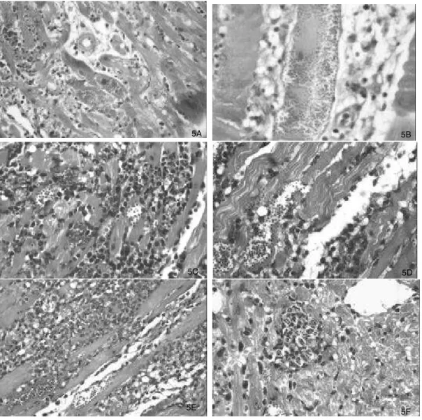
Histopathological study. Study of the tissue lesions and parasite tropism was comparatively performed for the parental strain and its clones. The parental strain determined progressive skeletal muscle and myocardium lesions, from the 14th day of infection, with slight mononuclear infiltration and low parasitism of
myocytes. By the 20th day of infection a progressive
parasitism of myocardiocytes was seen, with focal myocyte necrosis and diffuse mononuclear-cell infiltration (Figure 5A). Tissue parasitism and destruction of myocells with dense inflammatory infiltration were prominent in skeletal muscles and progressive until 30 days of infection (Figure 5B).
Histopathological lesions determined by each one of the seven clones, from 14 to 30 days of infection, showed the same evolution and histopathological aspects of the parental strain. For the seven clones, parasitism and inflammatory infiltration were present at 14 and 20 days p.i., varying from slight to moderate in keeping with the virulence of the clone. Necro-inflammatory lesions were more prominent in skeletal muscles and intensified up to the 30th day of infection. In general, this late phase exhibited parasite and myocell disintegration, together with severe mononuclear-cell infiltration and rare preserved parasites (Figures 5C,D, E and F).
Figure 5A and 5B - Cardiac and skeletal muscle lesions in mice infected with the Colombian parental strain: A – Diffuse and moderate myocarditis and presence of parasites in the cardiac myocytes (250X); B – necrosis of skeletal muscle cells with mononuclear infiltrate; presence of a large nest of amastigotes in a damaged skeletal muscle cell (400 X ); Figure CandD - Clone Cl-Col–C4: C -Intense myocarditis and presence of intracellular parasites in cardiac myocells (250X); D – Skeletal muscle cell with intracellular parasites associated with myocell disintegration and the presence of polymorphonuclear and mononuclear inflammatory cells infiltration (400X ); Figure EandF - Clone Cl-Col-C6: E - Intense destructive lesions of the skeletal muscle with parasite debris and intracellular amastigotes; intense polymorphonuclear and mononuclear inflammatory infiltration ( 250X); F – myocardium showing intense mononuclear infiltration and the presence of focal disintegration of parasitized myocell with intense polymorphonuclear infiltration (250X).
5A 5B
5C 5D
DISCUSSION
T. cruzi strains representmulticlonal populations, as described by Tibayrenc et al20. based on the multiloci isoenzymic profile of 121 strains from different geographical
areas. Macedo and Pena13 suggest that multiclonal
strains are formed by clonally propagating organisms with different tissue tropisms, correlated with complementary molecular interactions between the invading clones of T. cruzi and the host tissues. Clonal populations can be homogeneous or heterogeneous. It
has been admitted that a limited number of principal
clones could be circulating within different geographical
areas19. The present study has demonstrated a
homogeneity of the clones obtained from the Colombian strain, both considering its biological behavior and isoenzymic profiles. On analyzing the behavior of the Colombian strain after long period of maintenance in laboratory, by successive passages into mice, an increased virulence was detected, which was revealed by high mortality up to 30 days of infection. The
Colombian strain, as initially described3, was
characterized by a slow progression of parasitemia, that reached high levels by the 20th to 30th day of infection, with predominance of broad parasite forms from the initial phase of infection and low mortality up to 30 days p.i. Activation of parasite virulence, evaluated in infected mice, either after macrophage blockade with Indian Ink or treatment with corticoid, results in earlier increasing of parasitemia and the presence of slender parasitic forms6. However, considering the profiles of parasitemia, the tissue tropism and pathogenicity pattern, the biological behavior of the Colombian strain was maintained. The seven clones, with the same biological patterns of the parental strain, exhibited different degrees of virulence, as expressed by high levels of parasitemia and elevated mortality; however as has been previously demonstrated1, virulence is not a stable character, and therefore cannot independently be used for strain characterization. The same type of strain can present different degrees of virulence, evaluated by the levels of parasitemia and the rate of mortality of infected animals. The several strains isolated from São Felipe, BA classified as Biodeme Type II, could either exhibit
low virulence, without mortality by the 20th day of
infection; medium virulence, with 50% mortality or high virulence, with 100% mortality by 20 days of infection. However, comparative histopathological study revealed that lesions determined by the parental strain and its clones disclosed the same pattern of pathogenicity: same tissue tropism, with predominant involvement of skeletal muscles, intense myocarditis and high tissue parasitism. These findings suggest that when a predominant principal clone is present the histotropism of the strain and of the clones is similar, confirming the
isoenzymic patterns for GPI and PGM represent the same profile for the parental strain and clones, representing a homozygosis for these enzymes. However, the presence of a profile of three bands for ALAT in the clone C1 and two bands for ASAT in the clone C3 represents alternative patterns, as previously demonstrated7 14. According to Miles et al14, of the 11 enzymes which did contribute to the separation of the zymodemes, four gave alternative patterns within individual zymodemes; as for ALAT, up to four ancillary, prominent
bands were seen in zymodeme 1 (Z!)14; and ASAT has
been described with one or two bands, for Z17 14.
The present results, indicative of homogeneity for the clones isolated from the Colombian strain, support the suggestion that the presence of a
principal clone may be influencing the behavior of Type III strains that circulate in a given endemic area. The predominance of the same type of strain has
been recently confirmed by Rodriguez et al1 6,
studying 13 strains from Colombia, and showing that all of them belong to zymodeme Z1 and conform only
one cluster by analysis of the dendogram.
The zymodeme Z1 is recognized as representing a sylvatic origin of the parasite8 but with presence in the domestic environments. Humans infected with this type
of strain have already been identified5, and this can
REFERENCES
1. Andrade SG. Caracterização de cepas do Trypanosoma cruzi
isoladas Recôncavo baiano (Contribuição ao estudo da patologia geral da doença de Chagas em nosso meio). Revista de Patologia Tropical 3:65-121, 1974.
2. Andrade SG. Trypanosoma cruzi: clonal structure of parasites strains and the importance of principal clones. Memórias do Instituto Oswaldo Cruz94 (supl I): 185-187, 1999.
3. Andrade SG, Carvalho ML, Figueira RM. Caracterização morfobiológica e histopatológica de diferentes cepas do
Trypanosoma cruzi. Gazeta Médica da Bahia 70:32-42, 1970. 4. Andrade SG, Magalhães JB, Pontes AL. Evaluation of
chemotherapy with benzonidazole and nifurtimox in mice infected with Trypanosoma cruzi strains of different types. Bulletin of the World Health Organization63:721-726, 1985.
5. Andrade SG, Rassi A, Magalhães JB, Ferriolli Filho F, Luquetti, AO. Specific chemotherapy of Chagas’ disease: a comparison between the response in patients and experimental animals inoculated with the same strains. Transaction of the Royal Society of Tropical Medicine and Hygiene 86:624-626, 1992.
6. Andrade SG, Silva AA, Carvalho ML, Figueira, RM. Comportamento de uma cepa do Trypanosoma cruzi em hospedeiros de baixa resistência. Revista do Instituto de Medicina Tropical de São Paulo 14:154-161, 1972.
7. Andrade V, Brodskyn C, Andrade SG. Correlation between isoenzyme patterns and biological behaviour of different strains of Trypanosoma cruzi. Transaction of the Royal Society of Tropical Medicine and Hygiene 77:796-799, 1983.
8. Barrett TV, Hoff RH, Mott KE, Miles MA, Godfrey DG, Teixeira R, Almeida Souza JAA, Sherlock IA. Epidemiological aspects of three
Trypanosoma cruzi zymodemes in Bahia State, Brasil. Transaction of the Royal Society of Tropical Medicine and Hygiene 74:84-90, 1980.
9. Campos RMF, Andrade SG. Characterisation of subpopulations (clones and subclones) of the 21 SF strain of Trypanosoma cruzi
after long lasting maintenance in the laboratory. Memórias do Instituto Oswaldo Cruz91:795-800, 1996.
10. Campos RMF, Gonçalves MS, Reis EAG, Reis MG, Andrade SG. C omparative analysis by polymerase chain reaction ampliflied minicircles of kinetoplast DNA of a stable strain of Trypanosoma
cruzi from São Felipe, Bahia, its clones and subclones: possibility of predominance of a principal clone in this area. Memórias do Instituto Oswaldo Cruz 94:23-29, 1996.
11. Dvorak JA. Single cell isolates of Trypanosoma cruzi: How and Why? Revista da Sociedade Brasileira de Medicina Tropical 18 (supl):29-38, 1985.
12. Federici EE, Abelman WB, Neva FA. Chronic and progressive myocarditis and myositis in C3H mice infected with Trypanosoma cruzi. The American Journal of Tropical Medicine and Hygiene 13:272-280, 1964.
13. Macedo AM, Pena SDJ. Genetic variability of Trypanosoma cruzi : implications for the pathogenesis of Chagas’ disease. Parasitology Today 14:119-123, 1998.
14. Miles MA, Lanham SM, Souza AA, Póvoa M. Further enzymic characters of Trypanosoma cruzi and their evaluation for strain identification. Transaction of the Royal Society of Tropical Medicine and Hygiene 74:221-237, 1980.
15. Momem H. Taxonomy of Trypanosoma cruzi: a commentary on characterization and nomenclature. Memórias do Instituto Oswaldo Cruz94:181-184, 1999.
16. Rodriguez P, Montilla M, Nicholls S, Zarante I, Puerta C. Isoenzymatic Characterization of Colombian Strains of
Trypanosoma cruzi. Memórias do Instituto Oswaldo Cruz 93:739-740, 1998.
17. Silva LHP, Nussenzweig V. Sobre uma cepa de Trypanosoma cruzi altamente virulenta para o camundongo branco. Folia Clinica et Biologica20:191-208, 1953.
18. Tibayrenc M, Ayala FJ. Isozymic variability in Trypanosoma cruzi, the agent of Chagas’ disease: genetical, taxonomical, and epidemiological significance. Evolution 42:277-292, 1988.
19. Tibayrenc M, Brenière SF. Trypanosoma cruzi: major clones rather than zymodemes. Memórias do Instituto Oswaldo Cruz 83(supl 1): 249-255, 1988.
20. Tibayrenc M, Ward P, Moya A, Ayala FJ. Natural populations of