Late recognition and illness severity are
determi-nants of early death in severe septic patients
Flavia R. Machado,I,IIReinaldo Saloma˜o,II,IIIOtelo Rigato,IVElaine M. Ferreira,I,IIGuilherme Schettino,IV Tatiane Mohovic,V Carla Silva,IIIsac Castro,I Eliezer SilvaII,IV
IFederal University of Sa˜o Paulo, Department of Anesthesiology, Pain and Critical Care, Sa˜o Paulo/SP, Brazil.IILatin American Sepsis Institute, Sa˜o Paulo/SP,
Brazil.IIIFederal University of Sa˜o Paulo, Department of Infectious Diseases, Sa˜o Paulo/SP, Brazil.IVSı´rio Libaneˆs Hospital, Intensive Care Unit, Sa˜o Paulo/ SP, Brazil.VHospital Israelita Albert Einstein, Intensive Care Unit, Sa˜o Paulo/SP, Brazil.
OBJECTIVE: To identify the independent variables associated with death within 4 days after the first sepsis-induced organ dysfunction.
METHODS:In this prospective observational study, severe sepsis and septic shock patients were classified into 3 groups: Group 1, survivors; Group 2, late non-survivors; and Group 3, early non-survivors. Early death was defined as death occurring within 4 days after the first sepsis-induced organ dysfunction. Demographic, clinical and laboratory data were collected and submitted to univariate and multinomial analyses.
RESULTS:The study included 414 patients: 218 (52.7%) in Group 1, 165 (39.8%) in Group 2, and 31 (7.5%) in Group 3. A multinomial logistic regression analysis showed that age, Acute Physiology and Chronic Health Evaluation II score, Sepsis-related Organ Failure Assessment score after the first 24 hours, nosocomial infection, hepatic dysfunction, and the time elapsed between the onset of organ dysfunction and the sepsis diagnosis were associated with early mortality. In contrast, Black race and a source of infection other than the urinary tract were associated with late death. Among the non-survivors, early death was associated with Acute Physiology and Chronic Health Evaluation II score, chronic renal failure, hepatic dysfunction Sepsis-related Organ Failure Assessment score after 24 hours, and the duration of organ dysfunction.
CONCLUSION:Factors related to patients’ intrinsic characteristics and disease severity as well as the promptness of sepsis recognition are associated with early death among severe septic patients.
KEYWORDS: Prognosis; Mortality; Sepsis.
Machado FR, Saloma˜o R, Rigato O, Ferreira EM, Schettino G, Mohovic T, et al. Late recognition and illness severity are determinants of early death in severe septic patients. Clinics. 2013;68(5):586-591.
Received for publication onNovember 14, 2012;First review completed onDecember 17, 2012;Accepted for publication onJanuary 3, 2013 E-mail: [email protected]
Tel.: 55 11 5576-4650
& INTRODUCTION
Severe sepsis remains a challenge for healthcare providers because it carries a high mortality rate despite increased resource allocation from both health institutions and govern-ments (1–5). For these reasons, many researchers have devoted their efforts to finding efficient interventions (6,7).
There are few published data regarding the potential risk factors associated with early death in sepsis patients, and the concept of ‘‘early’’ is inconsistent (8–10). Some authors have attempted to construct models, which included patients’ general characteristics (11) or only coagulation factors (12), to predict short-term mortality in sepsis.
However, these authors used either 7 days or 14 days as the definition of short term. Another study only analyzed patients who were admitted to the emergency department without shock and reported a progression to shock in 17.8% of patients in the first 48 hours. However, they did not address early mortality (13).
Therefore, we hypothesized that some factors are linked to early death and that patients at risk for early death could be identified using clinical and laboratory data collected in the first days after intensive care unit (ICU) admission. Thus, the purpose of this study was to identify the variables that were independently associated with death within 4 days after the first sepsis-induced organ dysfunction.
& METHODS AND MATERIALS
Design and setting
This prospective cohort study was conducted at 3 mixed medical/surgical Brazilian intensive care units (1 public, 2 private; 87 total beds) from January 2007 through March 2009. All the study procedures were in accordance with the
Copyrightß2013CLINICS– This is an Open Access article distributed under the terms of the Creative Commons Attribution Non-Commercial License (http:// creativecommons.org/licenses/by-nc/3.0/) which permits unrestricted non-commercial use, distribution, and reproduction in any medium, provided the original work is properly cited.
No potential conflict of interest was reported.
ethical standards of the revised Helsinki Declaration (1983). The Research and Ethics Committee from each institution approved the study (Federal University of Sao Paulo: 1477/ 06; Sirio Libanes Hospital: HSL2006/27; Hospital Israelita Albert Einstein: 07/549). All patients or their closest relatives signed informed consent forms allowing data collection.
Inclusion/exclusion criteria and definitions
All patients older than 18 years of age diagnosed with severe sepsis or septic shock, as previously defined (14), were prospectively included. Briefly, patients were eligible for inclusion if they had 2 or more signs of systemic inflammatory response syndrome secondary to a known or suspected infection and at least 1 sepsis-induced organ dysfunction. Patients were excluded if they were pregnant or if their physicians were not committed to full life support. Sepsis-induced organ dysfunction was defined by one of the following symptoms: hypotension, arterial oxygen partial pressure/oxygen inspiratory fraction (PaO2/FiO2)
ratio#300, lactate level$1.5 times the reference value and base deficit.5, bilirubin level.2 times the reference value, urine output #0.5 ml/kg/hour after an adequate volume replacement or the need for renal replacement therapy, platelet count#100,000 mm3or a 50% decrease from the last 3 days’ values, and a reduced level of consciousness. Septic shock was defined as volume-refractory hypotension with a need for vasopressors. The time for sepsis diagnosis was defined as the number of hours elapsed between the onset of the first dysfunction and its recognition or management by the healthcare provider. We defined sepsis recognition as the record of a sepsis hypothesis in the patient’s chart. To identify the moment of organ dysfunction, the patient’s chart was carefully reviewed to determine the first record of hypotension, reduced level of consciousness or low urine output as well as the first laboratory sampling time whose results fulfilled the respiratory, metabolic, coagulation or hepatic criteria for organ dysfunction. For patients admitted from the emergency department who already had severe sepsis criteria, we used the time of triage.
Patients were classified into 3 groups according to their outcomes: survivors (Group 1), late death (Group 2), and early death (Group 3). Early death was defined as death occurring within 4 days after the onset of the first sepsis-induced organ dysfunction.
Data collection
Demographic data were collected at inclusion, as were the Acute Physiological and Chronic Health Evaluation (APACHE II) and Sequential Organ Failure Assessment (SOFA) scores (SOFA D0). SOFA scores were also determined on Day 1 (SOFA D1) and Day 3 (SOFA D3). The change in SOFA score was calculated by subtracting SOFA D0 from SOFA D3 (DSOFA D3) and SOFA D1 (DSOFA D1). Clinical and outcome features, including compliance to the 6-hour and 24-hour Surviving Sepsis Campaign (SSC) bundles, were registered as proposed by the Campaign (15). We did not record data on activated protein C or low-dose steroid adherence because of the controversial aspects related to their use. The patients were followed until hospital discharge or for 60 days, and ICU and hospital outcomes were recorded.
Statistics
Continuous data were presented as medians and inter-quartile ranges, and categorical variables were reported as
the number (%). The normality of continuous variables was determined with the Kolmogorov-Smirnov test. The 3 groups were compared regarding the mortality risk factors. The non-parametric Kruskal-Wallis test, with the Muller-Dunn post-test, were used for non-normally distributed variables. The chi-squared likelihood ratio test or Pearson’s chi-squared test was used, as appropriate, and partitioning of chi-square was performed if needed.
We performed a multinomial logistic regression analysis to allow for simultaneous comparisons among the 3 studied groups. For a variable to be included in the model, we transformed all the continuous variables into categorical variables according to the best cut-off point for early mortality prediction in the receiver operating characteristic (ROC) curve. For all the SOFA scores, we used the same cut-off value based on the ROC curve of SOFA scores at admission. For DSOFA D3 and DSOFA D1, any negative value was considered an improvement, while all positive or zero values were considered a worsening outcome. Variables associated with early death at the 0.2 level in the univariate analysis were included in the model, except for those with more than 50 missing cases or those not applicable for all patients. A step-by-step method was used to select the significant variables. To identify the risks factors for early and late death compared with the survivors, Group 1 was considered the reference group. To identify the risk factors related to early death among non-survivors, Group 2 (late death) was the reference group. A p-value ,0.05 for any of the groups being compared was considered to retain a specific variable in the analysis. After the selection, the results were presented as odds ratios and 95% confidence intervals (CI).
Statistical analyses were performed using PASW Statistics 18 for Windows (SPSS Inc., Chicago, USA), and all the tests were two tailed.
& RESULTS
Basic characteristics of septic patients
A total 414 septic patients were included in the study: 218 (52.7%) in Group 1, 165 (39.8%) in Group 2, and 31 (7.5%) in Group 3. We followed all the patients until hospital discharge or death. Among the deaths, 15.8% occurred in the first 4 days. All demographic and baseline character-istics are shown in Table 1.
Factors associated with early and late hospital mortality – univariate analysis
In the univariate analysis, the APACHE II and SOFA scores at admission were associated with both early and late mortality. However, more patients had high scores in the early mortality group compared to those with late mortality. The severity of organ dysfunction was higher in non-survivors, and the percentage of higher scores at Day 1 and Day 3 clearly distinguished between early and late death. The worsening of dysfunction, as measured by bothDSOFA D1 andDSOFA D3, occurred more frequently in the non-survivors, regardless of the time of death (Table 1). Respiratory dysfunction was a marker of early and late death, while hepatic dysfunction was more prevalent only in the early death group. We found higher lactate levels in the patients with hepatic dysfunction (17.4, 11.1–29.0 vs.
23.5, 14.2–47.3,p= 0.013). Late recognition of organ
were no differences between groups in compliance with any indicator of the SSC-6 h bundle or glycemic control (Table 2).
Factors associated with early and late hospital mortality – multinomial analysis
The covariance analysis using the Jaccard test detected a high degree of colinearity between the presence of shock and SOFA D1 (r = 0.764), as well as between respiratory
dysfunction and a pulmonary source of infection (r = 0.724). Thus, on the basis of their p values in the univariate analysis, we excluded both septic shock (p= 0.138) and a
pulmonary source of infection (p= 0.048) from the
multi-nomial analysis.
The multinomial analysis revealed that the patient age, SOFA score after the first 24 hours, nosocomial infection, and duration of organ dysfunction were associated with both early and late mortality. However, the odds ratios were
Table 1 -Patient demographics and infection characteristics according to the survival group.
Variable Group 1 (n = 218) Group 2 (n = 165) Group 3 (n = 31) p-value+
Age (years) 62.0 (46.0–77.0) 69.0 (54.0–78.0)*** 69.0 (54.0–77.0)* 0.069
Age$52 150 (68.8), 137 (83.0)*** 27 (87.1)* 0.001
Gender
Female 94 (43.1) 67 (40.6) 9 (29.0) 0.324
Male 124 (56.9) 98 (59.4) 22 (71.0)
Race
Black 22 (10.1) 35 (21.2)** 7 (22.6) 0.030
White 187 (85.8) 124 (75.2) 22 (71.0)
Other
APACHE II score 17.0 (13.0–21.0) 19.0 (15.0–24.0)* 22.0 (15.0–26.0)* 0.0002
APACHE$22 52 (23.9) 54 (32.7) 16 (51.6)*{
0.004 SOFA score
SOFA D0 6.0 (4.0–9.0) 8.0 (5.0–10.0)* 9.0 (8.0–11.0)* ,0.00001
SOFA D0$8 81 (37.5) 90 (54.5)*** 25 (80.6)***{{
,0.00001
SOFA D1 6.0 (4.0–8.0) 8.0 (6.0–11.0)* 10.0 (8.0–12.0)* ,0.0001
SOFA D1$8 73 (34.3) 101 (61.2)*** 23 (85.2)***{{
,0.0001
SOFA D3 5.0 (2.0–8.0) 8.0 (6.0–11.0)* 9.0 (8.0–12.0)* ,0.0001
SOFA D3$8 50 (26.5) 92 (56.8)*** 11 (84.6)***{
,0.0001
DSOFA D1 0.0 (22.0–1.0) 0.0 (21.0–2.0)* 1.0 (0.0–2.0)* ,0.0001
WorseningDSOFA D1 123 (57.7) 117 (70.9)** 21 (77.8)* ,0.009
DSOFA D3 21.0 (23.0–0.0) 0.0 (21.0–2.0)* 2.0 (0.0–4.0)* ,0.0001
WorseningDSOFA D3 68 (36.0) 92 (56.8)*** 10 (76.9)** ,0.0001
Lactate at admission 17.0 (11.0–30.0) 17.0 (11.9–28.8) 24.0 (16.0–55.0)*{ 0.035
Lactate$20 mg/dL 71 (47.7) 55 (49.1) 18 (66.7) 0.181
Comorbidities 158 (72.5) 134 (81.2) 25 (80.6) 0.116
COPD 32 (14.7) 23 (13.9) 5 (16.1) 0,944
Arterial hypertension 85 (39.0) 70 (42.4) 13 (41.9) 0.784
CRF 24 (11.00) 31 (18.8)* 2 (6.5) 0.043
Immunosuppression 40 (18.3) 42 (25.5) 10 (32.3) 0.100
Diabetes mellitus 50 (22.9) 47 (28.5) 8 (25.8) 0.465
Alcoholism 14 (6.5) 11 (6.7) 1 (3.2) 0.759
Admission category
Clinical 141 (64.7) 116 (70.3) 20 (64.5) 0.490
Surgical 77 (35.3) 49 (29.7) 11 (35.5)
Sepsis category
Severe sepsis 70 (32.1) 54 (32.7) 5 (16.1) 0.138
Septic shock 148 (67.9) 111 (67.3) 26 (83.9)
Infection category
Community acquired 137 (62.8) 56 (33.9)*** 12 (38.7)* ,0.0001
Hospital acquired 81 (37.2) 109 (66.1) 19 (61.3)
Source of infection
Pulmonary 100 (45.9) 95 (57.6)* 13 (41.9) 0.048
Abdominal 49 (22.5) 38 (23.0) 10 (32.3) 0.503
UTI 32 (14.7) 8 (4.8) *** 2 (6.5) 0.005
Organ dysfunction (n) 3.0 (2.0–3.0) 3.0 (2.0–3.0) 3.0 (2.0–4.0) 0.369
Dysfunction.2 113 (51.8) 91 (55.2) 20 (64.5) 0.386
Type of dysfunction
Cardiovascular 198 (90.8) 148 (89.7) 29 (93.5) 0.784
Respiratory 97 (44.5) 95 (57.6)* 21 (67.7)* 0.006
Renal 87 (39.9) 78 (47.3) 15 (48.4) 0.301
Metabolic 46 (21.1) 32 (19.4) 12 (38.7) 0.076
Hematologic 43 (19.7) 38 (23.0) 8 (25.8) 0.617
Hepatic 25 (11.5) 25 (15.2) 12 (38.7)***{{
,0.0001
Neurologic 59 (27.1) 60 (36.4) 10 (32.3) 0.150
APACHE: Acute Physiological and Chronic Health Evaluation; SOFA D0: Sequential Organ Failure Assessment at admission; ICU: intensive care unit; COPD: chronic obstructive pulmonary disease; CRF: chronic renal failure; UTI: urinary tract infection. The results are expressed as the number (%) or median (25%–75% percentile).+Chi-squared and Kruskal-Wallis tests. Chi-squared partition or Muller-Dunn post-test: *
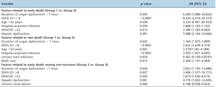
higher for early death. The illness severity at admission (APACHE II) and hepatic dysfunction were only correlated with early death, while Black race and a source of infection other than the urinary tract were only correlated with late death. When the risk factors for early death were analyzed among the non-survivors, APACHE II, chronic renal failure, hepatic dysfunction, and the SOFA score after 24 hours and
duration of organ dysfunction were identified. The results of the multinomial analysis are shown in Table 3. The lactate level was not included in the primary multinomial analysis, as there were too many missing values (n = 55). A secondary multinomial analysis that included the lactate levels categorized according to the ROC curve did not change the results (data not shown).
Table 2 -Characteristics related to adequate treatment and organ dysfunction evolution according to the survival group.
Variable Group 1 (n = 218) Group 2 (n = 165) Group 3 (n = 31) p-value+
Duration of organ dysfunction (hours)
2.2 (0.0–9.6) 6.0 (0.0–15.0)* 6.0 (1.6–9.0) 0.019
Duration of organ dysfunction
.1 hour
128 (59.3) 115 (70.1)* 26 (83.9)** 0.005
6-hour bundle compliance
Lactate sampling 137 (62.8) 106 (64.2) 24 (77.4) 0.283
Blood culture sampling 117 (53.9) 76 (46.1) 14 (45.2) 0.266
Antibiotics
Up to 1 hour 129 (59.2) 87 (52.7) 19 (61.3) 0.392
Time to antibiotics (h) 2.0 (0.62–6.0) 2.64 (1.0–6.0) 1.25 (0.5–5.0) 0.319
Volume/vasopressors 191 (97.0) 143 (96.6) 29 (96.7) 0.981
CVP achievement 67 (45.9) 51 (45.1) 17 (65.4) 0.154
SvcO2achievement 76 (52.1) 57 (50.4) 15 (57.7) 0.799
Compliance with all 6 h items 0 0 0 –
24 h bundle compliance Glycemic control
Median,180 mg/dL 160 (73.4) 121 (73.3) 20 (64.5) 0.567
Glycemia* 140.0 (120.0–162.0) 143.0 (120.0–167.0) 128.0 (119.0–169.0) 0.648
Hypoglycemia** 15 (6.9) 11 (6.7) 4 (12.9) 0.457
Plato pressure
Median,30 cmH2O 117 (89.3) 112 (91.1) 21 (87.5) 0.825
Median 24.0 (20.0–27.0) 25.5 (22.0–28.0)* 25.0 (22.0–28.0) 0.020
Median.24 cmH2O 67 (51.1) 82 (66.1)* 16 (69.6) 0.030
CVP: central venous pressure; SvcO2: central venous oxygen saturation; SOFA: Sequential Organ Failure Assessment; D: day; *median value between the
6thand 24thhours; ** patients with at least 1 episode of hypoglycemia. The results are expressed as the number (%) or median (25%–75% percentile). +Chi-squared and Kruskal-Wallis tests. Chi-squared partition or Muller-Dunn post-test: *
p#0.05vs. Group 1; **p#0.01vs. Group 1; ***p#0.001vs. Group 1;{p#0.05vs. Group 2;{{p#0.01vs. Group 2;{{{p#0.001vs. Group 2.
Table 3 -Risk factors related to early and late death – multinomial analysis.
Variable p-value OR (95% CI)
Factors related to early death (Group 1vs. Group 3)
Duration of organ dysfunction.1 hour 0.003 6.289 (1.886–20.833)
SOFA D1$8 ,0.0001 8.333 (2.610–26.315)
Age$52 years 0.058 3.225 (0.961–45.933)
Hospital-acquired infection 0.030 2.808 (1.102–7.142)
APACHE$22 0.013 3.389 (1.292–8.850)
Hepatic dysfunction 0.001 5.988 (2.164–16.666)
Factors related to late death (Group 1vs. Group 2)
Duration of organ dysfunction.1 hour 0.025 1.764 (1.075–2.899)
SOFA D1$8 ,0.0001 2.632 (1.639–4.219)
Age$52 years 0.003 2.370(1.345–4.184)
Hospital-acquired infection ,0.0001 2.950 (1.851–4.695)
Urinary tract infection 0.024 0.362 (0.150–0.8741)
Black race 0.013 2.304 (1.191–4.464)
Factors related to early death among non-survivors (Group 2vs. Group 3)
Duration of organ dysfunction.1 hour 0.024 3.953 (1.195–13.080)
SOFA D1$8 0.037 3.406 (1.077–10.771)
APACHE$22 0.043 2.610 (1.030–6.615)
Hepatic dysfunction 0.001 4.776 (1.833–12.445)
Chronic renal failure 0.040 0.188 (0.038–0.923)
& DISCUSSION
This study showed that both illness severity and early recognition were associated with early death. In the multi-nomial analysis, we demonstrated that the APACHE II score, presence of chronic renal failure, sepsis-induced hepatic dysfunction, duration of organ dysfunction, and SOFA score in the first day after initial organ dysfunction were able to distinguish early survivors from late non-survivors. We also demonstrated that compared with the survivors, early death was associated with age, illness severity at admission (APACHE II), illness severity on the first day (SOFA D1), and hospital-acquired infection. All of these issues are well-known risk factors for sepsis mortality. However, our study identified 2 other risk factors that have not been frequently described in previous studies: the presence of hepatic dysfunction and the duration of organ dysfunction. Both issues were not only risk factors for early mortality but also identified the patients who would die early among the non-survivors.
Only 3 studies have focused on early mortality risk factors in sepsis patients (8–10). A multicenter French study showed that 27% of all fatalities occurred in the first 2 days of severe sepsis, and the illness severity, as assessed by the SAPS II score, number of organ failures and the presence of shock or acidosis were associated with death (8). In their study, later mortality was associated with the underlying disease and chronic liver and heart failure. Blanco et al. analyzed risk factors related to death within the first 48 hours, during which 14.8% of all deaths occurred (9). Neither of these studies included variables related to the adequacy of treatment or the duration of organ dysfunction in their analysis. These studies included patients observed in 1993 and 2002, when the relevance of early detection and intervention was not as widely recognized. This early approach could have impacted early deaths, as we showed a lower proportion of early death compared to the previous authors.
The association between hepatic dysfunction and early death that was found in our study and by Blanco et al. is interesting. We assessed acute hepatic dysfunction related to sepsis and not chronic hepatic failure, which has already been described to be associated with late death (8). There are multiple possible explanations for this finding. First, liver dysfunction has been associated with the inability of the immune system to adequately react to host invasion by pathogenic organisms (16–18). Indeed, infected patients with chronic or acute liver dysfunction present with worse prognoses (19). Second, the liver is also responsible for coagulation factor synthesis, and its impairment is clearly related to prognosis (20,21). Activated prothrombin time and fibrinogen level can be predictors of early death (12). Finally, liver impairment is a neglected dysfunction in sepsis; thus, other studies might not have found this association simply because they did not evaluate it.
Non-compliance with the SSC bundles was not related to early or late death. This finding contrasts with other studies, which have shown a clear association between SSC bundle adherence and survival (22–27). A possible explanation is that patients who will die earlier could appear sicker at inclusion and are thus more prone to receive early and adequate care, which is not enough to permit survival in the face of severe disease. Another explanation is that we evaluated compliance at the time of sepsis diagnosis. In Brazil, the time between the onset of organ dysfunction and
a proper sepsis diagnosis can be long (28). In this context, the compliance measure after diagnosis would not reflect the appropriateness of the treatment.
One of the most important contributions of this study was to clearly demonstrate that the delay in diagnosis and, consequently, treatment was related to not only death but also early death among non-survivors. Although previous studies have already addressed this issue (28–30), they did not associate these delays with early death. Moreover, in our study, the best cut-off value for predicting death was 1 hour. The impact of small delays has already been demonstrated. Kumar et al. showed that the mortality of septic shock patients increased after delaying adequate antibiotic administration (31) by only 1 hour. Thus, it is unsurprising that even small delays in sepsis recognition could have substantial impacts on both global mortality and early mortality.
Another interesting finding was the association between lactate level and the risk of early death. Although this association could not be tested in an appropriate multi-nomial analysis because of excessive missing values, high lactate and its non-clearance are clearly associated with mortality (27,32–34). However, this is the first study to show that high lactate is associated with early mortality, even when only non-survivors are considered.
There are strengths and limitations to our study. One of its strengths is its multicentric nature, which comprised both private and public hospitals. Second, we used a strict definition of ‘‘early death’’ that considered the initiation of organ dysfunction in addition to diagnosing sepsis or ICU admissions. Third, we obtained robust data that was related to the specific therapeutic actions of the clinical team based on the SSC bundles. Although they were not related to mortality, most likely because of the long time between the initiation of organ dysfunction and adequate treatment, we provided an unprecedented analysis of the early mortality risk factors. Fourth, our multinomial analysis allowed us to clearly identify the risk factors for early or late death as well as the risk factors for early death among non-survivors.
One of our study limitations was the absence of information on withholding or withdrawal of life support, primarily in the early death group. However, as palliative care patients were not included, any initiative to stop life support would most likely reflect the severity of sepsis and not the severity of the underlying disease, for which palliative care would be advisable. Another weakness was the lack of information on causes of death. We should also consider the potential interaction between the presence of liver dysfunction and the SOFA score as a limitation. However, our covariance analysis using the Jaccard test did not show the presence of collinearity.
In summary, in a large, multicenter severe sepsis database, both the severity of disease and late recognition of sepsis were associated with early death.
& ACKNOWLEDGMENTS
We thank Dr. Ruy Guilherme Cal for helping with the electronic case report form and the database. This work was supported by grants from the Fundac¸a˜o de Amparo a Pesquisa do Estado de Sa˜o Paulo.
& AUTHOR CONTRIBUTIONS
Machado FR and Silva E drafted the manuscript. All the authors read and approved the final version of the manuscript.
& REFERENCES
1. Angus DC, Linde-Zwirble WT, Lidicker J, Clermont G, Carcillo J, Pinsky MR. Epidemiology of severe sepsis in the United States: analysis of incidence, outcome, and associated costs of care. Crit Care Med. 2001;29(7):1303-10, http://dx.doi.org/10.1097/00003246-200107000-00002. 2. Martin GS, Mannino DM, Eaton S, Moss M. The epidemiology of sepsis
in the United States from 1979 through 2000. N Engl J Med. 2003;348(16):1546-54.
3. Silva E, Pedro Mde A, Sogayar AC, Mohovic T, Silva CL, Janiszewski M, et al. Brazilian Sepsis Epidemiological Study (BASES study). Crit Care. 2004;8(4):R251-60, http://dx.doi.org/10.1186/cc2892.
4. Sogayar AM, Machado FR, Rea-Neto A, Dornas A, Grion CM, Lobo SM, et al. A multicentre, prospective study to evaluate costs of septic patients in Brazilian intensive care units. Pharmacoeconomics. 2008;26(5):425-34, http://dx.doi.org/10.2165/00019053-200826050-00006.
5. Beale R, Reinhart K, Brunkhorst FM, Dobb G, Levy M, Martin G, et al. Promoting Global Research Excellence in Severe Sepsis (PROGRESS): lessons from an international sepsis registry. Infection. 2009;37(3):222-32, http://dx.doi.org/10.1007/s15010-008-8203-z.
6. Dellinger RP, Levy MM, Carlet JM, Bion J, Parker MM, Jaeschke R, et al. Surviving Sepsis Campaign: international guidelines for management of severe sepsis and septic shock: 2008. Intensive Care Med. 2008;34(1):17-60, http://dx.doi.org/10.1007/s00134-007-0934-2.
7. Levy MM, Dellinger RP, Townsend SR, Linde-Zwirble WT, Marshall JC, Bion J, et al. The Surviving Sepsis Campaign: results of an international guideline-based performance improvement program targeting severe sepsis. Crit Care Med. 38(2):367-74.
8. Brun-Buisson C, Doyon F, Carlet J, Dellamonica P, Gouin F, Lepoutre A, et al. Incidence, risk factors, and outcome of severe sepsis and septic shock in adults. A multicenter prospective study in intensive care units. French ICU Group for Severe Sepsis. JAMA. 1995;274(12):968-74. 9. Blanco J, Muriel-Bombin A, Sagredo V, Taboada F, Gandia F, Tamayo L,
et al. Incidence, organ dysfunction and mortality in severe sepsis: a Spanish multicentre study. Crit Care. 2008;12(6):R158, http://dx.doi. org/10.1186/cc7157.
10. Vincent JL, Nelson DR, Williams MD. Is worsening multiple organ failure the cause of death in patients with severe sepsis? Crit Care Med. 2011;39(5):1050-5, http://dx.doi.org/10.1097/CCM.0b013e31820eda29. 11. Adrie C, Francais A, Alvarez-Gonzalez A, Mounier R, Azoulay E, Zahar
JR, et al. Model for predicting short-term mortality of severe sepsis. Crit Care. 2009;13(3):R72, http://dx.doi.org/10.1186/cc7881.
12. Lissalde-Lavigne G, Combescure C, Dorangeon E, Lefrant JY, Gris JC. Simple coagulation tests improve early mortality prediction for patients in intensive care units who have proven or suspected septic shock. J Thromb Haemost. 2007;5(5):1081-3, http://dx.doi.org/10.1111/j.1538-7836.2007.02492.x.
13. Glickman SW, Cairns CB, Otero RM, Woods CW, Tsalik EL, Langley RJ, et al. Disease progression in hemodynamically stable patients presenting to the emergency department with sepsis. Acad Emerg Med. 2010;17(4):383-90, http://dx.doi.org/10.1111/j.1553-2712.2010.00664.x. 14. Bone RC, Balk RA, Cerra FB, Dellinger RP, Fein AM, Knaus WA, et al.
Definitions for sepsis and organ failure and guidelines for the use of innovative therapies in sepsis. The ACCP/SCCM Consensus Conference Committee. American College of Chest Physicians/Society of Critical Care Medicine. Chest. 1992;101(6):1644-55. Epub 1992/06/01.
15. Surviving Sepsis Campaign - bundles of care. [cited 2011 October 22th]; Available from: http://www.survivingsepsis.org/Bundles/Pages/ BundlesforImprovement.aspx.
16. Eipel C, Hildebrandt A, Scholz B, Schyschka L, Minor T, Kreikemeyer B, et al. Mutation of mitochondrial ATP8 gene improves hepatic energy status in a murine model of acute endotoxemic liver failure. Life Sci. 2011;88(7-8):343-9, http://dx.doi.org/10.1016/j.lfs.2010.12.011.
17. Kim TH, Lee SH, Lee SM. Role of Kupffer cells in pathogenesis of sepsis-induced drug metabolizing dysfunction. FEBS J. 2011;278(13):2307-17, http://dx.doi.org/10.1111/j.1742-4658.2011.08148.x.
18. Romics L, Jr., Dolganiuc A, Kodys K, Drechsler Y, Oak S, Velayudham A, et al. Selective priming to Toll-like receptor 4 (TLR4), not TLR2, ligands by P. acnes involves up-regulation of MD-2 in mice. Hepatology. 2004;40(3):555-64, http://dx.doi.org/10.1002/hep.20350.
19. Umegaki T, Ikai H, Imanaka Y. The impact of acute organ dysfunction on patients’ mortality with severe sepsis. J Anaesthesiol Clin Pharmacol. 2011;27(2):180-4.
20. Lorente JA, Garcia-Frade LJ, Landin L, de Pablo R, Torrado C, Renes E, et al. Time course of hemostatic abnormalities in sepsis and its relation to outcome. Chest. 1993;103(5):1536-42, http://dx.doi.org/10.1378/chest.103.5.1536. 21. Walsh TS, Stanworth SJ, Prescott RJ, Lee RJ, Watson DM, Wyncoll D.
Prevalence, management, and outcomes of critically ill patients with prothrombin time prolongation in United Kingdom intensive care units. Crit Care Med. 2010;38(10):1939-46.
22. Gao F, Melody T, Daniels DF, Giles S, Fox S. The impact of compliance with 6-hour and 24-hour sepsis bundles on hospital mortality in patients with severe sepsis: a prospective observational study. Crit Care. 2005;9(6):R764-70, http://dx.doi.org/10.1186/cc3909.
23. Kortgen A, Niederprum P, Bauer M. Implementation of an evidence-based ‘‘standard operating procedure’’ and outcome in septic shock. Crit Care Med. 2006;34(4):943-9, http://dx.doi.org/10.1097/01.CCM.0000206112.32673.D4. 24. Ferrer R, Artigas A, Levy MM, Blanco J, Gonzalez-Diaz G,
Garnacho-Montero J, et al. Improvement in process of care and outcome after a multicenter severe sepsis educational program in Spain. JAMA. 2008;299(19):2294-303, http://dx.doi.org/10.1001/jama.299.19.2294. 25. Micek ST, Roubinian N, Heuring T, Bode M, Williams J, Harrison C, et al.
Before-after study of a standardized hospital order set for the manage-ment of septic shock. Crit Care Med. 2006;34(11):2707-13, http://dx.doi. org/10.1097/01.CCM.0000241151.25426.D7.
26. Nguyen HB, Corbett SW, Steele R, Banta J, Clark RT, Hayes SR, et al. Implementation of a bundle of quality indicators for the early manage-ment of severe sepsis and septic shock is associated with decreased mortality. Crit Care Med. 2007;35(4):1105-12, http://dx.doi.org/10.1097/ 01.CCM.0000259463.33848.3D.
27. Shapiro NI, Howell MD, Talmor D, Lahey D, Ngo L, Buras J, et al. Implementation and outcomes of the Multiple Urgent Sepsis Therapies (MUST) protocol. Crit Care Med. 2006;34(4):1025-32, http://dx.doi.org/ 10.1097/01.CCM.0000206104.18647.A8.
28. Freitas FG, Salomao R, Tereran N, Mazza BF, Assuncao M, Jackiu M, et al. The impact of duration of organ dysfunction on the outcome of patients with severe sepsis and septic shock. Clinics (Sao Paulo). 2008;63(4):483-8. 29. Wang JL, Chin CS, Chang MC, Yi CY, Shih SJ, Hsu JY, et al. Key process indicators of mortality in the implementation of protocol-driven therapy for severe sepsis. J Formos Med Assoc. 2009;108(10):778-87, http://dx. doi.org/10.1016/S0929-6646(09)60405-8.
30. Westphal GA, Koenig A, Caldeira Filho M, Feijo J, de Oliveira LT, Nunes F, et al. Reduced mortality after the implementation of a protocol for the early detection of severe sepsis. J Crit Care. 26(1):76-81.
31. Kumar A, Roberts D, Wood KE, Light B, Parrillo JE, Sharma S, et al. Duration of hypotension before initiation of effective antimicrobial therapy is the critical determinant of survival in human septic shock. Crit Care Med. 2006;34(6):1589-96, http://dx.doi.org/10.1097/01.CCM. 0000217961.75225.E9.
32. Howell MD, Donnino M, Clardy P, Talmor D, Shapiro NI. Occult hypoperfusion and mortality in patients with suspected infection. Intensive Care Med. 2007;33(11):1892-9, http://dx.doi.org/10.1007/ s00134-007-0680-5.
33. Trzeciak S, Dellinger RP, Chansky ME, Arnold RC, Schorr C, Milcarek B, et al. Serum lactate as a predictor of mortality in patients with infection. Intensive Care Med. 2007;33(6):970-7, http://dx.doi.org/10.1007/s00134-007-0563-9.