RESEARCH ARTICLE
Specific Depletion of Ly6C
hi
Inflammatory
Monocytes Prevents Immunopathology in
Experimental Cerebral Malaria
Beatrix Schumak1☯*, Katrin Klocke1☯, Janina M. Kuepper1, Aindrila Biswas2, Andrea Djie-Maletz3, Andreas Limmer4, Nico van Rooijen5, Matthias Mack6, Achim Hoerauf1, Ildiko Rita Dunay2*
1Institute of Medical Microbiology, Immunology and Parasitology, University of Bonn, Bonn, Germany, 2Institute of Medical Microbiology, University of Magdeburg, Magdeburg, Germany,3Department of Neurosurgery, University of Freiburg, Freiburg, Germany,4Institutes of Molecular Medicine and Experimental Immunology, University of Bonn, Bonn, Germany,5VUMC Department of Molecular Cell Biology, Faculty of Medicine Vrije Universiteit, Amsterdam, The Netherlands,6Department of Internal Medicine II, University Hospital Regensburg, Regensburg, Germany
☯These authors contributed equally to this work.
*[email protected](BS); [email protected](IRD)
Abstract
Plasmodium berghei ANKA(PbA) infection of C57BL/6 mice leads to experimental cerebral malaria (ECM) that is commonly associated with serious T cell mediated damage. In other parasitic infection models, inflammatory monocytes have been shown to regulate Th1 re-sponses but their role in ECM remains poorly defined, whereas neutrophils are reported to contribute to ECM immune pathology. Making use of the recent development of specific
monoclonal antibodies (mAb), we depletedin vivoLy6Chiinflammatory monocytes (by
anti-CCR2), Ly6G+neutrophils (by anti-Ly6G) or both cell types (by anti-Gr1) during infection with Ovalbumin-transgenic PbA parasites (PbTg). Notably, the application of Gr1 or anti-CCR2 but not anti-Ly6G antibodies into PbTg-infected mice prevented ECM development. In addition, depletion of Ly6Chiinflammatory monocytes but not neutrophils led to decreased IFNγlevels and IFNγ+CD8+T effector cells in the brain. Importantly, anti-CCR2 mAb injection
did not prevent the generation of PbTg-specific T cell responses in the periphery, whereas anti-Gr1 mAb injection strongly diminished T cell frequencies and CTL responses. In conclu-sion, the specific depletion of Ly6Chiinflammatory monocytes attenuated brain inflammation
and immune cell recruitment to the CNS, which prevented ECM followingPlasmodium
infec-tion, pointing out a substantial role of Ly6C+monocytes in ECM inflammatory processes.
Introduction
Malaria remains one of the most serious infectious diseases affecting 10% of the world's popu-lation. Although infections are endemic in over 100 countries, 90% of the deaths, most of
which affect children, occur in sub-Saharan Africa and South East Asia [1,2]. Malaria is elicited
OPEN ACCESS
Citation:Schumak B, Klocke K, Kuepper JM, Biswas A, Djie-Maletz A, Limmer A, et al. (2015) Specific Depletion of Ly6ChiInflammatory Monocytes Prevents Immunopathology in Experimental Cerebral Malaria. PLoS ONE 10(4): e0124080. doi:10.1371/ journal.pone.0124080
Academic Editor:Georges Snounou, Université
Pierre et Marie Curie, FRANCE
Received:September 3, 2014
Accepted:March 3, 2015
Published:April 17, 2015
Copyright:© 2015 Schumak et al. This is an open access article distributed under the terms of the
Creative Commons Attribution License, which permits unrestricted use, distribution, and reproduction in any medium, provided the original author and source are credited.
Data Availability Statement:All relevant data are within the paper and its Supporting Information files.
by various species of the protozoan parasite from the genusPlasmodiumand is transmitted to
humans through the bite of femaleAnophelesmosquitoes.P.falciparumis the most virulent of
the fivePlasmodiumspecies that cause disease in humans. Amongst the serious pathological
complications, cerebral malaria (CM) remains the greatest life-threatening risk. CM is a fatal neurological syndrome with multi-factorial, complex developmental stages and symptoms. It is generally acknowledged that CM results from immune-mediated pathology due to overwhelm-ing inflammatory processes and parasite sequestration [3]. Infections in C57BL/6 mice with Plasmodium berghei ANKA(PbA) infected red blood cells induce lethal experimental CM
(ECM) [4,5]. The resulting cerebral pathology in PbA-infected mice is induced by
pro-inflam-matory immune responses of CD8+T cells and subsequent IFNγproduction [6–8]. However, it
remains unclear how such strong immune responses are induced or regulated and the exact contribution of phagocytic cells in ECM is incompletely understood.
In the present study we addressed the question about the contribution of inflammatory mono-cytes in ECM development. In our previous studies, we demonstrated that a primary function of inflammatory monocytes (Gr1+Ly6ChiCCR2+CX3CR1lo), a subset of mononuclear cells, was to
drive strong Th1 responses within the host in the murine model ofToxoplasmosis[9,10].
Further-more, Ly6Chimonocytes were recruited to the site of infection and there, contributed to disease control via secretion of anti-microbial molecules [9,11,12]. Such Ly6ChiCCR2+monocytes emerge
from the bone marrow and populate non-lymphoid tissues [9,13,14]. They contribute to
orches-trate memory CD8+T cell and NK cell activation via the production of interleukin 18 and interleu-kin 15 [15]. In the absence of the CCR2 receptor, monocytes are unable to exit the bone marrow and in accordance, CCR2-/-mice display increased susceptibility to ListeriaandToxoplasma infec-tions[9,14]. In contrast to inflammatory Gr1+Ly6ChiCCR2+CX3CR1locells, the other major subset of monocytes, Gr1−Ly6C-CCR2−CX
3CR1hi, establish residency in the periphery where they
per-form important surveillance actions [16]. Similar subsets of monocytes have been described in
hu-mans; CD14+CD16−vs. CD14loCD16+cells which represent inflammatory and surveillance
populations, respectively [17,18]. In addition to monocytes, Gr1 is expressed on neutrophils and the most common antibody used to define this receptor, RB6, recognizes both Ly6C and Ly6G
iso-forms [11,19]. Recently, monoclonal antibodies (mAbs) detecting distinct Ly6 isoforms have been
developed allowing neutrophils (CD11b+F4/80−Ly6GhiLy6CintGr1hi) to be readily distinguished from inflammatory monocytes (CD11b+F4/80+Ly6ChiLy6G––CCR2+Gr1int) and selectively deplet-ed [11,19,20]. With regards to the role of Gr1+cells during malaria infection, previous depletion
studies conducted by Chenet al., using the anti-Gr1 antibody (clone RB6) in PbA infected mice,
concluded an important role for neutrophils in developing ECM pathology [21]. However, recent reports demonstrated broad elimination of Gr-1 positive cells including monocytes and T cells upon anti-Gr1 injection, thus questioning the validity of this clone. Here, we describe the impact of
Ly6Chiinflammatory monocytes on ECM pathology during experimentalPlasmodiuminfection
using the new selectively depleting antibodies anti-CCR2 and anti-Ly6G to analyse the impact of
Ly6Chiinflammatory monocytesversusneutrophils in the development of ECM.
Results
Depletion of phagocytic cells prevents ECM in PbTg-infected mice
Although ECM in PbA infected C57BL/6 mice is predominantly mediated by CD8+T cells and
IFNγ[6–8], the exact contribution of responding phagocytic cell subpopulations in developing
such Th1 responses remains insufficiently defined. Therefore, we examined the participation of
phagocytic cells in the development of ECM using a transgenic strain ofPlasmodium berghei
ANKAthat expresses ovalbumin (PbTg) [22]. Initially, groups of C57BL/6 mice were
intravenous-ly injected with clodronate liposomes (CloLip) that result in the depletion of all cells possessing
ukb.unibonn.de/bonfor/). IRD is supported by the German Research Foundation (DU1112/3-1). The funders had no role in study design, data collection and analysis, decision to publish, or preparation of the manuscript.
Competing Interests:The authors have declared
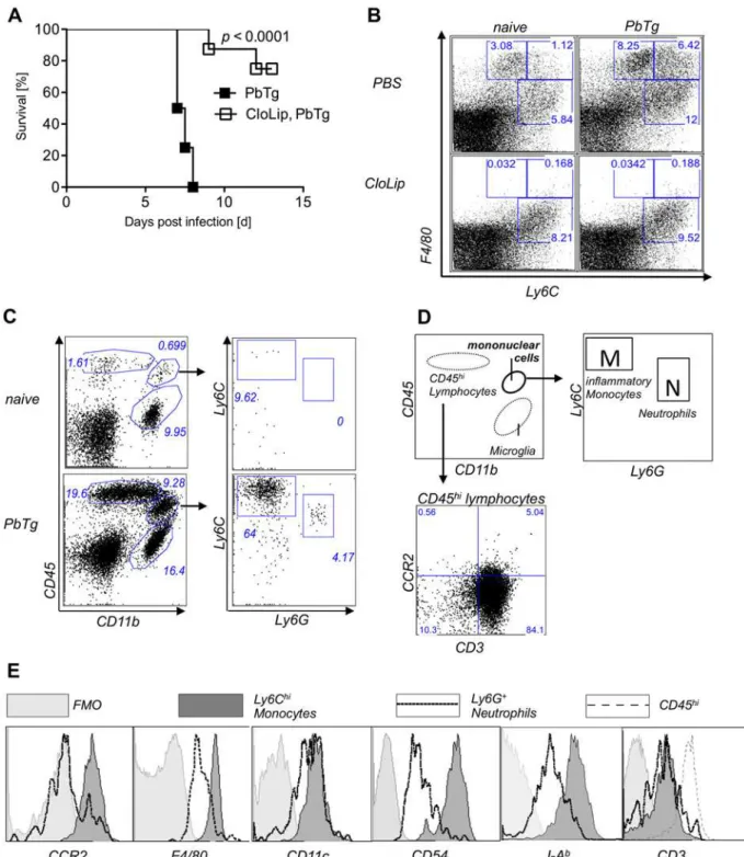
phagocytic activity [23] and then infected with PbTg. CloLip administration protected mice from developing lethal ECM and resulted in an 80% survival rate (Fig 1A). Parasitemia levels did not
differ significantly between the infected groups (Supplementary informationS1 Fig). When
com-pared to naïve mice, flow cytometric analysis of splenocytes from PbTg-infected C57BL/6 mice re-vealed increased frequencies of phagocytic cells, in particular more Ly6ChiF4/80+monocytes (6.42% vs. 1.12%), Ly6CintF4/80+monocytes (8.25% vs. 3.08%) and Ly6C+F4/80-neutrophils
(12% vs 5.84%) (Fig 1B, upper row). As expected, F4/80+cells were depleted upon injection of
CloLip, in both PbTg infected C57BL/6 mice and uninfected control mice (Fig 1Blower panel), in
comparison to non-depleted mice, that received PBS-loaded liposomes (Fig 1B, upper panel). Monocytes express high levels of both Ly6C and F4/80 and were depleted upon protective CloLip administration. On the other hand, there are data for a detrimental role of neutrophils inP. ber-ghei ANKAinduced ECM [21]. Therefore, we investigated the role of monocytes versus neutro-phils in the ECM model. First, we evaluated the composition of cellular infiltrates in brains of PbTg-infected mice at day 6 post infection (p.i.) and sought for the presence of lymphocytes,
in-flammatory monocytes (defined by Ly6ChiLy6G-, box M) and neutrophils (defined by Ly6C
in-tLy6G+, box N) (see gating scheme in Fig1Cand1D). We observed that in the brains of
PbTg-infected C57BL/6 mice with ECM, compared to naïve animals, there was a strong influx of
CD45hileukocytes, which were positive for CD3 (Fig 1D, lower panel), and CD45+CD11b+
mono-nuclear cells from the periphery that could be discriminated from CD45-CD11b+microglia. In
brains of PbTg-infected mice, but not in brains of naïve mice were both Ly6ChiLy6G-cells
(inflam-matory monocytes) and CD11b+Ly6G+Ly6Cintcells (neutrophils) among the CD45+CD11b+
mononuclear cells present (Fig 1Clower panel right). The inflammatory monocytes from brains
of PbTg infected mice also showed strong expression of CCR2 and F4/80, MHC class II, and
CD54, in contrast to Ly6G+neutrophils, which showed either intermediate expression of those
surface molecules or were devoid of these surface molecules (Fig 1E). Both mononuclear cell pop-ulations showed intermediate expression of CD11c, but were negative for CD3 (Fig 1E).
Early depletion of CD11b
+Ly6C
hiinflammatory monocytes in
Plasmodium
infection protects mice from ECM
To analyse the relevance of inflammatory monocytesversusneutrophils inP.berghei ANKA
in-duced ECM in C57BL/6 mice, we used mAb to either deplete (i) Ly6C+and Ly6G+cells by
anti-Gr-1 mAb as a comparison to previous studies [21], or (ii) to selectively deplete either Ly6G+
neutrophils (anti-Ly6G mAb) [19], or (iii) Ly6Chiinflammatory monocytes with the help of
anti-CCR2 mAb [20]. Groups of PbTg-infected mice received the respective depletion antibod-ies either directly upon PbTg infection (d0) or during infection (days 3 and 5 p.i.). The depletion
efficacy of inflammatory monocytes (seeFig 1Cfor gating, box M, upper left, defined by
CD11b+Ly6ChiLy6G-) or neutrophils (seeFig 1C, box N, upper right, defined by CD11b+Ly6Cint
Ly6G+) was confirmed in the blood of all groups in comparison to naïve animals and
control-in-fected mice 24h after mAb application (Fig 2A). These injection regimens led to reduction in
CD11b+leukocytes (Fig 2Aupper row). We observed an effective elimination of both desired
populations in the blood of anti-Gr1-administered mice (Fig 2A, lower row, middle plot). Ad-ministration of anti-Ly6G mAb, however, resulted in the depletion of neutrophils, but not of in-flammatory monocytes (Fig 2A, lower row, right). In contrast, administration of anti-CCR2
mAb resulted in a strong reduction of Ly6ChiCD11b+inflammatory monocytes, but not of
Ly6G+neutrophils (Fig 2A, lower row far right).
Next, we proceeded to monitor the effects of depletion on ECM development. Depletion
of Ly6Chimonocytes and Ly6G+neutrophils by the three different mAbs lasted for at least 48h
in all groups (S2 Fig). Therefore, we injected the appropriate mAbs either on the same day of
Fig 1. Depletion of Ly6C+F4/80+ cells, which are present in brains of ECM positive mice, prevents ECM.(A) Survival of PbTg-infected C57BL/6 mice. 24 hr before inoculation with iRBC (5*104) of PbTg, indicated C57BL/6 mice received 200μl CloLip i.v.. Graph shows survival rates of mice p.i. (n = 10 mice
per group) and represents 1 of 2 independent infection studies. Statistical analysis was performed using log-rank test. (B) Flow cytometric analysis of Ly6C+F4/80+cells in spleens of non-depleted mice (upper row) and CloLip depleted mice (lower row), which were either naïve (uninfected) (left panel) or
PbTg infected (right panel; 24 hours p.i.). Dot plots show representative stainings from 1 of 3 independent depletion studies in 3 mice per group. (C) Emergence of peripheral lymphocytes and mononuclear cells in brains of PbTg infected mice. Flow cytometric analyses show representative plots of naïve and PbTg infected mice, day 6 p.i.. Expression of CD45 and CD11b were used to discriminate lymphocytes (CD45hiCD11b-) and mononuclear cells
(CD45+CD11b+) from brain-resident microglia (CD45-CD11b+). (D) Scheme for discrimination of cell populations and gating of Ly6ChiLy6G-inflammatory
monocytes [M] versus neutrophils (Ly6CintLy6G+) [N] out of mononuclear cells as shown in data set of (B). CD45 hi cells were identified as CD3+T cells
(lower panel). (E) Ly6ChiLy6Gneginflammatory monocytes but not Ly6G+neutrophils from the brain of PbTg infected mice express CCR2, F4/80, CD54 and
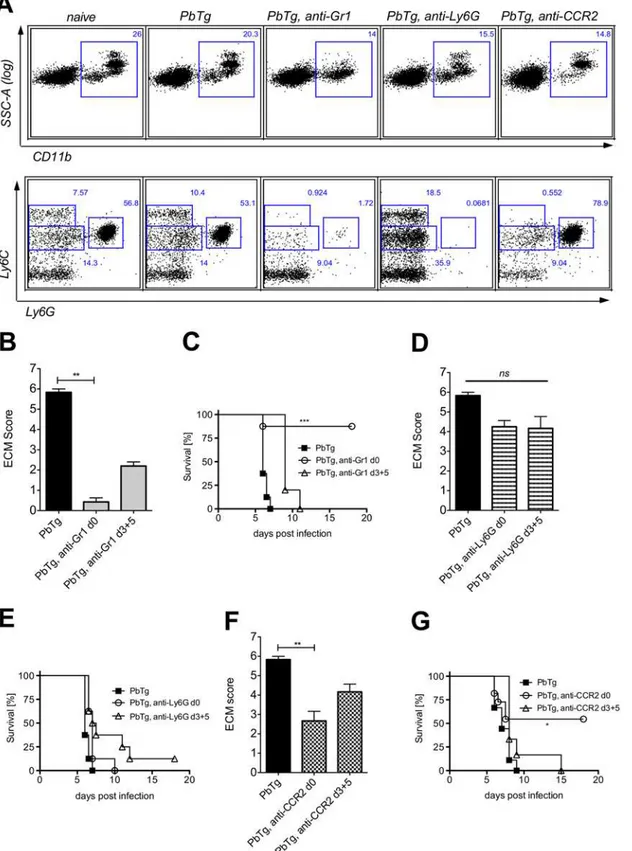
Fig 2. Early depletion of inflammatory monocytes protects PbTg infected mice against arising ECM.(A) Effective depletion of inflammatory
monocytes (Ly6ChiLy6G-) or neutrophils (Ly6CintLy6G+) in the blood of PbTg-infected C57BL/6 mice upon treatment with anti-Gr1 (middle), anti-Ly6G (right), or anti-CCR2 (far right) monoclonal antibodies, on d+1 post depletion. CD11b+leukocytes from the blood (upper row) were gated (squares) and analysed for
the presence of Ly6C+Ly6G-monocytes and Ly6intLy6G+neutrophils (lower row) The data of the lower row correspond to data of the upper row. n = 4 mice
PbTg infection (day 0) or on days 3 and 5 p.i. in order to evaluate the relevance of the different mononuclear cell populations at both the beginning of infection and during an ongoing infec-tion but before the onset of ECM.
Since the onset of ECM in the non-treated PbTg-infected C57BL/6 mice commonly occurs on day 6 p.i., we depicted the ECM score of all untreated and treated groups of PbTg-infected
mice on that day (Fig2B,2Dand2F). As expected, control-infected C57BL/6 mice, which did
not receive any depleting antibodies, presented strong ECM specific symptoms on day 6 p.i. (Fig2B,2Dand2F, black bars), and showed rapid disease progression (Fig2C,2Eand2G, closed boxes). Depletion of both inflammatory monocytes and neutrophils by mAb anti-Gr1
on the day of infection significantly prevented the severity of ECM (Fig 2Bc.fbars 1 and 2) and
led to continued survival of the infected mice (Fig 2Cc.fsquare symbols with open circles).
Similar significant results regarding the ECM score on day 6 p.i. were observed when anti-Gr1
mAb was injected during ongoing infection (d3+5) (Fig 2B). However, this“late”application
did not protect those mice, since we observed only a delay in ECM pathology (Fig 2C). Impor-tantly, selective depletion of neutrophils through administration of anti-Ly6G mAb only
mar-ginally influenced the developing pathology (Fig 2Dc.fbars 1 and 2) and survival (Fig 2E) and
this was regardless of the mAb administration time point. In agreement with anti-Gr1
injec-tion, only the early depletion of Ly6C+inflammatory monocytes through the administration of
anti-CCR2 mAb resulted in a significantly reduced ECM score at day 6 p.i. (Fig 2Fc.f. bars 1
and 2), as well as an enhanced survival (Fig 2G). In contrast, injection of anti-CCR2 mAb dur-ing an already ongodur-ing infection barely influenced developdur-ing pathology and did not protect
against ECM-related death (Fig 2Ec.f. bars 1 and 3). Importantly, parasitemia levels did not
differ significantly between the experimental groups with and without depletion approaches (S1 Fig). In conclusion, even if anti-Gr1 mAb-mediated depletion of both inflammatory mono-cytes and neutrophils resulted in the best protection of PbTg-infected mice from ECM, the se-lective depletion of inflammatory monocytes, but not neutrophils was sufficient to achieve significant protection against ECM.
Administration of anti-CCR2 or anti-Gr1 mAbs prevents lymphocyte
infiltration into the brains of PbTg infected mice
Next, we determined whether the selective depletion of these immune cell subsets using mAb also altered the amount of lymphocyte infiltration into the CNS since this has also been shown
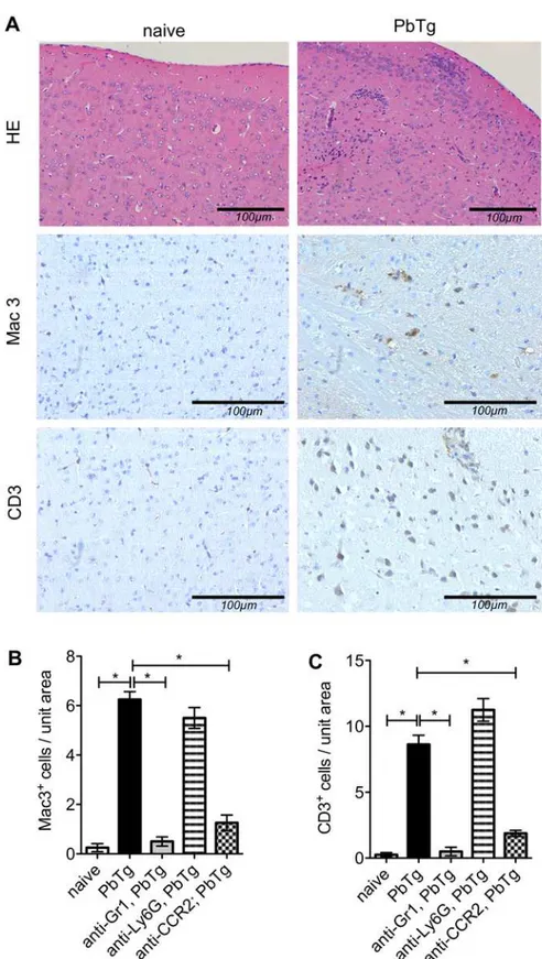
to play a decisive role in the outcome of murine malaria [6,8]. At the peak of ECM incidence
in PbTg infected C57BL/6 mice (day 6 p.i.), tissue sections from the brains of naïve and in-fected mice were analysed for the number of infiltrating mononuclear phagocytes (Mac3
stain-ing) and T cells (CD3 stainstain-ing).Fig 3Ashows representative stainings from brain frontal
cortex prepared from naïve and PbTg-infected mice, respectively. As expected, we detected a strong increase in infiltrating cells into the CNS parenchyma of infected mice compared to
naïve controls (representative images inFig 3A, upper right). These CNS infiltrates consisted
of increased numbers of CD11b+mononuclear cells and CD3+T cells (Fig3A,3Band3C).
Early depletion of inflammatory monocytes in the blood by using anti-Gr1 or anti-CCR2 mAb resulted in significantly reduced the numbers of both infiltrating mononuclear cells and T cells
in the frontal cortex (Fig3Band3Cc.f. bars 2 with 3 and 5). No differences in the number of
C57BL/6 mice were injected i.p. with anti-Gr1 mAb (B and C), anti-Ly6G mAb (D and E) or anti-CCR2 mAb (F and G) on the first day of infection (d0) or on days 3 and 5 p.i. (d3+5). Scores of cerebral pathology were determined on day 6 p.i. and bar graphs in B, D and F depict the mean and SEM of ECM score in individual mice (n = 8 mice per group). Statistical analysis was performed using Kruskal-Wallis test and Dunn’s Post test (**p<0.01). Survival data shown in C, E and G were analyzed with Mantel-Cox log-rank test.p<0.05 was considered significant (*p<0.05,**p<0.01,***p<0.001). n = 10 mice per group.
Fig 3. Histological analysis of brains of PbTg infected mice upon early depletion.C57BL/6 mice were infected i.v. with 5*104PbTg-iRBC and then subdivided into groups that received either anti-Gr1, anti-Ly6G or anti-CCR2 mAb on d0 p.i. (early depletion). On day 6 p.i., tissue sections from the brain parenchyma of individual mice were assessed for pathological changes. (A) Standard H&E staining is depicted in the upper panels whereas immunohistochemical staining for anti-Mac3 and anti-CD3 are shown in the middle and
infiltrating lymphocytes were observed when infected mice were subjected to treatment with
anti-Ly6G mAb (Fig3Band3Cc.f. bars 2 with 4). Depletion using anti-Gr1 and anti-CCR2
but not anti-Ly6G during ongoing PbTg infection on days 3 and 5 also reduced the influx of immune cells as shown by immunohistochemistry, but to a lesser extent (S2 Fig). Therefore, these data showing a massive reduction in lymphocyte influx into the brains of anti-Gr1 or anti-CCR2 mAb-injected mice strongly support our observations described above, that using
anti-Gr1 or anti-CCR2 mAb at the time point of infection prolonged the survival of
Plasmodi-uminfected mice (Fig2Cand2G).
To evaluate how the depletion of inflammatory cell subpopulations in the periphery could influence cellular infiltration into the CNS in more detail, we further determined on day 6 p.i. the frequency and composition of infiltrating cells within the brains of PbTg-infected mice by flow cytometry. When compared to naïve mice, PbTg infected animals exhibited in the brains
strongly elevated frequencies of CD45+CD11b-lymphocytes as well as CD45+CD11b+
mono-nuclear cells (Fig 4A, upper and lower panel). Within the CD11b+mononuclear cells in brains
of PbTg-infected mice, we found Ly6C+Ly6G-inflammatory monocytes and Ly6CintLy6G+
neutrophils (Fig 4A, compare upper and lower right plots). Upon anti-Gr1 mAb treatment, we detected a decrease in infiltrating cells which included 3 fold less infiltrating CD45+CD11b+
cells and a massive reduction in infiltrating CD11b-CD45hilymphocytes compared to WT
in-fected control mice (Fig 4B), which correlates to our findings in peripheral depletion (Fig 2A)
and the observed protection (Fig2Band2C) and previous studies [21]. Interestingly, the
fre-quency of inflammatory monocytes within the parental mononuclear CD45+CD11b+subset
was comparable to control infected animals (Fig4Aand4B; 63.8% vs 63.9.4%), but compared
to WT infected control mice, the overall frequency of mononuclear cells was strongly reduced
in brains of mAb treated mice (Fig4Eand4F). These population changes were even stronger
when infected mice were injected with anti-Gr1 mAb on days 3 and 5, demonstrating an en-during depletion effect upon administration of the mAb (S3 Fig).
Application of the neutrophil depleting mAb anti-Ly6G also resulted in the reduction of
in-filtrating mononuclear CD11b+cells as well as CD45hilymphocytes, which differs to our
find-ing with the help of immunohistochemistry from the brain frontal cortex (Fig 3C), although the overall infiltration of CD45hicells upon anti-Ly6G injection occurred to a lesser degree
compared to the other depletion approaches (Fig4Cand4E,S3 Fig). Finally, cell preparations
from the brains of mice that received anti-CCR2 mAb also contained lower amounts of
infil-trating CD45+cells. Here the proportion of CD45hilymphocytes was also strongly reduced
(Fig4Dand4F). Animals that received applications of the anti-CCR2 antibody on days 3 and 5
after infection, showed decreased amounts of lymphocytes and especially Ly6Chimonocytes
(S3 Fig). However, infiltrating neutrophils were barely detected in the brains and this was re-gardless of the therapy protocol.
Thus, either depletion approaches, anti-Gr1 or anti-CCR2 mAb, if performed early in infec-tion, inhibited the development of ECM, which is paralleled by decreased infiltration of periph-eral immune cells into the brains of PbTg infected mice. Even if the effects of late injections of the individual depleting antibodies on days 3 and 5 could be noticed in the brain, and also
anti-lower panels, respectively. Representative sections from naïve mice are shown on the left whereas those from PbTg infected mice are displayed on the right. (B, C) Quantification of Mac3+cells (B) and CD3+cells T
cells (C) in brain sections from the meninges and frontal cortex of individual mice. Scale bars indicate 100μm
in the magnifications. Bars show mean±SEM from n = 8 mice per group from 1 out of 3 independent depletion-infection experiments counted in 10 defined fields (High power fields, HPF) in the frontal cortex. Statistical analysis was performed using Kruskal-Wallis test and Dunn’s Post test and significant differences are indicated by the stars in brackets between the groups (*p<0.05).
Fig 4. Early monocyte depletion prevents lymphocyte infiltration into the brain.C57BL/6 mice were left either untreated or infected with 5*104PbTg
iRBC (A). In addition, groups of infected mice were treated either with anti-Gr1 (B), anti-Ly6G (C) or anti-CCR2 (D) mAb on the day of PbTg-infection (day 0). Six days later, cellular infiltrates from the brains of individual mice were prepared and analysed for the frequency of (E) infiltrating mononuclear cells (CD45+CD11b+)as well as (F) CD45hilymphocytes (CD45hiCD11b-). Bars show mean±SEM from n = 4–5 mice per group from 1 out of 3 independent depletion-infection experiments. Statistical analysis was performed using Kruskal-Wallis test and Dunn’s Post test and significant differences are indicated by the stars in brackets between the groups (*p<0.05).
doi:10.1371/journal.pone.0124080.g004
Ly6G mAb-injected mice showed less infiltrates as shown here by flow cytometry, nearly no one of these mice were protected against ECM. All late depletions and anti-Ly6G depletion ap-proaches that were analysed here showed a delay of the disease onset but no protection from ECM. Our data from early anti-CCR2 mAb administration show that the selective depletion of
Ly6Chimonocytes induced effects in the brain that were comparable to protective anti-Gr1
mAb administration, indicating an important role of Ly6Chimonocytes but not neutrophils in
ECM development during PbTg infection.
Depletion of Ly6C
hiinflammatory monocytes results in reduced
infiltration of IFN
γ
producing cells into the CNS
To determine whether the reduced number of CD3+T cells, which we observed in
immunohis-tochemistry in the frontal cortex of anti-Gr1 or CCR2 mAb PbTg-infected mice also affected different T cell populations that cross the blood brain barrier, we analysed the frequencies of
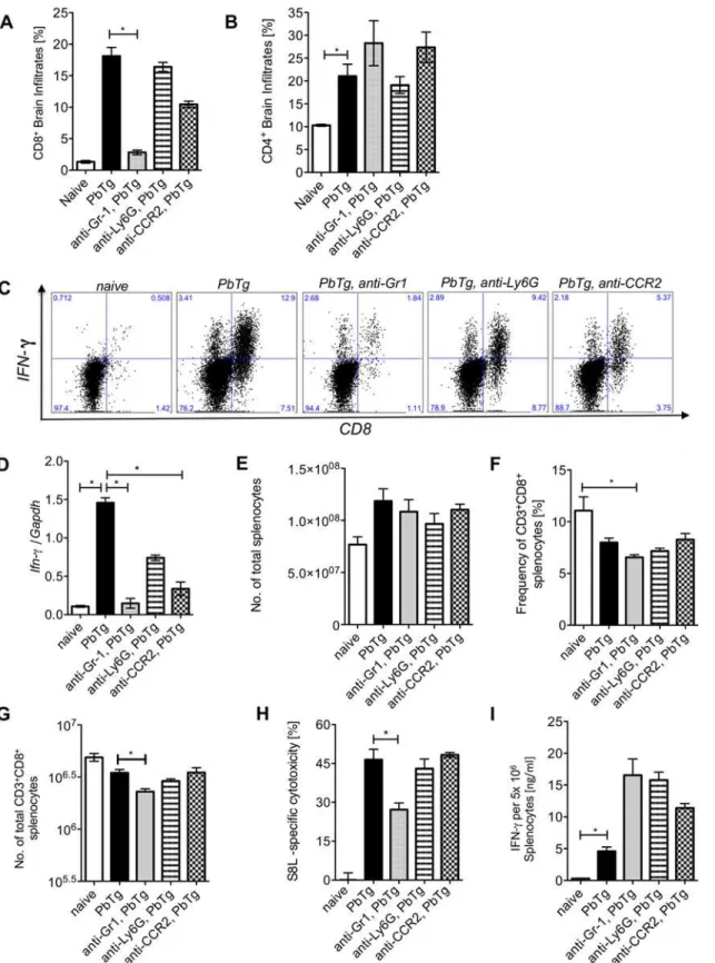
CD8+and CD4+T cells in the preparations from the whole brains by flow cytometry. In
addi-tion, we analysed the capacity of CD8+T cells to produce IFNγ. On day 6 p.i., PbTg infected
mice presented elevated frequencies of CD8+T cells and CD4+T cells in the brain when
com-pared to naïve mice (Fig5Aand5Bc.f. bars 1 and 2). More than half of the CD8+T cells that
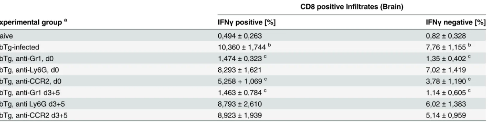
had infiltrated the brains of PbTg-infected mice, produced IFNγ(Table 1,Fig 5C).
Administra-tion of anti-Gr1 at the start of PbTg infecAdministra-tion resulted in a significant reducAdministra-tion of CD8+T
cells, including IFNγproducing and non-producing CD8+T cells (Table 1) but did not change
the influx of CD4+T cells within the brain (Fig5Aand5Bc.f. bars 2 with 3). These results were also observed when we administered antibodies on days 3 and 5 of infection (Table 1).
Impor-tantly, administration of anti-CCR2 mAb led to significantly reduced frequencies of IFNγ
pro-ducing CD8+T cells in the brains of the mice, whereas the frequencies of CD4+T cells were
not changed (Table 1). The depletion of neutrophils however, using anti-Ly6G mAb, did
nei-ther result in reduced frequencies of CD8+T or CD4+T cells nor in the changed proportion of
IFNγproducing CD8+T cells in the brain compared to non-depleted infected mice (Fig5A
and5Bc.f. bars 2 and 4,Fig 5C,Table 1). Thus, we conclude that the protection of
PbTg-in-fected mice from ECM upon depletion of Ly6Chimonocytes by anti-Gr-1 or anti-CCR2 mAb
was due to abrogated infiltration of lymphocytes into the CNS, mainly of IFN-γ-producing
CD8+T cells. Next, we analysed the expression of IFNγin the brains of PbTg-infected and
de-pleted mice. We detected elevated mRNA levels of IFNγin the brains of PbTg-infected mice
(Fig 5Dc.f. bars 1 and 2), which were 10-fold reduced in the brains of anti-Gr1 mAb treated
in-fected mice and five-fold reduced in the brains of anti-CCR2 treated inin-fected mice (Fig 5Dc.f.
bars 2 with 3 and 5). Administration of anti-CCR2 or anti-Gr-1 mAb during an ongoing PbTg infection also resulted in reduced IFNγlevels in the brains, which was significant in the case of
anti-Gr-1, demonstrating again a strong effect of this clone (S4 Fig). Interestingly, the d0
appli-cation of anti-Ly6G mAb to infected mice resulted in a two-fold decrease of IFNγlevels in the
brain, but remained a trend (Fig 5Dc.f. bars 2 and 4), similar to day 3 and 5 application of
anti-Ly6G (S4Fig 4). Taken together, we observed the strongest effects concerning influx of T cells
into the brain and block of IFNγproduction in anti-Gr-1 mAb injected mice, but importantly,
comparable effects were achieved with the selective depletion of Ly6Chiinflammatory
mono-cytes with the help of anti-CCR2-injection.
Specific depletion of Ly6C
hiinflammatory monocytes and neutrophils did
not prevent the generation of antigen-specific CTL responses
Fig 5. Early monocyte depletion prevents IFNγproducing T cell infiltration into the brain but does not affect peripheral PbTg-specific CTL
responses.C57BL/6 mice were infected with 5*10e4 PbTg iRBC i.v.. Simultaneously, groups of mice received either anti-Gr1, anti-Ly6G or anti-CCR2 mAb. Six days later, T cell subsets in the brain and spleen were analysed and cellular immune responses were evaluated. (A) Frequencies of CD8+cells and (B)
CD4+cells among brain infiltrates were determined by flow cytometry. Each group contained 4
–5 mice. (C) Leukocyte preparations from the brains of individual mice were re-stimulatedex vivoand intracellular stained to determine the frequency of IFNγproducing CD8+T cells. Representative images of
depletion interfered with the generation of functional T cell responses in the periphery. We de-termined first whether the cell subset depletion by the different mAbs had an impact on the total cell number in the spleen or on T cell frequencies and numbers during the early phase of infection and analysed all experimental groups on day 2 after PbTg infection with and without mAb depletion. We determined on day 2 after mAb-injection and PbTg infection in blood and
spleens of all PbTg-infected mice that received any depleting antibody, a reduction in CD11b+
leukocytes (S5 Fig). In spleens of mice that received anti-Ly6G mAb or anti-CCR2 mAb,
signif-icantly less Ly6G+neutrophils and Ly6C+monocytes (S5 Fig), respectively, were present
among those CD11b+cells. In contrast, anti-Gr1 mAb clone RB6 did not only result in
deple-tion of both Ly6C+monocytes and Ly6G+neutrophils, but also in significant reduction of
CD3+CD8+T cells, as observed on day 2 after infection and mAb injection (S5 Fig).
We analysed then the spleens of all experimental groups on day +6 after infection and ob-served, that the cell counts of splenocytes from all d0 depleted groups of mice were comparable to naïve and control-infected littermates (Fig 5E). In contrast, total splenocytes counts from d3+5 anti-Gr1 mAb depleted mice were significantly reduced (S4 Fig). Frequencies and total
numbers of CD8+T cells were reduced in all infected mice compared to naïve mice and reached
significance in the anti-Gr1 injected group of mice (Fig5Fand5G) as well as in the late deple-tion groups of anti-Gr1 mAb (S4 Fig). As this might have an impact also on parasite control
mechanisms in the periphery, we performed anin vivocytotoxicity assay using an adoptive
brain derived IFNγ+CD8+T cells by flow cytometry are shown. Calculated frequencies of CD8+IFNγ+T cells and CD8+IFNγ+T cells in the brains as analysed
from (A) are shown inTable 1. (D) Fold increase of IFNγmRNA levels relative to GAPDH in the brains of PbTg-infected mice±d0 depletion on day 6 p.i.
n = 6–8 per group, Kruskal Wallis test with Dunn’s Post test was performed. (E) Total cell count of splenocytes from all d0 depletion groups and controls at day 6 p.i. (F) Frequency of CD3+CD8+splenocytes in percent from all d0 mAb depletion groups and controls at day 6 p.i. (G) Calculated total amount of CD8+
splenocytes according to data from F and G. (H)In vivocytotoxicity assay analysing PbTg-specific T cells. Recipient PbTg-infected mice, that were or were not additionally treated with depletion antibodies, received an adoptive transfer of CSFE-labelled T cells, that were loaded with ovalbumin-derived MHC class I peptide SIINFEKL and unloaded control cells i.v. (1x10⁷/mouse) on day 5 p.i.. 18 hours later the level of lytic activity was measured by flow cytometry in the
spleen. (I) In addition, splenocytes from the same animals as in (H) were re-stimulated with SIINFEKLex vivofor 24 hours and IFNγproduction was
quantified by sandwich ELISA. (A-I) Bars show mean±SEM from n = 4–5 mice per group from 1 out of 3 independent depletion-infection experiments. Statistical analysis was performed using Kruskal-Wallis test and Dunn’s Post test and significant differences are indicated by the stars in brackets between the groups (*p<0.05,**p<0.01).
doi:10.1371/journal.pone.0124080.g005
Table 1. Impact of mononuclear cell subset depletion on IFNγproducing CD8+T cells (brain infiltrates).
CD8 positive Infiltrates (Brain)
Experimental groupa IFN
γpositive [%] IFNγnegative [%]
Naive 0,494±0,263 0,82±0,328
PbTg-infected 10,360±1,744b 7,76±1,155b
PbTg, anti-Gr1, d0 1,474±0,323c 1,35±0,402c
PbTg, anti-Ly6G, d0 8,293±1,621 7,02±1,419
PbTg, anti-CCR2, d0 5,258 + 1,069c 3,78±1,190c
PbTg, anti-Gr1 d3+5 1,463±0,784c 1,14±0,605c
PbTg, anti Ly6G d3+5 8,793±2,610 6,02±1,383
PbTg, anti-CCR2 d3+5 8,923±1,939 5,14±0,959
Six days after infection, cellular infiltrates from the brains of individual mice were prepared and analysed byflow cytometry for the frequency of infiltrating CD8+cells which produce IFN
γor not upon phorbol myristate acetate (PMA) /Ionomycin restimulation. an = 4
–5 per group
bSigni
ficant differences between PbTg infected group and naïve mice, (p<0.05, Mann-Whitney U test)
cSigni
ficant differences between mAb-depleted versus non-depleted PbTg infected mice, (p<0.05, Mann-Whitney U test)
transfer of fluorescent-labelled and OVA-peptide loaded spleen cells to evaluate the
antigen-specific cytotoxic capacity of CD8+T cells. Remarkably, functional CTL responses were present
in all groups, although infected mice treated with anti-Gr1 mAb on the day of infection
pre-sented a reduced lytic ability of Ag-specific CTLs (Fig 5Hc.f. bars 2 and 3), whereas the CTL
re-sponses of d3+5 depleted mice did not differ significantly from the control-infected group of mice (S4 Fig). Interestingly, we did not determine any deficits regarding the capacity of the T
cells to release IFNγupon peptide-specific restimulation between the different depleted groups
and the control-infected groups of mice, neither upon mAb administration at the time point of
infection nor during ongoing infection (Fig 5I,S4 Fig). Here, we concluded that all
PbTg-in-fected groups were equally able to generate functional antigen-specific T cell responses except anti-Gr1 treated mice, as here the IFNγresponses have to be set off against the strongly
re-duced CD8+T cell numbers. Thus, the depletion of inflammatory monocytes or neutrophils
with the help of selectively recognizing mAb did not prevent the generation of antigen-specific CTL responses in PbTg-infected mice, whereas application of anti-Gr1 significantly impaired CTL responses in the periphery.
Discussion
Cells of the innate immune system are essential for the detection and successful elimination of invading pathogens. They are also important for the induction of powerful adaptive immune responses, which aim at the complete clearance of pathogenic microbes. However, strong in-flammatory immune responses harbour the risk of harming the host and therefore should be
prevented or regulated. Infection withPlasmodiumparasite are a good example of the
possibili-ty of overwhelming inflammatory immune responses, sinceP.falciparuminfections in infants
and non-immune travellers are often fatal because they lead to cerebral malaria and other com-plications, whereas re-infected adults from endemic areas show successfully dampened im-mune responses and a mild course of disease. ECM, the pathological outcome of experimental
infection withP.berghei ANKAin C57BL/6 mice, is a result of IFNγproduction and CD8+T
cell infiltration into the CNS [6,8]. Here, we provide initial evidence that Ly6Chiinflammatory
monocytes are essential immune players in PbTg infection since specific depletion by injection of anti-CCR2 mAb protected PbTg-infected mice from ECM pathology. Moreover, depletion of this cell subset reduced both cellular infiltration to and IFNγlevels in the CNS. Importantly,
this selective elimination of Ly6Chimonocytes did not prevent the generation of
Plasmodium-specific immune responses in the periphery, as shown by effective CTL responses and IFNγ
production, which might be essential for parasite elimination.
The indispensable requirement of innate immune cells i.e. monocytes, macrophages and neutrophils, in a T-cell-driven disease such as malaria became apparent after the general deple-tion of phagocytic cells using CloLip, which resulted in prolonged survival of PbTg-infected C57BL/6 mice (Fig 1). Our analysis here revealed that upon clodronate liposome-mediated de-pletion mainly F4/80 positive cells were absent, which were also positive for Ly6C, strongly suggesting that these cells were inflammatory monocytes. However, data from previous studies
highlighted a role for F4/80-Ly6G+neutrophils in ECM [21].
Until recently, an exclusivein vivodepletion of these two immune cell populations, Ly6C+
monocytes and Ly6G+neutrophils, was not achievable. However, we previously reported the
necessity to properly distinguish between monocytes and neutrophils inT.gondiiinfections
using the anti-Ly6G clone 1A8 [9,11,12,24]. Indeed, those observations revealed the critical
importance of Ly6Chiinflammatory monocytes during an acute infection withT.gondiiand
clearly demonstrated that neutrophils are not protective as reported previously, but rather con-tribute to the pathology [9,11].
According to current studies, it is acknowledged that anti-Gr1 recognizes different Ly6 iso-forms, in particular Ly6G as well as Ly6C, resulting in a broad depletion spectrum, including Ly6C monocytes, but also certain lymphocyte subsets. Therefore, the question arose about how valid the commonly used anti-Gr1 clone RB6 was in selectively depleting neutrophils, as claimed in several previous reports. For this reason, in this study we utilized anti-Ly6G and anti-CCR2 mAbs to selectively deplete either neutrophils or inflammatory monocytes and compared those to mice that received anti-Gr1 mAb. To investigate whether and at which time point therapeutic intervention regarding ECM onset was possible, PbTg infected mice received the depleting mAb either on the day of infection or during infection. We show clearly that
Gr-1 mAb depletes both Ly6Chimonocytes and Ly6G+neutrophils, but however, injection of
anti-Gr1 did result also in significant reduction of CD3 T cells, which was observed during the early phase of infection, but lasted until day 6 after infection. From these data and the observations in regard to ECM development we conclude that the protective effect of anti-Gr1 mAb-mediat-ed depletion was probably mmAb-mediat-ediatmAb-mediat-ed by the early depletion of inflammatory monocytes but not of neutrophils and most likely also by the depletion of T cells. We come to this conclusion since anti-CCR2 injection, but not anti-Ly6G injection resulted in significant protection of PbTg infected mice. In addition, the protective effect of T cell depletion is well described in the literature [6]. This protection from ECM was characterized by dampened tissue-specific brain inflammation, but importantly, an anti-CCR2 mAb injection did not prevent peripheral im-mune responses. In contrast, anti-Gr1 mAb injection diminished the frequency and function of peripheral T cells.
We additionally provide immunohistochemical evidence showing that the number of
infil-trating CD3+T lymphocytes within the brain during infection was significantly reduced in
mice where inflammatory monocytes were absent. Moreover, on day 6p.i., T cells and also
functional immune responses could be detected in the spleens among all groups of mice which had received depleting antibodies either on day 0 of infection or days 3 and 5. Consequently,
we hypothesize that Ly6ChiCCR2posinflammatory monocytes are crucially involved in the
early phase ofPlasmodiumblood-stage infection. It remains to be analysed, at which step of
in-fection and how the monocytes interact with other immune cells to trigger T cell influx into the brain.
Importantly, an essential role for Ly6Chimonocytes in the control of blood-stage malaria
parasites was shown in the non-ECM murine model for malarial anaemia usingP.chabaudi
[25]. Sponaas et al. demonstrated CCR2-dependent egress of monocytes from the bone marrow and in infected CCR2-deficient mice a strong reduction of parasitemia in the acute stage of
in-fection due to an adoptive transfer of splenic Ly6Chimonocytes. The authors further concluded
that such control over the blood stage of infection was mediated through the production of re-active oxygen intermediates and inducible nitric oxide synthase by these monocytes [25]. Our data did not reveal significant differences in parasitemia levels between the experimental groups; although anti-Gr1 mAb injected mice presented slightly increased parasitemia levels compared to the control-infected mice, this remained a trend and did not reach significance.
In addition, our data suggest, that Ly6C+CCR2+inflammatory monocytes do not prime
par-asite-specific T cells, as the anti-CCR2 injected mice generated T cell responses in the periphery that were comparable to those of control-infected mice. In contrast, anti-Gr1 mAb injection did result in strongly diminished T cell frequencies and also numbers, which was also observed before. We show here for the first time, that anti-Gr1 treatment also reduced CTL responses. Several recent studies have reported that the administration of this mAb results in the
non-se-lective depletion of monocytes [19,26,27]. For example, Daley et al. showed loss of monocyte/
in a herpes simplex virus type 1 infection [26]. In addition, Johnson et al. used the
peptide-in-duced fatal syndrome (PIFS) model of CNS vascular permeability, which depends on CD8+T
cell-mediated break down of the blood-brain barrier. In their study, the depletion efficacy of the anti-Gr1 mAb RB6 clone was compared to the mAb 1A8 variant, which is anti-Ly6G.
There, Johnson et al. observed a strong reduction of CD45+Gr1+Ly6G+cells, but also
dimin-ished infiltration of Gr1+CD8+T cells upon administration of anti-Gr1 clone RB6-8C5,
where-as application of the anti-Ly6G mAb resulted in selective neutrophil depletion and did not affect T cell infiltration. They concluded that application of this anti-Gr1 mAb directly depleted infiltrating T cells and that BBB disruption in their model was independent of neutrophils [27]. However, they did not analyse the impact of depletion on migrating T cell populations or anti-gen-specific T cell responses in the periphery.
Monocytes require CCR2 to leave the bone marrow and therefore—to substantiate our
find-ings—we compared the outcome of PbTg infection using anti-CCR2 mAb as well as anti-Gr1.
This antibody has been used to specifically target CCR2+monocyte subsets in models of
toxo-plasmosis [28], arthritis [29,30] and gram negative bacterial meningitis [31]. The role of CCR2
in ECM has been analysed in previous studies which focused on infiltrating T cells [32]. Bel-noue and colleagues showed that PbA-infected CCR2 KO mice still developed ECM and no dif-ferences in infiltrating T cell numbers were observed in the brain. They did not observe any difference in brain infiltrating T cell numbers but instead found a reduced amount of macro-phages amongst brain-sequestered leukocytes, compared to infected WT. Contrary to our ob-servation that 50% of mice were protected against ECM by CloLip-mediated macrophage depletion at day 4 p.i. (data not shown), in their study, only depletion of CD8+T cells but not macrophages (at day 5 p.i.) did result in infected mice in protection against ECM [32]. In that study, Belnoue et al. showed an impact of CCR5 on T cell infiltration upon PbA infection, how-ever, a crucial analysis of monocyte subpopulations or functional T cell responses (i.e.
produc-tion of IFNγor antigen-specific lysis), which we have done here, was not performed. The
disparate observations between our results and what has been previously described by Belnoue et al. may be explained by the possible development of a compensatory mechanism in the ge-netically modified mice lacking CCR2 that would not be present in mAb immune cell depleted mice in our study. Importantly, we demonstrate that in our study, anti-CCR2 treatment re-sulted in diminished recruitment of CD8+T cells into the brain, but not in overall T cell deple-tion as we detected unaffected T cell frequencies 2 days after the depledeple-tion and also a robust T cell response in the spleen of CCR2-depleted mice at day 6 p.i., which was determined by CTL
activity and IFNγproduction upon peptide-restimulation. We assume that PbA—infected
CCR2-deficient mice have a different T cell pool that is recruited to the brain due to compensa-tory mechanisms, which may show a different migration pattern than in infected
WT littermates.
In conclusion, our data show that the selective depletion of Ly6ChiCCR2+monocytes is able
to prevent the development ofP.bergheiinfection-induced ECM. Their pathogenic phenotype
in this model is in agreement with the role these cells have been reported to play in other mod-els including atherosclerosis [33], parasitic infections [34], respiratory fungal infection [35], liver destructive processes [36], obesity [37] and Alzheimer’s disease [38,39]. Notably, in our
study, the selective depletion of these Ly6Chiinflammatory monocytes showed similar
protec-tive effects as anti-Gr1 mAb and prevented the crossing of pathogenic immune cells, including monocytes and T cells, into the brains of PbTg infected mice. But in contrast to anti-Gr1 mAb, anti-CCR2 mAb did not disrupt functional antigen-specific T cell responses in the periphery.
Moreover, the effects were more enhanced when depletion of Ly6ChiCCR2+monocytes
oc-curred early in PbTg infection, which has implications if such strategies were translated to ther-apeutic platforms. Future studies should determine whether individuals with immunity to
malaria have dampened inflammatory monocyte levels in the periphery and their immune-reg-ulatory properties should be analyzed.
Material and Methods
Mice
7 week old female C57BL/6N mice were purchased from Janvier (Le Genest Saint Isle, France). Mouse studies were approved by local regulatory agencies (Landesamt fuer Natur, Umwelt und
Verbraucherschutz Nordrhein-Westfalen (LANUV NRW) §84–02.04.2012.A264). Water and
food were providedad libitum.
Parasites, infection and disease
In all experiments, transgenicPlasmodium bergheiANKA expressing Ovalbumin (PbTg)
in-fected red blood cells (iRBCs) were used to infect mice [22]. All mice were inin-fected
intrave-nously (i.v.) with 5104pRBCs obtained from mice that had been previously infected
intraperitoneally (i.p.) with stock solution. Stock solution contained 1107iRBCs in glycerine
from liquid nitrogen storage. Stock mice were of the same background as experimental ani-mals. For evaluation of disease onset mice were monitored twice daily for ECM symptoms and parasitemia. ECM development was scored as described before according to the following symptoms: 0 = without symptoms, 1 = ruffled fur, 2 = hunching, 3 = wobbly gait, 4 = limb
pa-ralysis, 5 = convulsions, 6 = coma [40,41]. Mice reaching score 5 and ECM negative mice,
which developed anaemia, were sacrificed due to ethical guidelines. Parasitemia levels in the blood were determined as described [41].
Reagents and depleting antibodies
For cell culture RPMI 1640 medium (Sigma, Munich, Germany) supplemented with 10% FCS (PAA, Cölbe, Germany), 1% Penicillin/Streptomycin or Gentamicin (Lonza, Wuppertal, Ger-many), 1% L-Glutamine (PAA, Cölbe, Germany) was used. Directly conjugated monoclonal
an-tibodies used for FACS analysis (anti-mouse CD3, CD4, CD8α, CD11b, CD11c, CD45, CD54,
CCR2, I-Ab, F4/80, Ly6C, Ly6G,) were bought from BD Pharmingen (Heidelberg, Germany) and eBioscience (Frankfurt, Germany) and R and D. Phagocytic cells were depleted by i.v. injec-tion of 200μl clodronate liposomes containing 5mg clodronate per ml suspension [23]. Controls received 200μl PBS-filled liposomes i.v. Monocytes and neutrophils together were depleted using the monoclonal antibody (mAb) anti-Gr1 (clone RB6-8C5 from bioXcell, US, NH). Neu-trophils were depleted using anti-Ly6G mAb 1A8 [19] (bioXcell, US, NH). Inflammatory mono-cytes were depleted by the mAb anti-CCR2 (clone MC21) [20]. Monoclonal antibodies were administered by i.p. injection on either day 0 within 30 min of PbA infection (early), or on days
3 and 5 (late) post infection (p.i.). Per mouse, injections comprised of 500μg for mAb 1A8 (anti
Ly6G), 250μg for mAb RB6 (anti-Gr1) and 75μg for mAb MC21 (anti-CCR2).
Flow cytometry
For analysis by flow cytometry, mice were anaesthetized and then perfused intra-cardially with 1x PBS. Spleens and brains were isolated, cut into small pieces and digested with 0.5 mg/ml col-lagenase A (Roche, Basel, Switzerland), by incubation at 37°C for 25 min. Then the organs were gently pressed through a sieve to obtain a single cell suspension and washed with PBS.
Cells within the interphase were collected by aspiration through a pipette, transferred into a new tube and washed with 1x PBS / 1% FCS / 2mM EDTA.
Cells were stained in sterile 1x PBS containing 1% FCS for 20 min on ice. For intracellular
staining of IFNγ, cells were activated with ionomycin (1μg/ml), phorbol myristate acetate (50
ng/ml) and Golgi stop (BD Bioscience, Heidelberg, Germany) for 5h. Intracellular stainings were performed with the corresponding staining kits (BD Bioscience, Heidelberg, Germany) according to the manufacturer´s protocol. Acquisition was performed on a FACS Canto II flow cytometer and LSR Fortessa (BD, San Jose, USA). Data were analysed with FlowJo Software (Treestar Inc., Ashland, USA).
In vivo
cytotoxicity assay and peptide restimulation
CTL activity in PbTg infected animals was determinedin vivoon day 6 p.i. as described
else-where [42]. Briefly, splenocytes from syngenic donor mice were pulsed with 1μM of the specific
H-2kbpeptide SIINFEKL (Ovalbumin) for 30 min at 37°C and subsequently with 1μM of
CFSE for 15 min (CFSEhigh, specific target cells). Reference cells were not pulsed with peptide
and labeled with 0.1μM CFSE for 15 min (CFSElow, reference cells). After fluorochrome
label-ing, the cells were washed and the cell number was determined. Then, both cell populations were mixed at a 1:1 ratio (CFSEhigh/ CFSElow). Each recipient received 1x10⁷total cells into the tail vein on day 5. Mice were sacrificed 18 hours later on day 6 after infection and spleens were isolated to prepare single-cell suspensions. Lysis of peptide-loaded cells was quantified by mea-suring the ratio of CFSEhigh/ CFSElowcells via flow cytometry (Canto II, BD Biosciences). The percentage of specific lysis, termed SL8-specific lysis, was calculated using the following equa-tion: 100 - [(CFSEhigh/CFSElow)immunized/(CFSEhigh/CFSElow)naïve] x 100.
Cytokine ELISA
Spleens from experimental animals were isolated on day 6 p.i. and single cell suspensions were
prepared as described above. 106splenocytes were cultured in triplicate in 200μl RPMI medium
including 1μM SIINFEKL peptide over night. The supernatant was analysed for IFNγby
sand-wich ELISA according to the manufacture’s guide (eBioscience).
Histology
Animals were sacrificed on 6 days p.i. and the brains were removed and fixed in 4% neutral buffered formalin. Tissues were dehydrated in ethanol and embedded in paraffin, and 5-μm sections were stained with hematoxylin and eosin (H&E), Mac3 and CD3 as described previ-ously [9,11]. Quantification of cellular infiltrates in tissue sections of the frontal cortex was performed in 10 high power fields (HPF) as described before [43].
Real Time PCR
After removal, tissue samples from brains were immediately transferred to RNA later (QIAgen, Hilden, Germany) and kept on ice. They were stored at 4°C for at least 24h and then kept at -20°C until RNA isolation. For RNA isolation, the tissue sample was removed from RNA later and homogenized with 1mL of TriFast (peqGOLD, Erlangen, Germany) in BashingBeads tubes (Zymo Research, Freiburg, Germany). PeqGOLD HP Total RNA Kit was used for purification
according to the manufacturer’s instructions. Thereafter, on-membrane DNase I digestion
(peqGOLD, Erlangen, Germany) was performed. For qPCR, TaqMan RNA-to-Ct 1-Step Kit
(Life Technologies, Darmstadt, Germany) was used with 2μg total RNA in a reaction volume
of 10μL. The following TaqMan Gene Expression Assays (Life Technologies, Darmstadt,
Ger-many) were used to measure mRNA levels for IFNγ: (IFNγ: Mm01168134_m1).
Statistical analysis
Survival was performed with 10 mice per group and analysed with Log-rank sum test.Ex vivo
analyses of infection experiments were performed with 4–8 mice per group and statistical
anal-ysis was performed non-parametrically using Kruskal-Wallis test with Dunn’s Post test when
comparing more than 2 groups; Mann-Whitney U test was used to calculate differences
be-tween infected control mice and single antibody-depletion regiments in the analysis of IFNγ
production by T cells. If not otherwise stated, mean and standard error of the means are shown. Significant differences are indicated by the stars in brackets between the groups (p<0.05;p<0.01,p<0.001). All calculations were performed in GraphPad Prism.
Supporting Information
S1 Fig. No difference in parasitemia between infected controls and CloLip-depleted or mAb-depleted mice.Determination of parasitemia levels on day 6 after infection in the blood of mice that had received either (A) Clodronate liposomes or PBS-filled liposomes one day
be-fore infection with 5105PbTg-iRBCs or (B) mAb-injected mice on day 6 after infection with
5104PbTg-iRBCs. The indicated antibodies were injected on the day of infection as described
in the main text. N = 6–8 per group, Statistical analysis was performed by student’sttest (A) or Kruskal-Wallis test (B).
(TIF)
S2 Fig. Late depletion of Ly6C monocytes prevents lymphocyte infiltration into the brain.
C57BL/6 mice were infected i.v. with 510e4 PbTg-iRBC and then subdivided into groups that
received either anti-Gr1, anti-Ly6G or anti-CCR2 mAb on days 3 and day 5 p.i. (late deple-tion). On day 6 p.i., tissue sections from the brains of individual mice were assessed for patho-logical changes. Quantification of Mac3+cells (A) and CD3+T cells (B) in brain tissue sections of individual mice. Bars show mean ± SEM from n = 8 mice per group. Statistical analysis was
performed using Kruskal-Wallis test and Dunn’s Post test and significant differences are
indi-cated by the stars in brackets between the groups (p<0.05). HPF, High Power Field. (TIF)
S3 Fig. Monocyte depletion prevents lymphocyte infiltration into the brain.C57BL/6 mice were left either untreated or infected with 510e4 PbTg iRBC (see mainFig 4A). (A) In addition,
groups of infected mice were treated either with anti-Gr1 (upper plots), anti-Ly6G (middle plots) or anti-CCR2 mAb (lower plots) on day 3 and 5 during PbTg-infection. On day 6 p.i., cellular infil-trates from the brains of individual mice were prepared and analysed for the frequency of
infiltrat-ing lymphocytes (CD45hiCD11b-) and mononuclear cells CD45+CD11b+cells and therein the
amount of recruited monocytes (Ly6C+) and neutrophils (Ly6G+) by flow cytometry.
Representa-tive plots from one out of four mice are shown. (B) Frequency of CD11b+CD45+cells (upper
graph) and CD45hiCD11b- cellsamong the brain infiltrates (lower graph). (C, D) CD45+CD11b
-cells were then assessed for the expression of CD8 and CD4. Bars show mean ± SEM from n = 4–5
mice per group. Statistical analysis was performed using Kruskal-Wallis test and Dunn’s Post test and significant differences are indicated by the stars in brackets between the groups (p<0.05). (TIF)
S4 Fig. Impact of mononuclear cell subset depletion on cell counts and frequencies of T cells in the spleen.C57BL/6 mice were left either untreated or infected with 510e4 PbTg iRBC.
mAb (on day 3 and 5 during PbTg-infection. (A) Total cell count of splenocytes from all d3+5
depletion groups and controls at day 6 p.i. (B) Frequency of CD8+splenocytes in percent from
all d3+5 depletion groups and controls at day 6 p.i. (C) Calculated total amount of CD8+
spleno-cytes according to data from B and C. (D) Fold increase of IFN-γmRNA levels relative to
GAPDH in the brains of PbTg-infected mice ± d3+5 mAb depletion on day 6 p.i. n = 6–8 per
group, Kruskal Wallis test with Dunn’s Post test was performed. (E)In vivocytotoxicity assay analysing PbTg-specific T cells at day 6 in the spleens, using SIINFEKL loaded target cells which were adoptively transferred into infected and non-infected mice 18 hours before analysis. (F)
Splenocytes from the same animals as in E were re-stimulated with SIINFEKLex vivofor 24
hours and IFN-γproduction was quantified by sandwich ELISA.(A-F) Bars show mean ± SEM
from n = 4–5 mice per group. Statistical analysis was performed using Kruskal-Wallis test and
Dunn’s Post test and significant differences are indicated by the stars in brackets between the groups (p<0.05).
(TIF)
S5 Fig. Analysis of specific depletion in the spleen on day 2 after PbTg infection.C57BL/6
mice were left either untreated or infected with 510e4 PbTg iRBC. In addition, groups of
in-fected mice were treated either with anti-Gr1, anti-Ly6G or anti-CCR2 mAb on the day of PbTg-infection. Two days later, mice were sacrificed for analysis. (A) The diagram illustrates on the left panel the gating strategy for leukocytes from spleen and blood used in flow cytometric analysis.
The right panel shows further analysis of CD11b+gated splenocytes for expression of Ly6C and
Ly6G to identify monocytes and neutrophils, respectively, as well as further analysis of CD3+
gated cells for expression of CD4 and CD8. The data show splenocytes from a naïve C57BL/6 mouse. (B) According to the gating scheme shown in (A), splenocytes from all experimental
groups were analyzed for the expression of CD3 versus CD11b (left panel). Ly6C+monocytes
and Ly6G+neutrophils among the previously gated CD11b+cells are shown in the middle panel,
whereas CD3+gated cells were further analyzed for the expression of CD4 and CD8 (right
panel). Representative data of each experimental group are shown, one out of four mice. (C) Total splenocytes count (D-O) Flow cytometric analysis of frequencies and calculation of total amounts of splenic subpopulations, which were gated according to the scheme shown in (A). (D)
Frequency and (E) total amount of CD11b+splenocytes; (F) Frequency and (G) total amount of
Ly6C+monocytes gated from CD11b+splenocytes; (H) Frequency and (I) total amount of
Ly6G+neutrophils gated from CD11b+splenocytes; (J) Frequency and (K) total amount of CD3+
splenocytes; (L) Frequency and (M) total amount of CD8+T cells gated from CD3+splenocytes,
(N) Frequency and (O) total amount of CD4+T cells gated from CD3+splenocytes. N = 4 per
group, Statistical analysis was performed by Kruskal-Wallis test with Dunn’s Post test. Significant differences are indicated by the stars in brackets between the groups (p<0.01).
(TIF)
Acknowledgments
We thank Laura E. Layland for critical reading the manuscript and helpful discussions. We thank the flow cytometry Core Facility team (IMMEI, University of Bonn) for excellent techni-cal support.
Author Contributions
Conceived and designed the experiments: BS IRD. Performed the experiments: BS KK JMK AB ADM. Analyzed the data: BS KK JMK IRD. Contributed reagents/materials/analysis tools: AL NvR MM AH. Wrote the paper: BS MM AH IRD.
References
1. WHO. World Malaria Report 2013. Report. World Health Organization, 2013.
2. Murray CJ, Rosenfeld LC, Lim SS, Andrews KG, Foreman KJ, Haring D, et al. Global malaria mortality between 1980 and 2010: a systematic analysis. Lancet. 2012; 379(9814):413–31. Epub 2012/02/07. doi:10.1016/S0140-6736(12)60034-8PMID:22305225.
3. Hunt NH, Golenser J, Chan-Ling T, Parekh S, Rae C, Potter S, et al. Immunopathogenesis of cerebral malaria. Int J Parasitol. 2006; 36(5):569–82. Epub 2006/05/09. doi: S0020-7519(06)00073-7 [pii] doi:
10.1016/j.ijpara.2006.02.016PMID:16678181.
4. Greenberg J, Kendrick LP. Some characteristics of Plasmodium berghei passed within inbred strains of mice. J Parasitol. 1957; 43(4):420–7. Epub 1957/08/01. PMID:13463687.
5. Greenberg J, Kendrick LP. Parasitemia and survival in inbred strains of mice infected with Plasmodium berghei. J Parasitol. 1957; 43(4):413–9. Epub 1957/08/01. PMID:13463686.
6. Belnoue E, Kayibanda M, Vigario AM, Deschemin JC, van Rooijen N, Viguier M, et al. On the pathogen-ic role of brain-sequestered alphabeta CD8+ T cells in experimental cerebral malaria. J Immunol. 2002; 169(11):6369–75. Epub 2002/11/22. PMID:12444144.
7. Belnoue E, Potter SM, Rosa DS, Mauduit M, Gruner AC, Kayibanda M, et al. Control of pathogenic CD8+ T cell migration to the brain by IFN-gamma during experimental cerebral malaria. Parasite Immu-nol. 2008; 30(10):544–53. Epub 2008/07/31. doi: PIM1053 [pii] doi:10.1111/j.1365-3024.2008.01053.x
PMID:18665903.
8. Yanez DM, Manning DD, Cooley AJ, Weidanz WP, van der Heyde HC. Participation of lymphocyte subpopulations in the pathogenesis of experimental murine cerebral malaria. J Immunol. 1996; 157(4):1620–4. Epub 1996/08/15. PMID:8759747.
9. Dunay IR, Damatta RA, Fux B, Presti R, Greco S, Colonna M, et al. Gr1(+) inflammatory monocytes are required for mucosal resistance to the pathogen Toxoplasma gondii. Immunity. 2008; 29(2):306–17. Epub 2008/08/12. doi: S1074-7613(08)00326-9 [pii] doi:10.1016/j.immuni.2008.05.019PMID:
18691912; PubMed Central PMCID: PMC2605393.
10. Mohle L, Parlog A, Pahnke J, Dunay IR. Spinal cord pathology in chronic experimental Toxoplasma gondii infection. European journal of microbiology & immunology. 2014; 4(1):65–75. Epub 2014/03/29. doi:10.1556/EuJMI.4.2014.1.6PMID:24678407; PubMed Central PMCID: PMC3955833.
11. Dunay IR, Fuchs A, Sibley LD. Inflammatory monocytes but not neutrophils are necessary to con-trol infection with Toxoplasma gondii in mice. Infect Immun. 2010; 78(4):1564–70. Epub 2010/02/ 11. doi: IAI.00472-09 [pii] doi:10.1128/IAI.00472-09PMID:20145099; PubMed Central PMCID: PMC2849397.
12. Dunay IR, Sibley LD. Monocytes mediate mucosal immunity to Toxoplasma gondii. Curr Opin Immunol. 2010; 22(4):461–6. Epub 2010/06/12. doi: S0952-7915(10)00072-5 [pii] doi:10.1016/j.coi.2010.04.008
PMID:20537517; PubMed Central PMCID: PMC2922417.
13. Geissmann F, Jung S, Littman DR. Blood monocytes consist of two principal subsets with distinct mi-gratory properties. Immunity. 2003; 19(1):71–82. Epub 2003/07/23. doi: S1074761303001742 [pii]. PMID:12871640.
14. Serbina NV, Pamer EG. Monocyte emigration from bone marrow during bacterial infection requires sig-nals mediated by chemokine receptor CCR2. Nat Immunol. 2006; 7(3):311–7. Epub 2006/02/08. doi:
10.1038/ni1309PMID:16462739.
15. Soudja SM, Ruiz AL, Marie JC, Lauvau G. Inflammatory monocytes activate memory CD8(+) T and in-nate NK lymphocytes independent of cogin-nate antigen during microbial pathogen invasion. Immunity. 2012; 37(3):549–62. Epub 2012/09/04. doi: S1074-7613(12)00368-8 [pii] doi:10.1016/j.immuni.2012. 05.029PMID:22940097; PubMed Central PMCID: PMC3456987.
16. Auffray C, Fogg D, Garfa M, Elain G, Join-Lambert O, Kayal S, et al. Monitoring of blood vessels and tis-sues by a population of monocytes with patrolling behavior. Science. 2007; 317(5838):666–70. Epub 2007/08/04. doi: 317/5838/666 [pii] doi:10.1126/science.1142883PMID:17673663.
17. Geissmann F, Auffray C, Palframan R, Wirrig C, Ciocca A, Campisi L, et al. Blood monocytes: distinct subsets, how they relate to dendritic cells, and their possible roles in the regulation of T-cell responses. Immunol Cell Biol. 2008; 86(5):398–408. Epub 2008/04/09. doi: icb200819 [pii] doi:10.1038/icb.2008. 19PMID:18392044.
18. Schenk M, Mueller C. Adaptations of intestinal macrophages to an antigen-rich environment. Semin Immunol. 2007; 19(2):84–93. Epub 2006/10/24. doi: S1044-5323(06)00107-2 [pii] doi:10.1016/j.smim. 2006.09.002PMID:17055292.
20. Mack M, Cihak J, Simonis C, Luckow B, Proudfoot AE, Plachy J, et al. Expression and characterization of the chemokine receptors CCR2 and CCR5 in mice. J Immunol. 2001; 166(7):4697–704. Epub 2001/ 03/20. PMID:11254730.
21. Chen L, Zhang Z, Sendo F. Neutrophils play a critical role in the pathogenesis of experimental cerebral malaria. Clin Exp Immunol. 2000; 120(1):125–33. Epub 2000/04/12. doi: cei1196 [pii]. PMID:
10759773; PubMed Central PMCID: PMC1905604.
22. Lundie RJ, de Koning-Ward TF, Davey GM, Nie CQ, Hansen DS, Lau LS, et al. Blood-stage Plasmodi-um infection induces CD8+ T lymphocytes to parasite-expressed antigens, largely regulated by CD8al-pha+ dendritic cells. Proc Natl Acad Sci U S A. 2008; 105(38):14509–14. Epub 2008/09/19. doi:10. 1073/pnas.0806727105PMID:18799734; PubMed Central PMCID: PMC2567226.
23. Van Rooijen N, Sanders A. Liposome mediated depletion of macrophages: mechanism of action, prep-aration of liposomes and applications. J Immunol Methods. 1994; 174(1–2):83–93. Epub 1994/09/14. PMID:8083541.
24. Karlmark KR, Tacke F, Dunay IR. Monocytes in health and disease—Minireview. European journal of microbiology & immunology. 2012; 2(2):97–102. Epub 2012/06/01. doi:10.1556/EuJMI.2.2012.2.1
PMID:24672677; PubMed Central PMCID: PMC3956957.
25. Sponaas AM, Freitas do Rosario AP, Voisine C, Mastelic B, Thompson J, Koernig S, et al. Migrating monocytes recruited to the spleen play an important role in control of blood stage malaria. Blood. 2009; 114(27):5522–31. Epub 2009/10/20. doi:10.1182/blood-2009-04-217489PMID:19837977.
26. Wojtasiak M, Pickett DL, Tate MD, Londrigan SL, Bedoui S, Brooks AG, et al. Depletion of Gr-1+, but not Ly6G+, immune cells exacerbates virus replication and disease in an intranasal model of herpes simplex virus type 1 infection. J Gen Virol. 2010; 91(Pt 9):2158–66. Epub 2010/06/12. doi:10.1099/vir. 0.021915-0PMID:20538903.
27. Johnson HL, Chen Y, Jin F, Hanson LM, Gamez JD, Pirko I, et al. CD8 T cell-initiated blood-brain barri-er disruption is independent of neutrophil support. J Immunol. 2012; 189(4):1937–45. Epub 2012/07/ 10. doi:10.4049/jimmunol.1200658PMID:22772449; PubMed Central PMCID: PMC3411871. 28. Biswas A, Bruder D, Wolf SA, Jeron A, Mack M, Heimesaat MM, et al. Ly6Chigh Monocytes Control
Ce-rebral Toxoplasmosis. J Immunol. 2015. Epub 2015/02/25. doi:10.4049/jimmunol.1402037PMID:
25710908.
29. Bruhl H, Cihak J, Plachy J, Kunz-Schughart L, Niedermeier M, Denzel A, et al. Targeting of Gr-1+, CCR2+ monocytes in collagen-induced arthritis. Arthritis Rheum. 2007; 56(9):2975–85. Epub 2007/09/ 01. doi:10.1002/art.22854PMID:17763443.
30. Wang B, Zinselmeyer BH, Runnels HA, LaBranche TP, Morton PA, Kreisel D, et al. In vivo imaging im-plicates CCR2(+) monocytes as regulators of neutrophil recruitment during arthritis. Cell Immunol. 2012; 278(1–2):103–12. Epub 2012/11/06. doi: S0008-8749(12)00154-2 [pii] doi:10.1016/j.cellimm. 2012.07.005PMID:23121982; PubMed Central PMCID: PMC3760198.
31. Ribes S, Regen T, Meister T, Tauber SC, Schutze S, Mildner A, et al. Resistance of the brain to Escher-ichia coli K1 infection depends on MyD88 signaling and the contribution of neutrophils and monocytes. Infect Immun. 2013; 81(5):1810–9. Epub 2013/03/13. doi:10.1128/IAI.01349-12PMID:23478323; PubMed Central PMCID: PMC3648016.
32. Belnoue E, Costa FT, Vigario AM, Voza T, Gonnet F, Landau I, et al. Chemokine receptor CCR2 is not essential for the development of experimental cerebral malaria. Infect Immun. 2003; 71(6):3648–51. Epub 2003/05/23. PMID:12761155; PubMed Central PMCID: PMC155705.
33. Tacke F, Alvarez D, Kaplan TJ, Jakubzick C, Spanbroek R, Llodra J, et al. Monocyte subsets differen-tially employ CCR2, CCR5, and CX3CR1 to accumulate within atherosclerotic plaques. J Clin Invest. 2007; 117(1):185–94. Epub 2007/01/04. doi:10.1172/JCI28549PMID:17200718; PubMed Central PMCID: PMC1716202.
34. Sheel M, Engwerda CR. The diverse roles of monocytes in inflammation caused by protozoan parasitic diseases. Trends Parasitol. 2012; 28(10):408–16. Epub 2012/09/07. doi: S1471-4922(12)00126-2 [pii] doi:10.1016/j.pt.2012.07.008PMID:22951424.
35. Hohl TM, Rivera A, Lipuma L, Gallegos A, Shi C, Mack M, et al. Inflammatory monocytes facilitate adap-tive CD4 T cell responses during respiratory fungal infection. Cell Host Microbe. 2009; 6(5):470–81. Epub 2009/11/18. doi: S1931-3128(09)00349-7 [pii] doi:10.1016/j.chom.2009.10.007PMID:
19917501; PubMed Central PMCID: PMC2785497.
36. Helk E, Bernin H, Ernst T, Ittrich H, Jacobs T, Heeren J, et al. TNFalpha-mediated liver destruction by Kupffer cells and Ly6Chi monocytes during Entamoeba histolytica infection. PLoS Pathog. 2013; 9(1): e1003096. Epub 2013/01/10. doi: 10.1371/journal.ppat.1003096 PPATHOGENS-D-12-01755 [pii]. PMID:23300453; PubMed Central PMCID: PMC3536671. doi:10.1371/journal.ppat.1003096
37. Maffei M, Funicello M, Vottari T, Gamucci O, Costa M, Lisi S, et al. The obesity and inflammatory mark-er haptoglobin attracts monocytes via intmark-eraction with chemokine (C-C motif) receptor 2 (CCR2). BMC
Biol. 2009; 7:87. Epub 2009/12/19. doi:10.1186/1741-7007-7-87PMID:20017911; PubMed Central PMCID: PMC2809058.
38. Naert G, Rivest S. Age-related changes in synaptic markers and monocyte subsets link the cognitive decline of APP(Swe)/PS1 mice. Front Cell Neurosci. 2012; 6:51. Epub 2012/11/06. doi:10.3389/fncel. 2012.00051PMID:23125823; PubMed Central PMCID: PMC3485573.
39. Naert G, Rivest S. A deficiency in CCR2+ monocytes: the hidden side of Alzheimer's disease. J Mol Cell Biol. 2013. Epub 2013/07/31. doi:10.1093/jmcb/mjt028PMID:23892208.
40. Amante FH, Stanley AC, Randall LM, Zhou Y, Haque A, McSweeney K, et al. A role for natural regulato-ry T cells in the pathogenesis of experimental cerebral malaria. Am J Pathol. 2007; 171(2):548–59. Epub 2007/06/30. doi: S0002-9440(10)61988-8 [pii] doi:10.2353/ajpath.2007.061033PMID:
17600128; PubMed Central PMCID: PMC1934517.
41. Schmidt KE, Schumak B, Specht S, Dubben B, Limmer A, Hoerauf A. Induction of pro-inflammatory me-diators in Plasmodium berghei infected BALB/c mice breaks blood-brain-barrier and leads to cerebral malaria in an IL-12 dependent manner. Microbes Infect. 2011; 13(10):828–36. Epub 2011/05/26. doi: S1286-4579(11)00114-6 [pii] doi:10.1016/j.micinf.2011.04.006PMID:21609776.
42. Feuerer M, Beckhove P, Garbi N, Mahnke Y, Limmer A, Hommel M, et al. Bone marrow as a priming site for T-cell responses to blood-borne antigen. Nat Med. 2003; 9(9):1151–7. Epub 2003/08/12. doi: 10.1038/nm914 nm914 [pii]. PMID:12910264.