KLHL12 Promotes Non-Lysine Ubiquitination
of the Dopamine Receptors D
4.2
and D
4.4
, but
Not of the ADHD-Associated D
4.7
Variant
Kamila Skieterska, Pieter Rondou¤, Béatrice Lintermans¤, Kathleen Van Craenenbroeck*
Laboratory of GPCR Expression and Signal Transduction (L-GEST), Ghent University, Ghent, Belgium
¤ Current address: Center for Medical Genetics Ghent, Ghent University, Ghent, Belgium
*kathleen.vancraenenbroeck@ugent.be
Abstract
Dopamine D
4Receptor Polymorphism
The dopamine D4receptor has an important polymorphism in its third intracellular loop that is intensively studied and has been associated with several abnormal conditions, among others, attention deficit hyperactivity disorder.
KLHL12 Promotes Ubiquitination of the Dopamine D
4Receptor on
Non-Lysine Residues
In previous studies we have shown that KLHL12, a BTB-Kelch protein, specifically interacts with the polymorphic repeats of the dopamine D4receptor and enhances its ubiquitination, which, however, has no influence on receptor degradation. In this study we provide evi-dence that KLHL12 promotes ubiquitination of the dopamine D4receptor on non-lysine resi-dues. By using lysine-deficient receptor mutants and chemical approaches we concluded that ubiquitination on cysteine, serine and/or threonine is possible.
Differential Ubiquitination of the Dopamine D
4Receptor Polymorphic
Variants
Additionally, we show that the dopamine D4.7receptor variant, which is associated with a predisposition to develop attention deficient hyperactivity disorder, is differentially ubiquiti-nated compared to the other common receptor variants D4.2and D4.4. Together, our study suggests that GPCR ubiquitination is a complex and variable process.
Introduction
Dopamine is an important neurotransmitter in the mammalian brain that controls many basic processes, such as reward-motivated behavior, emotion, movement, sexual behavior and endo-crine regulation, by binding to the specific receptors at the postsynaptic and presynaptic
a11111
OPEN ACCESS
Citation:Skieterska K, Rondou P, Lintermans B, Van Craenenbroeck K (2015) KLHL12 Promotes Non-Lysine Ubiquitination of the Dopamine Receptors D4.2
and D4.4, but Not of the ADHD-Associated D4.7
Variant. PLoS ONE 10(12): e0145654. doi:10.1371/ journal.pone.0145654
Editor:Aldrin V. Gomes, University of California, Davis, UNITED STATES
Received:August 24, 2015
Accepted:December 7, 2015
Published:December 30, 2015
Copyright:© 2015 Skieterska et al. This is an open access article distributed under the terms of the
Creative Commons Attribution License, which permits unrestricted use, distribution, and reproduction in any medium, provided the original author and source are credited.
Data Availability Statement:All relevant data are within the paper and its Supporting Information files.
Funding:This work was supported by FWO (Fonds voor Wetenschappelijk onderzoek-Research Foundation Flanders), G010909N,http://www.fwo.be/. KS has a pre-doctoral FWO scholarship. PR and KVC have postdoctoral FWO fellowships.
terminal. Dopamine receptors (DR) belong to the superfamily of G protein-coupled receptors (GPCR) and are divided into two classes: dopamine D1-like receptors (including D1R and
D5R) that bind to Gsproteins and activate adenylyl cyclase and dopamine D2-like receptors
(including D2R, D3R and D4R) that inhibit adenylyl cyclase via Gi/oproteins [1–3].
The dopamine D4receptor (D4R) has an important polymorphism in its third intracellular
loop (IC3) where a 16-amino acid sequence is repeated 2 to 11 times resulting in different receptor variants referred to as D4.2R to D4.11R [4]. Although many variants exist, three of
them are the most frequent in the human population, respectively D4.4R (64%), D4.7R (21%)
and D4.2R (8%) [5]. The functional role of this polymorphism is not well characterized but
sev-eral studies suggest association of the 7-repeat variant (D4.7R) with a predisposition to develop
attention deficit hyperactivity disorder (ADHD) [6,7]. In our previous study we have identified by yeast two-hybride screening a BTB-Kelch protein, KLHL12, which specifically interacts with the polymorphic repeats of the D4R and functions as adaptor in the Cullin3-based E3
ligase complex promoting ubiquitination of the receptor [8].
Ubiquitination is a posttranslational modification involving a covalent attachment of a 76-amino acid polypeptide, ubiquitin, predominantly to lysine residues in the substrate pro-tein. Ubiquitin can be attached to a single lysine residue resulting in monoubiquitination or to multiple lysines in the substrate protein resulting in multi-monoubiquitination. Ubiquitin itself contains seven lysine residues and all of them can serve as ubiquitin binding sites. Addition of an extra ubiquitin to the previous substrate-bound ubiquitin molecule leads to the formation of polyubiquitin chains of different configuration [9,10].
Unconventional ubiquitination is the phenomenon suggested for the first time by Berejtschopf et al. in 1998 [11] and refers to ubiquitin binding to non-lysine residues. Until now, besides on lysine, ubiquitination on the N-terminus of the polypeptide backbone [11–13], on cysteine [14–16], on serine and threonine [17–19] residues has been described. Different types of ubiquitination can lead to different functional effects. The best characterized role of ubiquitination is to mark its target proteins for proteasomal degradation, which is mainly mediated by lysine 48-linked polyubiquitination, and lysosomal degradation. Additionally, ubi-quitination can also be involved in many other processes, such as protein trafficking, DNA repair, inflammation and protein translation [9,20]. The unconventional ubiquitination seems to have similar functions as typical lysine-mediated ubiquitination and works mainly as a sig-nal for protein degradation [14,17–19,21].
Another interesting feature of conventional and unconventional ubiquitination is their redundancy. When the most preferred ubiquitin binding site is not available, ubiquitin can be attached to another lysine or non-lysine residue in the substrate protein [17–19,21]. Finally, ubiquitination is also a reversible process in which ubiquitin can be removed from the substrate protein by deubiquitinating enzymes.
Ubiquitination also plays an important role in GPCR regulation. Two major types of GPCR ubiquitination can be identified. The first one, agonist-dependent ubiquitination of plasma membrane receptors, functions as a sorting signal for lysosomal degradation. This mechanism is important for signal termination of receptors after prolonged agonist stimulation or irrevers-ibly activated receptors [22]. In this case, stimulated receptors are targeted to lysosomes via the highly conserved endosomal-sorting complex required for transport (ESCRT) pathway [22–
28].
However, there are examples described of properly folded GPCRs e.g. Adenosine receptor A2A
that are directed from the ER to the proteasome and after deubiquitination are transported to the cell surface [30].
We have investigated before the role of KLHL12-mediated ubiquitination in D4R
degrada-tion but no influence of KLHL12 on the expression level of D4R or on the half-life of the
recep-tor were observed, allowing us to conclude that this specific ubiquitination does not lead to receptor degradation [31].
In this current work we investigate which specific residues in the sequence of the D4R are
ubiquitinated and provide evidence for the ubiquitination on non-lysine residues. This poorly studied phenomenon has so far only once been suggested for a GPCR [32]. Moreover, we also show for the first time that the D4.7R variant, which is associated with a predisposition to
develop ADHD, is differentially ubiquitinated compared to the other common receptor poly-morphic variants, namely D4.2R and D4.4R, suggesting the possibility of different downstream
signaling effects.
Materials and Methods
Plasmids and antibodies
Plasmids encoding HA D4.2R, HA D4.4R, HA D4.7R, HA D4.0R, Flag Ubiquitin, Etag KLHL12,
Cmyc Cullin3, were described before [8]. The D4.2R contains only four lysine (K) residues in its
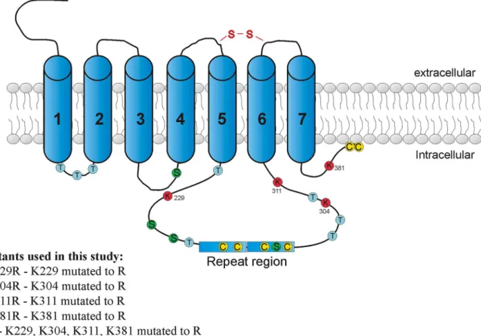
whole primary structure, namely at sites 229 (in the third intracellular loop before the poly-morphic region), 304, 311 (both in the third intracellular loop after the polymorphism) and 381 (in the C-terminal tail). The HA D4.2R construct was used as a template for the application
of single-nucleotide mutations (encoding arginine instead of lysine) using the QuickChange1 Site-Directed Mutagenesis Kit from Stratagene (La Jolla, CA, USA).
The four individual K-R mutants were made separately, and are further denoted as D4.2R
K229R, K304R, K311R, and K381R. The K304R mutant was used as a template for the construc-tion of the quadruple mutant D4.2R K229,304,311,381R (further denoted as HA D4.2 4KRR).
HAβ2AR was a kind gift from Prof. Dr. R. Lefkowitz (Duke University, Durham, NC). HA CXCR4 was purchased from pCDNA.org.
Primary antibodies used were mouse monoclonal anti-HA (clone16B12; Covance Research Products, cat. no. MMS-101-P-1000), rabbit polyclonal anti-Etag (Abcam, cat. no. ab3397-250), rabbit polyclonal anti-c-myc (Sigma, cat. no. C3956), rabbit polyclonal anti-HA (Gene-Tex, cat. no. GTX29110), rat monoclonal anti-HA (clone 3F10; Roche, cat. no. 11867423001), horseradish peroxidase (HRP)-conjugated mouse monoclonal anti-Flag M2 (Sigma, cat. no. A8592), rabbit polyclonal phospho-p44/42 MAPK (Cell Signaling, cat. no. 9101L) and mouse monoclonal p44/42 MAPK (clone L34F12; Cell Signaling, cat. no. 4696S).
Secondary antibodies used were goat anti-rabbit IRDye680 LT (cat. no. 926–68021), goat anti-mouse IRDye680 LT (cat no. 926–68020), goat anti-mouse IRDye680 RD (cat. no. 926– 68070), goat anti-rabbit IRDye800 (cat. no. 926–32211), goat anti-mouse IRDye800 (cat. no. 926–32210), goat anti-rat IRDye680 LT (cat. no. 926–68029) and were all purchased from LI-COR Biosciences.
Cell culture and transfection
HEK293T cells were cultured in Dulbecco's modified Eagle's medium (DMEM; Invitrogen), supplemented with 10% fetal calf serum, penicillin (100 U/ml), and streptomycin (100μg/ml)
in a controlled environment (37°C, 98% humidity, 5% CO2). HEK293T cells were transfected using the Polyethylenimine (PEI) method as described before [8]. A total amount of 10μg of
HEK293S cells parental were a kind gift of Prof. Dr. Nico Callewaert [33]. Cells were stably transfected with calcium phosphate method with pHA D4.2R or pHA D4.2 4KRR and were
grown in DMEM/F12 (Gibco, Invitrogen) supplemented with 10% fetal calf serum, penicillin (100 U/ml), streptomycin (100μg/ml), and 0.5 mg/ml G418 (Geneticin, Gibco) in a controlled
environment (37°C, 98% humidity, 5% CO2).
Co-immunoprecipitation
Forty eight hours after transfection cells were washed twice with cold phosphate-buffered saline (PBS), harvested and the cell pellet was frozen at−70°C for at least 1 h before lysis. Cell
lysates were subjected to immunoblot analysis, or to immunoprecipitation (IP) followed by immunoblotting (IB), as described before [34].
Ubiquitination assay
—
sequential double immunoprecipitation
Ubiquitination assay was described before [35]. Briefly, cells were lysed in radioimmunopreci-pitation buffer (RIPA) supplemented additionally with the inhibitor of deubiquitinating enzymes (10 mM), N-ethylmaleimide (NEM). For the first IP 2μg of HA (16B12)
anti-body was used and samples were incubated overnight with antianti-body and agarose beads. Next, three wash steps with the lysis buffer were performed and proteins bound to the beads (recep-tor and interacting partners) were eluted under denaturing conditions, and a quarter of the elu-ate was used to confirm the first IP. The rest of the eluelu-ates, containing denatured proteins, were diluted with lysis buffer and subjected to a second IP to remove receptor-interacting proteins from the first IP and specifically isolate the protein of interest. Finally, the eluates from the sec-ond IP were subjected to immunoblotting for the detection of ubiquitinated receptor.
Chemical cleavage of ubiquitin with sodium hydroxide and dithiothreitol
A modified protocol of sequential double immunoprecipitation was used to detect the influ-ence of highly reducing conditions (dithiothreitol-DTT) or high pH (NaOH) on the ubiquiti-nation status of the receptor. High pH treatment was performed by resuspendingimmunoprecipitates in 0.5% SDS, boiling for 3 min, cooling to room temperature, adding NaOH to a final concentration of 50 mM, and then incubating the samples at 32°C for 1 h. Mock-treated samples were incubated with PBS. Samples were then neutralized via the addi-tion of 0.5 M Tris/HCl, pH 6.8. 5x Laemmli sample buffer (5% SDS; 50% glycerol; 0.2% bromo-phenol blue; 65 mM Tris/HCl, pH 6.8) without reducing agents was added, and samples were boiled for 3 min. Next, eluates were transferred to the new tubes, diluted with lysis buffer and a second round of IP was performed as described before.
Reducing treatments were performed by resuspending immunoprecipitates in 5x Laemmli sample buffer supplemented with 100 mM DTT followed by boiling for 3 min. Next, eluates were transferred to the new tubes, diluted with the lysis buffer and a second round of IP was performed.
All protein samples were separated via SDS-PAGE, transferred to the nitrocellulose mem-brane and subjected to immunodetection with primary and secondary antibodies.
Detection of p44/42 mitogen-activated protein kinase (MAPK)
phosphorylation by in cell western
HEK293S cells stably transfected with HA D4.2R or HA D4.2 4KRR were grown on poly-D-lysine
37°C. Incubation was stopped by removing the culture medium, followed by addition of fixing solution (3.7% formaldehyde in PBS) for 20 min at room temperature (RT). Next, cells were permeabilized by washing 4 times for 5 min with Triton washing solution (0.1% Triton X-100 in PBS). Subsequently, cells were blocked with a blocking buffer (LI-COR Biosciences) for 90 min and finally, cells were incubated overnight at 4°C with two primary antibodies: rabbit phospho-p44/42 MAPK and mouse p44/42 MAPK diluted in the blocking buffer. The next day, the plate was washed 4 times for 5 min at RT with Tween washing solution (0.1% Tween-20 in PBS) and incubated with fluorescently labeled secondary antibodies: goat anti-rabbit IRDye800 and goat anti-mouse IRDye680 RD for 1 hour at RT. After final washing with Tween washing solution, fluorescent signal was detected with the Odyssey Infrared Imaging system. In the analysis, background values of the secondary antibodies are subtracted and the phospho-p44/42 MAPK signal is normalized against the total p44/42 MAPK signal.
Immunofluorescence microscopy
HEK293S cells stably expressing HA D4.2R or HA D4.2 4KRR were seeded in wells with
cover-slips. Twenty four hours after seeding, cells were fixed with fixing solution (3.7% formaldehyde in PBS) for 20 min at RT. After washing, cells were permeabilized with 0.1% Triton X-100 in blocking buffer (3% newborn calf serum, 1% bovine serum albumin in PBS) for 20 min at RT. Subsequently, cells were incubated for one hour with blocking buffer, followed by incubation for one hour with rabbit anti-HA diluted in blocking buffer (1:1000). Next, secondary antibody anti-rabbit Alexa Fluor 488 diluted in blocking buffer (1:500) was applied and incubation was performed in the dark for 1 hour. Cell nuclei were stained with DAPI (1μg/ml). After each
step, mentioned above, coverslips were washed three times with PBS. Mounting was performed with Mowiol and samples were analysed using the Axiocam 200 microscope (Zeiss, Thorn-wood, NY).
Statistical analyses
To determine significance, results were compared with control conditions by means ofttest
(for two groups) or by One-way analysis of variance (ANOVA) with Bonferroni post-test (for more than two groups). All statistical analyses were performed using Prism software (version 5; GraphPad Software). P<0.05 at the 95% confidence level was considered significant.
Results
The dopamine D
4receptor is ubiquitinated on non-lysine residues
In previous studies we have shown that KLHL12, a BTB-Kelch protein, functions as an adaptor in the Cullin3-based E3 ubiquitin ligase complex and promotes ubiquitination of the D4R [8].
In this study, we aim to characterize the ubiquitination pattern of the D4R into more detail.
The D4.2R contains only four lysine (K) residues in its whole primary structure, namely at
unable to undergo ubiquitination. First, the expression profile of these mutants and their ability to interact with KLHL12 was tested. Upon transient transfection of the HA-tagged receptor constructs and Etag KLHL12, lysates were tested for expression of all receptors and further sub-jected to immunoprecipitation with anti-HA. From these experiments, it can be concluded that all K-R receptor mutants are clearly expressed, and are able to interact with KLHL12 (S1 Fig).
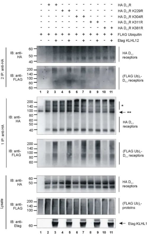
Subsequently, ubiquitination of all individual mutant receptors was tested by ubiquitination assays in HEK293T cells (Fig 2). Surprisingly, comparable ubiquitination levels could be observed for all four single K-R mutant receptors (lanes 4, 6, 8, 10). KLHL12 promoted ubiqui-tination of the WT HA D4.2R (lane 3) but also of all four single K-R mutant receptors (1 and 2
IP: lanes 5, 7, 9, 11). These observations could reflect the possibility that upon mutation of one lysine residue, another lysine residue serves as an alternative acceptor site for ubiquitin-conju-gation. Therefore, these results suggest redundancy of the lysine residues in the D4R to serve as
attachment sites for basal or KLHL12-promoted ubiquitination.
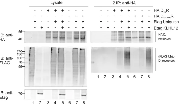
In the next step, the membrane expression and functionality of the quadruple HA D4.2 4KRR
mutant was verified by examining its ability to phosphorylate MAP kinase in response to ago-nist stimulation (S2 Fig). In a second stage, the ubiquitination level of this mutant receptor was investigated. In these experiments, a very strong increase in ubiquitination of the mutant
Fig 1. Approximate localization of all possible ubiquitin binding sites in the intracellular part of D4.2R.The figure shows all possible ubiquitin binding sites: lysines (K), cysteines (C), serines (S) and threonines (T) that are present in the intracellular part of the D4.2R. Additionally, information about receptor mutants used in this study is given.
Fig 2. Ubiquitination of all four single K-R mutants of D4R is increased by KLHL12.HEK293T cells were transiently transfected as indicated. 48 h post-transfection, cells were harvested and lysed. 5% of the lysate was used for IB to visualize HA-tagged WT D4.2R and all single K-R mutants, (Flag Ub)n-proteins, and Etag KLHL12, respectively (bottom panels). The rest of the lysate was subjected to a sequential double IP with anti-HA (16B12). Specific purification of the HA D4.2R WT and all K-R mutants was confirmed after the first (middle panels) and second IP (upper panels) upon IB with anti-HA (16B12, 1:2000), whereas receptor ubiquitination was revealed upon IB with anti-Flag-HRP (1:2000).*D4R specific signal.**Association of two heavy chains of mouse anti-HA antibody (each 50 kDa). The results shown are representative of three independent experiments.
receptor was again detected in the ubiquitination assay upon overexpression of Etag KLHL12 (Fig 3) which suggests that D4R can be ubiquitinated on other residues than lysine.
Up to now, besides on lysine, ubiquitination on the N-terminus [11–13], on cysteine [14–
16], on serine and threonine [17–19] was described. Because of its extracellular location, KLHL12-mediated ubiquitination of the N-terminus of D4R is unlikely. Therefore, in further
investigations we focused only on the possible ubiquitination of the residues which are located in the intracellular parts of the receptor.Fig 1shows the estimated localization of all possible ubiquitination sites in the intracellular part of the D4R.
KLHL12 can promote ubiquitination on cysteine residues of D
4R
As shown inFig 1there are two conserved cysteine residues in each polymorphic repeat in the third intracellular loop and an additional two are present at the C-terminus of D4R. To
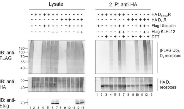
investi-gate a possible attachment of ubiquitin to cysteine we decided to use a specific chemical approach, in which we rely on the fact that the thioester bond which is formed between ubiqui-tin and cysteine is susceptible to strong reducing conditions (for example high concentration of DTT). For this assay, HA-tagged WT D4.2R or mutant D4.2 4KRR were immunoprecipitated
from transiently transfected HEK293T cells. The resulting IP pellets containing ubiquitinated receptors were treated with 100 mM DTT, which has been shown to cleave Ub chains attached to cysteine residues [15,16,36]. Next, eluates were diluted with lysis buffer and a second IP was performed, as before, to prevent detection of a ubiquitination signal originating from D4R
interacting proteins instead of the receptor itself (Fig 4). Treatment with the strong reducing agent DTT after performing the ubiquitination assay resulted in a very strong decrease in ubi-quitination signal of WT D4.2R and of the lysine-deficient D4.2 4KRR mutant. When Etag
KLHL12 was overexpressed, the detected ubiquitination signal was much stronger also in the samples treated with DTT (e.g. compare inFig 4lane 11 and lane 13 for WT D4.2R and
com-pare lane 5 and 7 for the mutant D4.2 4KRR), but DTT treatment again decreased the
ubiquitina-tion signal very efficiently. These results indicate that KLHL12 can promote cysteine-linked D4R ubiquitination. The ubiquitination signal is still visible after DTT treatment in case of D4.2
4KRR when Etag KLHL12 is overexpressed and therefore, these results suggest that there are
also other residues, besides cysteine and lysine, that can be ubiquitinated in the D4R
KLHL12 promotes ubiquitination on serine and/or threonine residues of
D
4R
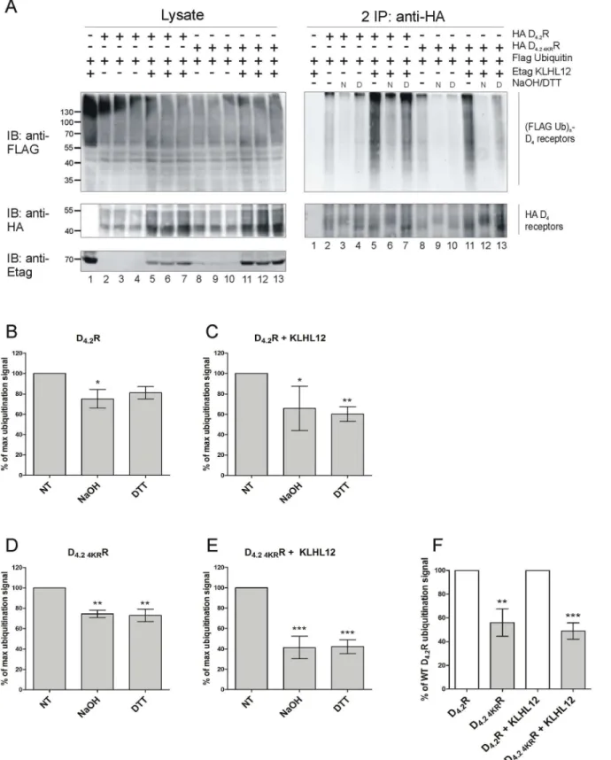
When ubiquitin is attached to serine or threonine an oxyester bond is formed. This kind of bond can be destroyed by treatment with a strong base (50–100 mM NaOH) [36,37]. To exam-ine the possibility of ubiquitination of the D4R on serine and threonine residues, IP eluates
were treated with 50 mM NaOH for 1h at 32°C as described before [37] (Fig 5A). In the same experiment we also examined the influence of highly reducing conditions on the same samples to remove ubiquitin from cysteine residues (as described above). This facilitated the compari-son of the results of the two different chemical treatments, indicative for serine/threonine and cysteine ubiquitination, respectively. After the first IP samples were diluted with lysis buffer and a second IP was performed similar as before.
As shown inFig 5Atreatment with NaOH led to a significant decrease in basal ubiquitina-tion as well as a strong decrease in KLHL12-enhanced ubiquitinaubiquitina-tion signal in case of both WT D4.2R and D4.2 4KRR mutant. In this experiment the influence of DTT can also be clearly
equal in all of the samples and therefore, we decided to quantify the detected ubiquitination sig-nal and normalize it against the receptor levels to identify trends in obtained data (Fig 5B–5E). The results presented inFig 5Asuggest that NaOH is more potent than DTT in removing ubi-quitin moieties from D4R, although the performed quantification from two to seven
indepen-dent experiments does not support this observation. However, and most importantly, all these results together suggest that ubiquitination of cysteine, serine and/or threonine residues in the D4R is possible. Moreover, from this quantification we can conclude that the decrease in
ubi-quitination signal caused by the chemical treatments is bigger in case of KLHL12-enhanced ubiquitination especially for the D4.2 4KRR (compare bars representing percentage of maximal
ubiquitination signal detected upon treatment inFig 5B and 5Cfor WT D4.2R andFig 5D and
5Efor D4.2 4KRR). Another interesting observation that can be made based on experiments
involving chemical treatments is the overall lower ubiquitination level of D4.2 4KRR mutant
compared to the WT D4.2R (Fig 5F). This suggests that lysines which are present in WT
recep-tor can also be ubiquitinated and represent a fraction of the total ubiquitination signal, but it is not possible to conclude whether they are the preferred ubiquitin-binding sites in the
KLHL12-promoted ubiquitination. As suggested by Wang et al. [38] before making a conclu-sion about non-lysine ubiquitination based on chemical treatment experiments one should be aware that too long alkaline treatment can affect also amide bonds formed between lysine and ubiquitin. Therefore, an additional control experiment was performed in which the influence of alkaline treatment on known lysine-linked ubiquitination samples was tested. We have examined the effect of NaOH treatment on ubiquitination of two other GPCRs for which
Fig 3. Lysine lacking D4.2 4KRR mutant is ubiquitinated and KLHL12 enhances its ubiquitination.HEK293T cells were transiently transfected as indicated. 48 h post-transfection, cells were harvested and lysed. 5% of the lysate was used for IB to visualize HA D4R, (Flag Ub)n-proteins, and Etag KLHL12, respectively (left panels). The rest of the lysates were subjected to a sequential double IP with anti-HA (16B12). Specific purification of the receptor after the second IP was confirmed upon IB with rat anti-HA (1:2000), whereas receptor ubiquitination was revealed upon IB with anti-Flag-HRP (right panels, 1:2000). The results shown are representative of four independent experiments.
ubiquitination on lysine residues was described, namely the chemokine CXCR4 receptor [26,
39] and theβ2-adrenoceptor (β2AR) [28].S3 Figclearly demonstrates that NaOH strongly affects basal and KLHL12-enhanced ubiquitination of D4R but seems to have no effect on
ubi-quitination of the other two receptors. This indicates that the NaOH treatment conditions used in our experiments are not strong enough to affect typical isopeptide bonds which are formed between ubiquitin and lysine residues in substrate proteins.
KLHL12 promotes differential ubiquitination of the D
4R polymorphic
variants
As mentioned in the introduction D4R has an important polymorphism in the third
intracellu-lar loop where a 16-amino acid sequence is repeated from 2 to 11 times. The most common variants in the human population are D4.4R (64%), D4.7R (21%) and D4.2R (8%), respectively
[4]. Not much is known about functional differences between different polymorphic variants but several studies suggest an association between the D4.7R allele and ADHD [6].
Previously, we have demonstrated that KLHL12 binds specifically to the polymorphic region of D4R and promotes ubiquitination of the receptor. For these reasons, we were
inter-ested to explore whether KLHL12 differentially binds to the most frequent polymorphic vari-ants and to study the effect of KLHL12 on the ubiquitination of these receptor varivari-ants.
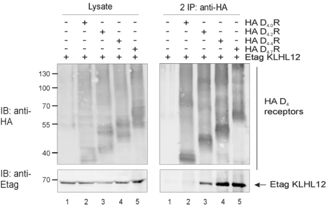
First, we have examined using co-immunoprecipitation if KLHL12 binds to all common D4R polymorphic variants, namely D4.2R, D4.4R and D4.7R. As a negative control we used an
Fig 4. Treatment with a highly reducing agent decreases ubiquitination of D4.2R WT and D4.2 4KRR mutant.HEK293T cells were transiently transfected as indicated. 48 h post-transfection, cells were harvested and lysed. 5% of the lysates were used for IB to visualize (Flag Ub)n-proteins, HA D4R, and Etag KLHL12, respectively (left panels). The rest of the lysates were subjected to IP with anti-HA (16B12). After IP proteins were eluted at 95°C with or without addition of 100 mM DTT. Next, a second round of IP with anti-HA antibody was performed. Specific purification of the receptor after the second IP was confirmed upon IB with rat anti-HA (1:2000), whereas receptor ubiquitination was revealed upon IB with anti-Flag-HRP (right panels, 1:2000). Shown are results from a single experiment, representative of three independent experiments.
Fig 5. Treatment with NaOH decreases ubiquitination of D4.2R WT and D4.2 4KRR mutant.A) HEK293T cells were transiently transfected as indicated. 48 h post-transfection, cells were harvested and lysed. 5% of the lysates were used for IB to visualize (Flag Ub)n-proteins, HA D4R, and Etag KLHL12,
artificial receptor variant from which the polymorphic region was removed (D4.0R).Fig 6
clearly shows that KLHL12 binds to all common polymorphic variants D4.2R, D4.4R and D4.7R
and as expected no interaction with D4.0R was detected.
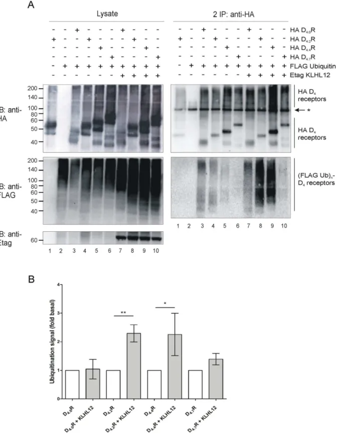
Next, ubiquitination assays were performed in HEK293T cells overexpressing different HA-tagged D4R polymorphic variants with or without co-expression of Etag KLHL12. A strong
increase in ubiquitination signal was observed in case of D4.2R and D4.4R variants when Etag
KLHL12 was overexpressed. Surprisingly, we could hardly detect an increase in ubiquitination of the D4.7R variant (Fig 7).
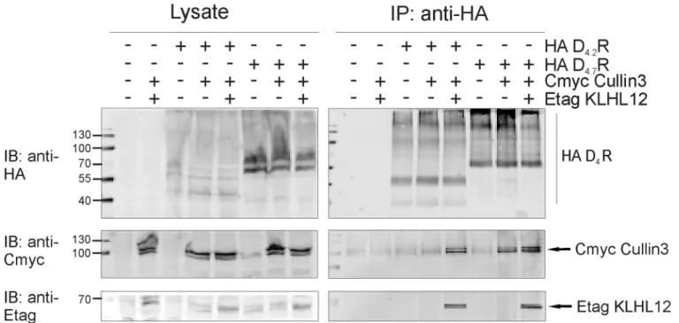
The ubiquitination process requires integrated action of many proteins at the same time and therefore, we have also examined if the D4.7R variant is still capable to form a complex
with E3 ligase, Cullin3. The results from the co-immunoprecipitation studies indicate that D4.7R is able to form a complex with Cullin3 (Fig 8), although we cannot rule out the possibility
that this interaction could be mediated by adaptor proteins other than KLHL12.
Discussion
Previously, we have shown that KLHL12 interacts with the polymorphic repeats of the D4R
and enhances its ubiquitination [8] but this has no influence on receptor degradation [31]. In the present study we investigated which specific residues of the D4R are ubiquitinated by the
KLHL12-Cullin3 E3 ligase complex. In the whole sequence of D4R there are only four lysine
residues and all of them can potentially serve as ubiquitin binding sites as they are localized in the intracellular domains of the receptor. Using a site-directed mutagenesis approach each lysine was separately mutated to arginine. Next, sequential double IP experiments demon-strated that all four mutants are ubiquitinated and overexpression of KLHL12 increases their ubiquitination level (Fig 2). This suggests that ubiquitination occurs on multiple lysines or that the ubiquitination system is flexible and when the most preferred ubiquitin binding site is unavailable it can ubiquitinate another available lysine. Next, we created the quadruple mutant HA D4.2 4KRR in which all four lysines were mutated to arginines. This mutant should be
unable to undergo lysine-mediated ubiquitination. Surprisingly, we could still detect basal quitination of this mutated receptor and upon co-expression of KLHL12 an increase in its ubi-quitination was visualized (Fig 3). This finding encouraged us to search for other possible ubiquitination sites in the D4R. Ubiquitination on non-lysine residues is still a poorly studied
phenomenon and until now ubiquitination on the N-terminus [11–13,40], cysteine [14–16], serine and threonine [17–19] residues was described, but it has only been suggested once for a GPCR unfortunately without characterizing the precise amino acid residue [32]. In our study we investigated the possibility of ubiquitin binding to cysteine, serine and threonine residues within the intracellular domains of the D4R. One of the most common approaches to verify
ubiquitination on a non-lysine residue is a chemical cleavage of ubiquitin [38]. Ubiquitin attached to a lysine residue forms an isopeptide bond which is very stable, whereas a thioester bond connecting ubiquitin with cysteine can be destroyed during treatment with a highly
Specific purification of the receptor after the second IP was confirmed upon IB with rat anti-HA (1:2000), whereas receptor ubiquitination was revealed upon IB with anti-Flag-HRP (right panels, 1:2000). N: samples treated with 50 mM NaOH, D: samples treated with 100 mM DTT. Experiment with two different chemical treatments was performed three times. Additionally, two more experiments were performed with NaOH treatment alone. B-E) Quantification of the ubiquitination signal detected in four to seven independent experiments was performed using Fiji software. Values were normalized to the receptor level in the corresponding lanes. Graph bars show percentage of maximal ubiquitination signal (mean±SEM) where maximum is equal to 100% and represents the signal detected in samples which did not undergo any chemical treatment (NT- non-treated) in each independent experiment. Compared with NT:***, P<0.001;**, P<0.01;*, P<0.05. F) Quantification of the ubiquitination signal detected in five to seven independent experiments was performed using Fiji software. Values were normalized to the receptor level in the corresponding lanes. Graph bars show percentage of ubiquitination signal detected for WT D4.2R (mean±SEM). Compared with the ubiquitination signal detected for WT D4.2R without or with overexpression of KLHL12, respectively:***, P<0.001;
**, P<0.01.
reducing agent e.g. a high concentration of DTT [15,16,36]. Our results clearly indicate that upon treatment with a very high concentration of DTT the ubiquitination signal detected after the second IP is significantly reduced for both WT D4.2R and D4.2 4KRR, suggesting
ubiquitina-tion on cysteine residues (Fig 4).
Next, we examined the possibility of ubiquitination on serine and/or threonine residues. When ubiquitin is attached to a serine or threonine residue an oxyester bond is formed which is susceptible for cleavage in a strong alkaline environment [36,37]. We have performed a ubi-quitination assay in which, after the first IP, the samples were treated with NaOH and after the second IP a strong decrease in ubiquitination signal was observed (Fig 5A). These results thus suggest that D4R ubiquitination on cysteine, serine and/or threonine residues can be promoted
by KLHL12. It is, however, not possible to say which residues are the most preferred for KLHL12-induced ubiquitination as quantification of the results from multiple independent experiments indicates a comparable decrease in ubiquitination levels for both types of treat-ments (Fig 5B–5E). The decrease is more explicit when Etag KLHL12 is co-expressed which suggests that during KLHL12-promoted ubiquitination more non-lysine residues in the D4R
are ubiquitinated compared to the basally ubiquitinated receptor. This decrease is even more striking in case of the D4.2 4KRR mutant in which no lysine residues are available. This indicates
that in the WT receptor KLHL12 promotes ubiquitination on lysine and non-lysine residues and in the D4.2 4KRR mutant, in which lysines are not available, only non-lysine residues are
ubiquitinated. The fact that in all experiments we detect an overall lower ubiquitination signal from D4.2 4KRR (Fig 5F) compared to WT D4.2R further supports the hypothesis about the
Fig 6. All common D4R polymorphic variants interact with KLHL12 while D4.0R, lacking repeats in the third intracellular loop, does not interact. HEK293T cells were transiently transfected as indicated. 48 h post-transfection, cells were harvested and lysed. Part of the lysate was used for IB to verify expression of HA-tagged D4R variants and Etag KLHL12 (left panels). The rest of the lysate was subjected to IP with anti-HA (16B12). Specific purification of the receptor after IP was confirmed upon IB with rat anti-HA (1:2000). Interaction of Etag KLHL12 with the D4R polymorphic variants was verified by IB with anti-Etag (1:2000). Data shown are representative of three independent experiments. Expected molecular weights of different D4R variants: D4.0R (44–50 kDa), D4.2R (47-53kDa), D4.4R (51–57 kDa), D4.7R (56–62 kDa).
involvement of lysine residues in KLHL12-mediated ubiquitination of D4R. To exclude the
possibility that the alkaline treatment affects amide bonds which are normally formed between ubiquitin and lysine residues we have also checked the influence of NaOH on ubiquitination of other GPCRs for which lysines were described as ubiquitin binding sites. We have used the CXCR4 receptor for which three lysines in the C-terminal tail were shown to be critical for receptor ubiquitination [26]. As a second receptor we used theβ2AR for which lysines in the
third intracellular loop and in the C-terminal tail were presented to be important for ubiquiti-nation and subsequent lysosomal degradation [28]. In this control experiment (S3 Fig), we have seen again a very strong effect of NaOH treatment on basal and KLHL12-induced ubiqui-tination of D4.2R and no effect on CXCR4 andβ2AR ubiquitination. This result allows us to
conclude that the conditions used for alkaline treatment are not affecting amide bonds which are formed during the typical lysine-linked ubiquitination process. Most papers that study ubi-quitination on non-lysine residues identified several different residues as possible ubiquitina-tion sites. Some of them are most preferred by the ubiquitinaubiquitina-tion machinery but when those are not available ubiquitin can bind to other amino acids. Such situation was described for example for NS-1 nonsecreted immunoglobulin light chain (NS-1 LC) which is predominantly ubiquitinated on serine/threonine residues but when these amino acids were mutated then lysine residues could serve as ubiquitin-binding sites. Mutation of all three types of amino acids (serines, threonines and lysines) was required to efficiently reduce ubiquitination and
experiments. B) Quantification of the ubiquitination signal detected in three to five independent experiments was performed using Fiji software. Values were normalized to the receptor level in the corresponding lanes. Graph bars show ubiquitination signal (mean±SEM, fold basal) where basal ubiquitination represents the signal detected in samples without overexpression of KLHL12, for the specific D4R variant, in each independent experiment. Compared with the D4.XR without KLHL12 overexpression:**, P<0.01;*, P<0.05.
doi:10.1371/journal.pone.0145654.g007
Fig 8. D4.7R forms a complex with Cullin3.HEK293T cells were transiently transfected as indicated. 48 h post-transfection, cells were harvested and lysed. 5% of the lysate was used for IB to verify expression of HA-tagged receptors, Cmyc Cullin3 and Etag KLHL12 (left panels). The rest of the lysate was subjected to IP with anti-HA (16B12). Specific purification of the receptor after IP was confirmed upon IB with rat anti-HA (1:2000). Interaction of Etag KLHL12 with D4R was verified by IB with anti-Etag (1:2000) and the presence of Cmyc Cullin3 was visualized with anti-Cmyc (right panels, 1:1000).
stabilize the NS-1 LC [37]. Another example represents the T-cell antigen receptorα-chain (TCRα) for which mutation of two conserved serine residues to alanines in the cytosolic tail inhibited significantly receptor ubiquitination. However, when serine residues were replaced with lysine, cysteine or threonine residues the protein was still ubiquitinated [17]. Our findings seem to be in agreement with this previously described phenomenon showing that the nation machinery is a very flexible system and can easily change from one to another ubiquiti-nation site if necessary.
To obtain a direct proof of ubiquitination on a non-lysine residue we decided to perform mass spectrometry (MS) analysis. Detection of ubiquitination by MS is possible due to the di-glycine remnant that remains attached to the ubiquitinated residue after tryptic digestion [41]. We have performed MS/MS analysis using a Fourier transform mass spectrometer but no ubi-quitination was detected. Unfortunately, during tryptic digestion D4R is cut to single amino
acid or very large fragments making it very hard to analyze. The computational prediction of cleavage of D4R with other enzymes, commonly used to prepare samples for MS analysis, did
not suggest any good alternative to trypsine that could resolve this problem. According to our knowledge non-conventional ubiquitination was never proved with MS analysis probably due to the labile nature of thio-/oxy-ester bonds which are formed between ubiquitin and non-lysine residues. So far, only one study described the use of a novel peptide-based SILAC method to identify non-conventional ubiquitination of T-cell receptorα[42]. The authors could identify modified peptide which did not contain lysine in its sequence, however, they were unable to pinpoint the specific residue that undergoes ubiquitination.
Therefore, it is not very surprising that the detection of ubiquitination of D4R with MS/MS
was not successful.
However, and most importantly, all the obtained data allow us to conclude that KLHL12 can promote ubiquitination of D4R on non-lysine residues and that ubiquitination on cysteine,
serine and/or threonine is possible. The functional role of this non-lysine ubiquitination is hard to study because it would require the creation of a receptor with all possible ubiquitination sites mutated. Mutation of all intracellular cysteines, serines and threonines increases the chances of disrupted protein folding and it can also interfere with other posttranslational modi-fications. GPCRs are tightly regulated by phosphorylation which mainly occurs on serine and threonine residues and also by palmitoylation on cysteine residues (conserved cysteines located on the C-terminus of many GPCR) and also D4R shows these posttranslational modifications.
Additionally, this can also inhibit binding of some interacting proteins, e.g.β-arrestins for which often phosphorylation of the receptor is required and which also play a critical role in the regulation of GPCR signaling. Therefore, mutation of all of these residues increases the pos-sibility of creating of a mutant with completely different signaling properties compared to the wild type receptor. In most of the examples of non-lysine ubiquitination, which were described up to now, this modification seems to serve as a signal for protein degradation [14,17–19,21]. Only in the case of Pex5p receptor (cytosolic receptor for peroxisome matrix proteins) ubiqui-tination of conserved cysteine11was shown to be involved in regulation of the recycling of the receptor form the peroxisomes to cytosol [15]. To the best of our knowledge, ubiquitination of the non-lysine residue was suggested only once for a GPCR. The group of Shenoy has shown thatβ2AR is probably ubiquitinated on a non-lysine residue in response to the treatment with
In the second part of our study we investigated the ubiquitination status of the three most common D4R polymorphic variants. First, the interaction of KLHL12 with these variants was
examined. After IP of HA-tagged receptors co-precipitation of Etag KLHL12 with D4.2R, D4.4R
and D4.7R was detected (Fig 6). As a negative control we used D4.0R which is an artificial
recep-tor variant lacking the polymorphic repeats. Next, a double sequential IP was performed to investigate the ubiquitination status of the different polymorphic variants. Ubiquitination of D4.2R and D4.4R was clearly increased when Etag KLHL12 was co-expressed, but surprisingly
we hardly observed increased D4.7R ubiquitination (Fig 7A and 7B), although this receptor
var-iant is still able to form a complex with KLHL12 and Cullin3 (Fig 8). The differential ubiquiti-nation pattern of the D4.7R, compared to the other D4R variants, is highly interesting as until
now there is hardly evidence for pharmacological differences between this variant and the other receptor variants [43–45], although behavioral research showed that the D4.7R is linked
with a predisposition to develop ADHD [6], and also its association with increased sexual behavior, alcohol and cigarette craving was suggested. The functional role of differential KLHL12-mediated ubiquitination of the D4R is still under investigation. This finding opens a
lot of new questions for future research on the possible role of ubiquitination in receptor sig-naling and suggests that ubiquitination can regulate GPCRs in a much more complicated way than previously expected.
Supporting Information
S1 Fig. All K-R D4R mutants interact with KLHL12.HEK293T cells were transiently
trans-fected as indicated. 48 h post-transfection, cells were harvested and lysed. 5% of the lysate was used for IB to visualize HA-tagged D4receptors and Etag KLHL12. The rest of the lysate was
subjected to IP with anti-HA (16B12). Specific purification of the receptors after IP was con-firmed upon IB with anti-HA (1:2000), whereas co-precipitation of Etag KLHL12 was verified by IB with anti-Etag (1:2000).Association of two heavy chains of mouse anti-HA antibody
(each 50 kDa). (TIF)
S2 Fig. D4.2 4KRR is expressed on the membrane and its stimulation leads to the
phosphory-lation of p44/42 MAPK.A) HEK293S cells stably expressing HA D4.2R and HA D4.2 4KRR
were treated with dopamine in concentration ranging from 10−8.5to 10−4.5M. Anin cell
west-ernassay was performed and phosphorylated p44/42 MAP kinase was detected with rabbit
anti-phospho p44/42 MAPK (1:1000). Values were normalized against the signal detected with mouse anti-p44/42 MAPK (1:800). B) HEK293S cells stably expressing HA D4.2R and HA D4.2
4KRR were seeded in wells with coverslips. Cells were stained using primary antibody rabbit
anti-HA (1:1000) and secondary antibody anti-rabbit Alexa Fluor 488 (1:500). Cell nuclei were colored with DAPI.
(TIF)
S3 Fig. Treatment with NaOH decreases ubiquitination of D4.2R but has no influence on
ubiquitination of CXCR4 receptor orβ2-adrenoceptor (β2AR).HEK293T cells were
with rabbit HA (1:2000), whereas receptor ubiquitination was revealed upon IB with anti-Flag-HRP (right panel, 1:2000). N: samples treated with 50 mM NaOH.
(TIF)
Acknowledgments
We thank Griet Debyser and Bart Devreese for performing MS analysis.
Author Contributions
Conceived and designed the experiments: KS PR KVC. Performed the experiments: KS PR BL. Analyzed the data: KS PR KVC. Wrote the paper: KS PR KVC.
References
1. Beaulieu JM, Gainetdinov RR. The physiology, signaling, and pharmacology of dopamine receptors. Pharmacological Reviews. 2011; 63(1):182–217. doi:10.1124/pr.110.002642PMID:21303898
2. Iversen SD, Iversen LL. Dopamine: 50 years in perspective. Trends in Neurosciences. 2007; 30 (5):188–93. PMID:17368565
3. Vallone D, Picetti R, Borrelli E. Structure and function of dopamine receptors. Neuroscience & Biobe-havioral Reviews. 2000; 24(1): 125–32.
4. Oak JN, Oldenhof J, Van Tol HH. The dopamine D4 receptor: one decade of research. European Jour-nal of Pharmacology. 2000; 405(1):303–27.
5. Helmeste DM, Tang SWa. Dopamine D4 receptors. The Japanese Journal of Pharmacology. 2000; 82 (1):1–14.
6. Faraone SV, Doyle AE, Mick E, Biederman J. Meta-analysis of the association between the 7-repeat allele of the dopamine D4 receptor gene and attention deficit hyperactivity disorder. American Journal of Psychiatry. 2001; 158(7):1052–57. PMID:11431226
7. Li D, Sham PC, Owen MJ, He L. Meta-analysis shows significant association between dopamine sys-tem genes and attention deficit hyperactivity disorder (ADHD). Human Molecular Genetics. 2006; 15 (14):2276–84. PMID:16774975
8. Rondou P, Haegeman G, Vanhoenacker P, Van Craenenbroeck K. BTB Protein KLHL12 targets the dopamine D4 receptor for ubiquitination by a Cul3-based E3 ligase. Journal of Biological Chemistry. 2008; 283(17):11083–96. doi:10.1074/jbc.M708473200PMID:18303015
9. Ikeda F, Dikic I. Atypical ubiquitin chains: new molecular signals. EMBO reports. 2008; 9(6):536–42. doi:10.1038/embor.2008.93PMID:18516089
10. Komander D. The emerging complexity of protein ubiquitination. Biochemical Society Transactions. 2009; 37(Pt 5):937–53. doi:10.1042/BST0370937PMID:19754430
11. Breitschopf K, Bengal E, Ziv T, Admon A, Ciechanover A. A novel site for ubiquitination: the N-terminal residue, and not internal lysines of MyoD, is essential for conjugation and degradation of the protein. The EMBO Journal. 1998; 17(20):5964–73. PMID:9774340
12. Ciechanover A, Ben-Saadon R. N-terminal ubiquitination: more protein substrates join in. Trends in Cell Biology. 2004; 14(3):103–6. PMID:15055197
13. Fajerman I, Schwartz AL, Ciechanover A. Degradation of the Id2 developmental regulator: targeting via N-terminal ubiquitination. Biochemical and Biophysical Research Communications. 2004; 314(2):505– 12. PMID:14733935
14. Cadwell K, Coscoy L. Ubiquitination on nonlysine residues by a viral E3 ubiquitin ligase. Science's STKE. 2005; 309(5731):127–30.
15. Okumoto K, Misono S, Miyata N, Matsumoto Y, Mukai S, Fujiki Y. Cysteine ubiquitination of PTS1 receptor Pex5p regulates Pex5p recycling. Traffic. 2011; 12:1067–83. doi:10.1111/j.1600-0854.2011. 01217.xPMID:21554508
16. Williams C, van den Berg M, Sprenger RR, Distel B. A conserved cysteine is essential for Pex4p-dependent ubiquitination of the peroxisomal import receptor Pex5p. Journal of Biological Chemistry. 2007; 282(31):22534–43. PMID:17550898
unassembled protein. Journal of Biological Chemistry. 2010; 285(31):23916–24. doi:10.1074/jbc. M110.127936PMID:20519503
18. Tokarev AA, Munguia J, Guatelli JC. Serine-threonine ubiquitination mediates downregulation of BST-2/tetherin and relief of restricted virion release by HIV-1 Vpu. Journal of Virology. 2011; 85(1):51–63. doi:10.1128/JVI.01795-10PMID:20980512
19. Wang X, Herr RA, Chua WJ, Lybarger L, Wiertz EJHJ, Hansen TH. Ubiquitination of serine, threonine, or lysine residues on the cytoplasmic tail can induce ERAD of MHC-I by viral E3 ligase mK3. The Jour-nal of Cell Biology. 2007; 177(4):613–24. PMID:17502423
20. Haglund K, Dikic I. Ubiquitylation and cell signaling. The EMBO Journal. 2005; 24(19):3353–9. PMID: 16148945
21. Tait SW, de Vries E, Maas C, Keller AM, D'Santos CS, Borst J. Apoptosis induction by Bid requires unconventional ubiquitination and degradation of its N-terminal fragment. The Journal of Cell Biology. 2007; 179(7):1453–66. doi:10.1083/jcb.200707063PMID:18166654
22. Marchese A, Paing MM, Temple BRS, Trejo JA. G protein-coupled receptor sorting to endosomes and lysosomes. Annual Review of Pharmacology and Toxicology. 2008; 48:601–29. PMID:17995450
23. Dores MR, Trejo JA. Ubiquitination of G Protein-Coupled Receptors: Functional Implications and Drug Discovery. Molecular Pharmacology. 2012; 82(4):563–70. PMID:22700696
24. Hislop JN, von Zastrow M. Role of Ubiquitination in Endocytic Trafficking of G Protein-Coupled Recep-tors. Traffic. 2005; 12(2):137–48.
25. Jacob C, Cottrell GS, Gehringer D, Schmidlin F, Grady EF, Bunnett NW. c-Cbl mediates ubiquitination, degradation, and down-regulation of human protease-activated receptor 2. Journal of Biological Chem-istry. 2005; 280(16):16076–87. PMID:15708858
26. Marchese A, Benovic JL. Agonist-promoted ubiquitination of the G protein-coupled receptor CXCR4 mediates lysosomal sorting. Journal of Biological Chemistry. 2001; 276(49):45509–12. PMID: 11641392
27. Martin NP, Lefkowitz RJ, Shenoy SK. Regulation of V2 vasopressin receptor degradation by agonist-promoted ubiquitination. Journal of Biological Chemistry. 2003; 278(46):45954–9. PMID:12960162
28. Xiao K, Shenoy SK.β2-adrenergic receptor lysosomal trafficking is regulated by ubiquitination of lysyl residues in two distinct receptor domains. Journal of Biological Chemistry. 2011; 286(14):12785–95. doi:10.1074/jbc.M110.203091PMID:21330366
29. Petaja-Repo UE, Hogue M, Laperriere A, Bhalla S, Walker P, Bouvier M. Newly synthesized humanδ -opioid receptors retained in the endoplasmic reticulum are retrotranslocated to the cytosol, deglycosy-lated, ubiquitinated, and degraded by the proteasome. Journal of Biological Chemistry. 2001; 276 (6):4416–23. PMID:11054417
30. Milojevic T, Reiterer V, Stefan E, Korkhov VM, Dorostkar MM, Ducza E, et al. The ubiquitin-specific pro-tease Usp4 regulates the cell surface level of the A2A receptor. Molecular Pharmacology. 2006; 69 (4):1083–94. PMID:16339847
31. Rondou P, Skieterska K, Packeu A, Lintermans Ba, Vanhoenacker P, Vauquelin G, et al. KLHL12-mediated ubiquitination of the dopamine D4 receptor does not target the receptor for degradation. Cel-lular Signalling. 2010; 22(6):900–13. doi:10.1016/j.cellsig.2010.01.014PMID:20100572
32. Han S-o, Xiao K, Kim J, Wu J-H, Wisler JW, Nakamura N, et al. MARCH2 promotes endocytosis and lysosomal sorting of carvedilol-boundβ2-adrenergic receptors. The Journal of Cell Biology. 2012; 199 (5):817–30. doi:10.1083/jcb.201208192PMID:23166351
33. Reeves PJ, Callewaert N, Contreras R, Khorana HG. Structure and function in rhodopsin: high-level expression of rhodopsin with restricted and homogeneous N-glycosylation by a tetracycline-inducible N-acetylglucosaminyltransferase I-negative HEK293S stable mammalian cell line. Proceedings of the National Academy of Sciences. 2002; 99(21):13419–24.
34. Skieterska K, Duchou J, Lintermans B, Van Craenenbroeck K. Detection of G protein-coupled receptor (GPCR) dimerization by coimmunoprecipitation. Methods in Cell Biology. 2013; 117:323–40. PMID: 24143985
35. Skieterska K, Rondou P, Van Craenenbroeck K. Ubiquitination of Dopamine Receptor Studied by Sequential Double Immunoprecipitation. In: Tiberi M, editor. Dopamine Receptor Technologies. Neuro-methods. 96. Springer; 2015. pp. 139–56.
36. Gustin JK, Douglas JL, Bai Y, Moses AV. Ubiquitination of BST-2 Protein by HIV-1 Vpu Protein Does Not Require Lysine, Serine, or Threonine Residues within the BST-2 Cytoplasmic Domain. Journal of Biological Chemistry. 2011; 287(18):14837–50.
38. Wang X, Herr RA, Hansen TH. Ubiquitination of substrates by esterification. Traffic. 2012; 13(1):19–24. doi:10.1111/j.1600-0854.2011.01269.xPMID:21883762
39. Marchese A, Raiborg C, Santini F, Keen JH, Stenmark H, Benovic JL. The E3 ubiquitin ligase AIP4 mediates ubiquitination and sorting of the G protein-coupled receptor CXCR4. Developmental Cell. 2003; 5(5):709–22. PMID:14602072
40. Coulombe P, Rodier Gv, Bonneil E, Thibault P, Meloche S. N-Terminal ubiquitination of extracellular signal-regulated kinase 3 and p21 directs their degradation by the proteasome. Molecular and Cellular Biology. 2004; 24(14):6140–50. PMID:15226418
41. Jeram SM, Srikumar T, Pedrioli PG, Raught B. Using mass spectrometry to identify ubiquitin and ubi-quitin-like protein conjugation sites. Proteomics. 2009; 9(4):922–34. doi:10.1002/pmic.200800666 PMID:19180541
42. Anania VG, Bustos DJ, Lill JR, Kirkpatrick DS, Coscoy L. A Novel Peptide-Based SILAC Method to Identify the Posttranslational Modifications Provides Evidence for Unconventional Ubiquitination in the ER-Associated Degradation Pathway. International Journal of Proteomics. 2013; 2013:857918. doi: 10.1155/2013/857918PMID:23431445
43. Asghari V, Sanyal S, Buchwaldt S, Paterson A, Jovanovic V, Van Tol HHM. Modulation of intracellular cyclic AMP levels by different human dopamine D4 receptor variants. Journal of Neurochemistry. 1995; 65(3):1157–65. PMID:7643093
44. Asghari V, Schoots O, van Kats S, Ohara K, Jovanovic V, Guan H-C, et al. Dopamine D4 receptor repeat: analysis of different native and mutant forms of the human and rat genes. Molecular Pharmacol-ogy. 1994; 46(2):364–73. PMID:8078498