Cell-free H-cluster Synthesis and [FeFe] Hydrogenase
Activation: All Five CO and CN
2
Ligands Derive from
Tyrosine
Jon M. Kuchenreuther1, Simon J. George2,3, Celestine S. Grady-Smith3, Stephen P. Cramer2,3, James R. Swartz1,4*
1Department of Chemical Engineering, Stanford University, Stanford, California, United States of America,2Physical Biosciences Division, Lawrence Berkeley National Laboratory, Berkeley, California, United States of America,3Department of Applied Science, University of California Davis, Davis, California, United States of America, 4Department of Bioengineering, Stanford University, Stanford, California, United States of America
Abstract
[FeFe] hydrogenases are promising catalysts for producing hydrogen as a sustainable fuel and chemical feedstock, and they also serve as paradigms for biomimetic hydrogen-evolving compounds. Hydrogen formation is catalyzed by the H-cluster, a unique iron-based cofactor requiring three carbon monoxide (CO) and two cyanide (CN2) ligands as well as a dithiolate bridge. Three accessory proteins (HydE, HydF, and HydG) are presumably responsible for assembling and installing the H-cluster, yet their precise roles and the biosynthetic pathway have yet to be fully defined. In this report, we describe effective cell-free methods for investigating H-cluster synthesis and [FeFe] hydrogenase activation. Combining isotopic labeling with FTIR spectroscopy, we conclusively show that each of the CO and CN2ligands derive respectively from the carboxylate and amino substituents of tyrosine. Such in vitro systems with reconstituted pathways comprise a versatile approach for studying biosynthetic mechanisms, and this work marks a significant step towards an understanding of both the protein-protein interactions and complex reactions required for H-cluster assembly and hydrogenase maturation.
Citation:Kuchenreuther JM, George SJ, Grady-Smith CS, Cramer SP, Swartz JR (2011) Cell-free H-cluster Synthesis and [FeFe] Hydrogenase Activation: All Five CO and CN2Ligands Derive from Tyrosine. PLoS ONE 6(5): e20346. doi:10.1371/journal.pone.0020346
Editor:Anna Kristina Croft, University of Wales Bangor, United Kingdom
ReceivedFebruary 15, 2011;AcceptedApril 29, 2011;PublishedMay 31, 2011
Copyright:ß2011 Kuchenreuther et al. This is an open-access article distributed under the terms of the Creative Commons Attribution License, which permits unrestricted use, distribution, and reproduction in any medium, provided the original author and source are credited.
Funding:This work was supported by funding from the DOE BioEnergy Science Program (JRS & SPC), the DOE Office of Biological and Environmental Research (SPC), the National Health Institute (NIH-GM-65440, SPC), and the National Science Foundation (NSF-CHE-0745353, SPC). The funders had no role in study design, data collection and analysis, decision to publish, or preparation of the manuscript.
Competing Interests:The authors have declared that no competing interests exist.
* E-mail: [email protected]
Introduction
Hydrogenase enzymes are efficient biocatalysts for the most fundamental of chemical reactions, the reversible combination of protons and electrons to form molecular hydrogen (2H+
+2e2OH2).
With catalytic rates comparable to those of expensive platinum catalysts [1], hydrogenases hold great promise for use in fuel cells [2], for photosynthetic H2 evolution [3], for H2 production from
carbohydrates [4], and as paradigms for synthetic catalysts [5]. They are also important for energy exchange in many ecological systems [6] and were probably key enzymes in the development of primordial biology [7].
Hydrogenases contain complex [FeFe]-, [NiFe]-, or [Fe]-based catalytic cofactors that are stabilized by multiple non-protein ligands [8]. [FeFe] hydrogenases are the fastest H2producers and require the
H-cluster, a catalytic cofactor comprised of two iron-based clusters connected via a cysteinyl sulfur atom (Fig. 1). The cubane Fe–S cluster ([4Fe]H) presumably delivers electrons to the catalytic 2Fe unit
([2Fe]H), which contains three carbon monoxide (CO) and two
cyanide (CN2) adducts as well as a dithiol bridging group of disputed composition [9,10]. Three proteins called the HydE, HydF, and HydG maturases participate in the synthesis of the H-cluster and the activation of [FeFe] hydrogenases [11]. The final maturation step presumably occurs when the HydF maturase transfers the [2Fe]H
cluster to the hydrogenase [12,13], likely through a positively charged channel as proposed by Mulderet al.[14].
One of the most intriguing mysteries has been the origin of the H-cluster CO and CN2ligands, both of which are highly reactive toxins in their free states. Glycine was first considered as a plausible substrate [15], although recent and informative studies on HydG-catalyzed radical chemistry indicated that CO and CN2 could be generated from tyrosine [16,17,18]. These studies, however, were by no means definitive in showing that each of the five CO and CN2ligands derive from tyrosine. The coordination of CO and CN2 to a hydrogenase-bound or a maturase-bound metal cluster was not demonstrated (i.e. formation of the H-cluster or a precursor thereof), and an active [FeFe] hydrogenase was not produced. Rather, the CO and CN2 molecules were indepen-dently detected using separate non-physiological assays. In the work by Drieseneret al., 20% perchloric acid was used to denature
HydG and release protein-bound products, and CN2 was
subsequently identified by derivatization methods [16]. In the work by Shepardet al., CO production was detected by measuring carboxyhemoglobin, although the detectable quantities (10mM
Hb–CO) were substantially lower than the measured CN2
H-cluster CO and CN2ligands [16,17,18], the required methods and nature of the results highlight the need for approaches in which the complete H-cluster biosynthetic pathway is reconstruct-ed. Such methods would provide more flexibility in experimental design and enable detailed analyses of active [FeFe] hydrogenases. The in vitro reconstitution of pathways for activating complex biological catalysts has historically been crucial for gaining insights into the underlying biochemistry [19]. For example, a detailed under-standing of the nitrogenase accessory proteins and the synthesis of the iron-molybdenum cofactor (FeMo-co) only came after the develop-ment of cell-free approaches for nitrogenase activation [20,21,22]. Enabled by the discovery of the HydE, HydF, and HydG maturases [11], we previously reported the first example of in vitro [FeFe] hydrogenase maturation methods that could be used to examine the required substrates [23]. Although suggested substrates such as carbamoyl phosphate and glycine had no observable effects [15,24], S-adenosyl methionine (SAM), cysteine, and tyrosine were essential for hydrogenase activation [23]. In our previous study, however, the maturases had been co-expressed inE. coli. This can lead to thein vivo synthesis of H-cluster precursors that associate with the HydF maturase [12,13,25], thereby complicatingin vitroinvestigations.
In this work, we improved our previousin vitrosystem by employing separately produced maturases. Hydrogenase maturation is thus entirely dependent on the cell-free synthesis of the H-cluster. We demonstrate the utility of such methods by using tyrosine either fully or selectively labeled with 13C and 15N to generate milligram
quantities of active and isotopically labeled [FeFe] hydrogenases, which are subsequently examined using Fourier Transform Infrared (FTIR) spectroscopy. In doing so, we prove that each of the H-cluster CO and CN2 ligands are synthesized from the carboxylate and amino substituents of tyrosine.
Results and Discussion
Our newin vitrosystem includes inactiveClostridium pasteurianum [FeFe] hydrogenase (CpI) apoenzyme combined with three Escherichia colicell lysates, each containing one of the maturases native to Shewanella oneidensis (Fig. 1). SAM, cysteine, tyrosine, ferrous ammonium sulfate (Fe+2
), sodium sulfide (S22), dithiothre-itol (DTT), guanosine-59-triphosphate (GTP), pyridoxal-59 -phos-phate (PLP), and sodium dithionite are added to this mixture of proteins to reconstitute the pathway for H-cluster synthesis and hydrogenase activation.
The work in this report would not have been possible without scalable methods for making large quantities of active [FeFe] hydrogenases in a cell-free environment. We recently improved thein vivoexpression of active hydrogenases inE. coli[26], and we extended those methods for high-yield expression of the individual maturases and CpI apoenzyme. The maturase lysates used forin vitro hydrogenase maturation (Fig. 1) therefore contained high concentrations of HydE, HydF, or HydG, which we estimated to be 3–15 mg?mL21 (Fig. 2). This was crucial to achieve nearly
Figure 1.In vitro[FeFe] hydrogenase activation for FTIR spectroscopic analysis.(Fig. 1A) A ball and stick representation of the hydrogenase H-cluster. The catalytic [2Fe]Hcluster is joined to the cubane [4Fe]Hcluster, colored with the following scheme: brown (Fe), yellow (S), gray (C), red (O),
blue (N), and magenta (unknown). (Fig. 1B) The chemical structure for L-tyrosine, with carbon atoms numbered 1–9. (Fig. 1C) Thein vitrohydrogenase maturation process. For cell-free H-cluster synthesis, (1) CpI apoenzyme (PDB ID 3C8Y) as well as (2) exogenous substrates are added to (3) a mixture of three lysates containingE. coliproteins (yellow ovals) and individually produced maturases. HydE, HydF, and HydG are expressed separately to avoid H-cluster synthesis duringin vivomaturase expression. Following hydrogenase maturation, (4) the CpI holoenzyme is re-purified, and (5) the active hydrogenase is examined using FTIR spectroscopy.
complete activation of the CpI hydrogenase (Table 1) at concentrations of,200 mg?L21, more than 300-fold higher than with methods that lackin vitroH-cluster synthesis [12,27]. By using non-purified maturation proteins, the activation reaction volumes could be increased to more than 100 mL, which allowed us to produce and re-purify the milligram quantities of CpI hydrogenase required for spectroscopic analysis.
Active hydrogenases with either non-labeled or isotopically labeled H-clusters were produced in vitro (Table 1) [28], subsequently isolated, and then characterized using FTIR spectroscopy. The coordinated CO and CN2 ligands provide well-defined absorption bands that indicate the different chemical states of the H-cluster [29]. Moreover, labeling of CO and CN2
with 13C and 15N alters the observed vibrational energies, providing distinctive fingerprints for tracing which atoms originate from labeled substrates [29,30].
The IR spectrum of CpI hydrogenase activated in vitro with natural abundance tyrosine (Fig. 3, CpItyr) is characteristic for an H-cluster in the oxidized state (Hox) [30]. Two peaks at
2082 cm21 and 2070 cm21 derive from the terminal CN2 vibrational (n(CN)) stretches. Peaks at 1970 cm21 and
1947 cm21 correspond to the terminal CO (n(CO)) stretches,
while the peak at 1801 cm21indicates the bridging CO (n(m-CO))
stretch. A nearly identical spectrum has been reported for the CpI hydrogenase isolated fromC. pasteurianum[30].
IR spectra were next recorded for CpI activated in the presence of tyrosine uniformly labeled with13C and 15N isotopes (Fig. 3, CpIU-13C-15N-tyr). The peaks for all fiven(CO) andn(CN) modes
unambiguously shift to lower vibrational energies. Both n(CN)
modes decrease by 75–76 cm21as expected for a two mass unit increase. Both terminaln(CO) modes decrease by 45–46 cm21as
expected for a one mass unit increase. Finally, the bridgingn(m
-CO) mode decreases by 39 cm21also indicating a one mass unit increase. These changes indicate the presence of both the13C and
15
N isotopes and confirm that all five of the CO and CN2ligands derive from tyrosine.
We then used tyrosine with selectively labeled 13C atoms to identify the precise source of the CO and CN2ligands. Reasoning that the CN2 ligands originate from the amino group, we produced active CpI using tyrosine labeled only at the amino carbon (Fig. 3, CpI2-13C-tyr). The IR spectrum shows that both
n(CN) modes decrease by 43–45 cm21, matching the predicted
change for terminally coordinated 13CN2 moieties; all n(CO)
modes are unchanged. Therefore, the H-cluster CN2 ligands derive from the amino substituent in tyrosine.
Tyrosine contains two carbon atoms with bound oxygen atoms that are plausible sources of the CO ligands: the carboxylic C1 and phenolic C7. CpI was activated in the presence of [1-13
C]-tyrosine to determine if the CO ligands derive from the carboxylic acid group. The IR spectrum for CpI1-13C-tyrshows that all threen(CO) modes
decrease by 40–45 cm21, as previously observed for CpIU-13C-15N-tyr, while bothn(CN) modes are unchanged. Hence, the IR spectrum for
CpI1-13C-tyr clearly illustrates that the H-cluster CO adducts are synthesized from the tyrosine carboxylate substituent.
We also examined the IR spectra for each CpI sample mixed with exogenous CO, which binds to the H-cluster distal Fe atom. The CO binding causes well-characterized changes in the spectrum [29,30],
Figure 2. SDS-PAGE and Coomassie staining of purified CpI apoenzyme andE. colilysates with heterologous maturases.All proteins were identified using the Mark12TMprotein ladder (Invitrogen),
and the 36, 55, and 66 kD protein standards are indicated. The control lysate from E. coli strain BL21(DE3) DiscR (lane 1) has no proteins produced from recombinant DNA plasmids. Maturase lysates with soluble HydE (40 kD), HydF (45 kD), or HydG (54 kD) are shown in lanes 2–4, respectively. We estimated that the cell lysates (0.25mL of lysate loaded per lane) contained 3–15 mg?mL21 of each maturase, and
approximately 2.5mg of CpI–Strep-tag II apoenzyme (64 kD) is shown in lane 5.
doi:10.1371/journal.pone.0020346.g002
Table 1.Specific activities of CpI activatedin vitrousing natural abundance or isotopically labeled tyrosine.
Tyrosine substrate CpI (mg?L21) MV reduction assay{
H2evolution assay
{
L-tyrosine 25 683648 ND*
50 674643 ND*
100 675679 16496223
200 660645 ND*
L-[1-13C]-tyrosine 100 658
650 18406124
L-[2-13C]-tyrosine 100 657
630 18306392
L-[U-13C-15N]-tyrosine 100 672
623 14756375
{
CpI activities (mmol H2?min21?mg21) were measured using the methyl viologen (MV) reduction assay or the H2evolution assay. The exogenous tyrosine substrates and the CpI apoenzyme concentrations used forin vitro[2Fe]Hsynthesis are provided. Substituting natural abundance L-tyrosine with each isotopically labeled tyrosine analog had no effect on final CpI activities. H2evolution rates, which were measured at the Kmfor MV (6.25 mM), were nearly half the reportedVmaxof 4000mmol H2?min21?mg21for CpI isolated fromC. pasteurianum[28]. Also, CpI activities did not decrease when more CpI apoenzyme was added to the reaction mixture. Taken together, these observations indicate nearly complete hydrogenase activation. Data are the average of 3–6 measurements6standard deviations.
*ND, not determined.
doi:10.1371/journal.pone.0020346.t001
and the shifts in then(CO) and then(CN) modes that we observed
support our previous assignments and interpretations (Fig. 4). Reconstituting the H-cluster biosynthetic pathway using a Clostridial hydrogenase, Shewanella maturases, and E. coli lysates highlights the modularity of the hydrogenase maturation system and suggests that the mechanisms for CO and CN2ligand synthesis for [FeFe] hydrogenases may be broadly conserved. Questions still remain, however, as to how CO and CN2 are synthesized from tyrosine and subsequently coordinate to an iron cluster. The formation of a radical at the tyrosine C7 hydroxyl group could lead to either a glycyl radical or a reactive dehydroglycine intermediate [31], and such radical SAM chemistry has precedence given the requirement for the para-hydroxyl substituent of tyrosine forin vitro H-cluster synthesis [23]. Recent investigations comparing the wild-type and a mutant HydG maturase have provided further insights into the mechanism for CO and CN2synthesis, and the authors proposed that a glycyl radical is the more likely intermediate derived from tyrosine [17].
As we have shown, reconstituting biosynthetic pathways using cell lysates can lead to new insights, yet establishingin vitrosystems containing purified enzymes and a defined set of substrates can also be important for understanding biochemical conversions [20]. Interestingly, the hydrogenase maturation pathway could not be reconstituted when using purified HydE–Strep-tag II, HydF–Strep -tag II, and Strep-tag II–HydG combined with Fe+2
, S22, SAM, cysteine, tyrosine, DTT, GTP, PLP, and dithionite. AnE. colicell lysate without any maturases was also required with these constituents to activatein vitroH-cluster synthesis and hydrogenase maturation. This difference indicates that uncharacterized
com-ponents of the E. coli lysates are necessary, perhaps proteins involved in Fe–S cluster synthesis.
The roles of the small molecule substrates also require further investigation. Compared to our previous system, four additional chemicals were beneficial for high-yield CpI activation. These include two reducing agents (DTT and sodium dithionite), GTP, and PLP. Dithionite is likely an electron source for the maturase-based radical SAM chemistry [16,18,32,33]. A GTP requirement is also not unexpected as HydF is a GTPase, although high concentrations of this nucleotide (.10 mM) were needed when maturing micromolar concentrations of the [FeFe] hydrogenases. We also observed that GTP could be replaced by ATP, though nucleoside diphosphate kinase activity from the E. coli lysates might be regenerating GTP from GMP and GDP. The third substrate, PLP, may be a cofactor of the maturases, although it is more likely contributing as a cofactor for cysteine desulfurases such as NifS and IscS, which may be facilitating cell-free Fe–S cluster synthesis [34]. This interpretation is supported by the observation that cysteine also enhancesin vitrohydrogenase activation [23].
The in vitro system we have described can also be used for studying the maturases. For example, we replaced the HydF lysate with one containing an affinity-tagged maturase (HydF–Strep-tag II). Following cell-free H-cluster synthesis in the absence of the CpI hydrogenase, we purified the HydF protein to greater than 95% purity and hypothesized that it could have a bound H-cluster precursor [13,25,35]. Interestingly, the purified HydF showed hydrogenase-like activity, with the ability to evolve hydrogen (1.5mmol H2produced?min21?mg21 HydF) as well as to reduce
methyl viologen in the presence of 2% H2 (1.2mmol MV Figure 3. FTIR spectroscopic analysis for active CpI producedin vitrousing natural abundance tyrosine or isotopically labeled tyrosine analogs.The IR spectra are for the as-isolated active CpI hydrogenase containing the H-cluster produced in the presence of L-tyrosine (CpItyr), L-[2-13C]-tyrosine (CpI2-13C-tyr), L-[1-13C]-tyrosine (CpI1-13C-tyr), and L-[U-13C-15N]-tyrosine (CpIU-13C-15N-tyr). The shifts in vibrational energies
correlate with expected changes forn(13CO),n(13CN), andn(13C15N) modes, confirming that the CO and CN2ligands are synthesized from tyrosine.
Labels indicating the assignedn(CO) andn(CN) vibrational modes are provided at the top of the figure, with the13CN2/13C15N2and13CO ligands
shown in red and green, respectively, in the molecular diagrams. Vertical scale bars provided at 1740 cm21
represent a difference of 0.5 milliabsorbance units. Table 2 summarizes the vibrational energies and corresponding assignedn(CN) andn(CO) modes for the Hoxclusters.
reduced?min21?mg21HydF, likely by H2 uptake). The catalytic
rates are less than 1% of those from the active CpI hydrogenase, but identical reaction mixtures lacking both HydF and the CpI
apoenzyme showed no detectable activity. Therefore, the HydF activities indicate that this maturase contained an in vitro synthesized H-cluster precursor.
Figure 4. Infrared spectra for CpI hydrogenase with isotopically labeled H-cluster containing exogenously bound CO.The infrared spectra are for the CO-inhibited CpI enzyme harboring an H-cluster produced in the presence of L-tyrosine (CpItyr), L-[2-13C]-tyrosine (CpI2-13C-tyr),
L-[1-13C]-tyrosine (CpI1-13C-tyr), and L-[U-13C-15N]-tyrosine (CpIU-13C-15N-tyr). Natural abundance COexowas added to CpItyrand CpI2-13C-tyr, which have
intrinsic CO ligands. Conversely,13CO
exowas added to CpI1-13C-tyrand CpIU-13C-15N-tyr, which have intrinsic13CO ligands. Comparing the Hox–COexo
spectrum for each CpI sample to its respective Hoxspectrum (Fig. 3), shifts of 5–10 cm21were observed for then(CN) modes and then(m–CO) mode
in all four cases. Then(CO) mode for the Fep–CO ligand did not change. Meanwhile, then(CO) mode for the Fed–CO moiety was replaced with two
peaks resulting from symmetric and asymmetric coupled vibrational stretches, as two CO molecules of equal mass are coordinated to the Fedatom.
The peak for then(CO)symmetricmode is visible at 2015/1970 cm21for CO/13CO. Then(CO)asymmetricmode, however, cannot be distinguished because
its vibrational energy is similar to the n(CO) mode at 1972/1928 cm21for the Fep–CO/Fep–13CO adducts. The changes in vibrational energies,
indicated by the dashed lines, correlate with expected changes forn(13CO),n(13CN), andn(13C15N) modes, again confirming that the CO and CN2
ligands are synthesized from tyrosine. Labels indicating the assignedn(CO) andn(CN) vibrational modes are provided. The13CN/13C15N and13CO
ligands are shown in red and green, respectively, in the molecular diagrams. Vertical scale bars shown at 1740 cm21
represent a difference of 0.5 milliabsorbance units. Table 3 summarizes the vibrational energies and corresponding assignedn(CN) andn(CO) modes for the Hox–COexoclusters.
doi:10.1371/journal.pone.0020346.g004
Table 2.Summary of energies of the assigned CO and CN2vibrational modes for the CpI hydrogenase H-cluster.
Tyrosine substrate H-cluster ligand and IR vibrational energy (cm21)
(Fe)–CN (Fe)–CN (Fep)–CO (Fed)–CO m–CO
L-tyrosine 2082 2070 1970 1947 1801
L-[2-13C]-tyrosine 2037 (13CN) 2027 (13CN) 1968 1947 1801
L-[1-13C]-tyrosine 2081 2070 1925 (13CO) 1902 (13CO) 1761 (
m–13CO)
L-[U-13C-15N]-tyrosine 2006 (13C15N) 1995 (13C15N) 1924 (13CO) 1902 (13CO) 1762 (m–13CO)
The vibrational energies and correspondingn(CN) andn(CO) mode assignments are provided for each Hoxcluster from active CpI produced with either unlabeled or isotopically labeled tyrosine. Energies were determined from spectra measured using FTIR spectroscopy (Fig. 3). The spectrum for each isotopically labeled sample also contains low intensity bands indicating trace amounts of unlabeled CO and CN2incorporated into the H-cluster. The intensities of these bands vary from sample to
sample, and they do not depend on the location of either CO or CN2on the H-cluster. We thus attribute these features to either adventitious free tyrosine present in the
cell lysates or possibly to low quantities of an iron cluster with CO and CN2ligands that is pre-assembled by a single Hyd maturase duringin vivoexpression. Each
spectrum also shows evidence for CpI with reduced H-cluster (Hred), characterized in the CpItyrcase by bands located at 2053 cm21, 2039 cm21, 1961 cm21, 1914 cm21, and 1899 cm21.
doi:10.1371/journal.pone.0020346.t002
This report provides the first example of cell-free H-cluster synthesis and hydrogenase activation using individually expressed maturases, and it also clearly details the origin of all five H-cluster CO and CN2 ligands. Furthermore, our results underscore the utility of thisin vitroapproach for follow-up studies such as57Fe labeling for Mossbauer spectroscopy as well as attempts to determine the origin of the H-cluster dithiolate ligand. One hypothesis is that the bridge also derives from tyrosine [32], and we are now in a position to directly examine this possibility.
Materials and Methods
Materials and Chemical Solutions
Isotopically labeled L-[1-13C]-tyrosine, L-[2-13C]-tyrosine, and L-[U-13C-15N]-tyrosine were obtained from Cambridge Isotope Laboratories, Inc. Fresh solutions of SAM, L-tyrosine, L-cysteine, GTP, sodium dithionite, and PLP were routinely prepared with anaerobic buffers before allin vitrostudies. SAM was dissolved in 10% ethanol and 5 mM sulfuric acid. All other additives were dissolved in 50 mM Hepes buffer, and the final pH was adjusted to 7.0–8.0.
Expression Constructs
S. oneidensis maturase genes hydE, hydF, and hydG were PCR amplified from the pACYCDuet-1–hydGX–hydEFconstruct [23], and the [FeFe] hydrogenase gene hydA from C. pasteurianum, previously codon-optimized for expression inE. coli, was amplified
from the pK7 shydA vector [36]. All PCR products were
subsequently cloned into the pACYC plasmid (Novagen) or the pET-21(b) plasmid (Novagen), and the following constructs were made: pACYChydE, pET-21(b)hydE–Strep-tag II, pET-21(b)hydF, pET-21(b)hydF–Strep-tag II, pACYChydG, pET-21(b)Strep-tag II– hydG, pET-21(b) hydG–Strep-tag II, pET-21(b) Strep-tag II–shydA, and pET-21(b) shydA–Strep-tag II. Proteins containing an N-terminal or C-N-terminalStrep-tag IIHaffinity tag (IBA GmbH) with a two residue linker have the added peptide sequence 59 -WSHPQFEKSA-39 or 59-SAWSHPQFEK-39, respectively. All expression constructs were individually transformed into E. coli strain BL21(DE3)DiscR::kan. The engineeredDiscRstrain has been shown to improve recombinant expression of Fe–S proteins [37], and more recently to produce high yields of active [FeFe] hydrogenases [26].
Expression ofStrep-tag II–CpI did not result in the production of soluble full-length hydrogenase. The maturase HydG–Strep-tag II expressed as a soluble protein, but did not function with HydE and HydF to activate the [FeFe] hydrogenasein vitro. Prior to this work, the HydF–Strep-tag II maturase was expressed in E. coli from the plasmid pACYCDuet-1–hydGX–hydEF–Strep-tag II, and then
purified. Edman degradation of HydF–Strep-tag II revealed an N-terminal sequence and translation start site different than previously suggested (Accession # AAN56901). The protein sequence of the HydF maturase used in this work is provided in the supporting information as Figure S1 (also Accession #
ADK73963). Sequences for the HydE, HydF, and HydG maturases have been deposited in the National Center for Biotechnology Information GenBank (accession codes HM357715, HM357716, and HM357717).
Maturase Lysate and Hydrogenase Apoenzyme Preparations
Batch fermentations were performed using a 5 L BioFlo 3000 fermentor (New Brunswick Scientific) as described previously [26]. 4 L of LB Miller complex growth medium also contained 50 mM MOPS buffer, 25 mM glucose, 500 mg?L21 ferric ammonium citrate, and the appropriate antibiotics (pH 7.4). Cells were aerobically grown (25uC, 4 SLPM airflow) until the OD600reached 0.5–0.7. At this time, gas flow was changed to
100% N2at 2 SLPM, agitation speed was changed from 500 to
100 rpm, and both 10 mM sodium fumarate and 2 mM L-cysteine were added to the culture. After 15 min, strict anoxic expression of heterologous protein was induced with 0.5 mM IPTG for 12 hr. The final OD600 of cultures ranged from
1.6 to 2.4.
Following maturase or hydrogenase apoenzyme expression, cells were harvested, pelleted, and lysed while maintaining anaerobic conditions. An anaerobic glove box (Coy Laboratory Products) containing 98% N2and 2% H2, generally at 25–27uC, was used
for allin vitrowork. Cells were resuspended in BugBusterHMaster Mix lysis solution (4 mL per gram of wet-cell paste) supplemented with 50 mM Hepes buffer (pH 8.2), 50 mM KCl, 2 mM dithionite, and 2mM resazurin. After 30 min, lysates were clarified
at 20,0006g. Maturase lysates (HydElysate, HydFlysate, and HydGlysate) were sealed anaerobically, flash frozen using liquid N2, and stored at280uC.
Purification of the CpI apoenzyme and the maturases was done following lysate clarification usingStrep-TactinHSuperflowHhigh capacity resin (IBA GmbH) equilibrated with 50 mM Hepes buffer (pH 7.8) and 100 mM KCl. CpI yields after purification were 10– 20 mg?L21culture, and apoenzyme solutions were concentrated to 3–6 mg?mL21 (50–100mM) using a stirred cell concentrator
and a 5 kD membrane (Amicon). Concentrated apoenzyme was subsequently buffer exchanged using PD-10 desalting columns (GE Healthcare) to remove the D-desthiobiotin. Solutions of purified proteins were sealed anaerobically, flash frozen using liquid N2, and stored at280uC.
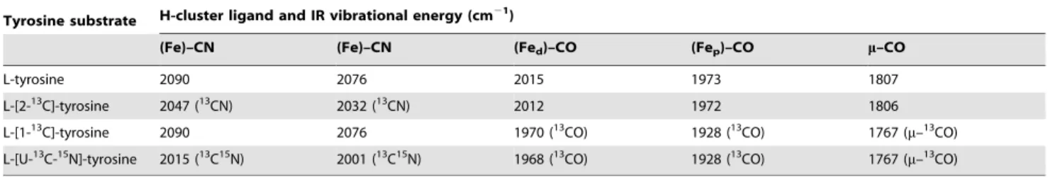
Table 3.Summary of energies of the assigned CO and CN2vibrational modes for the CpI hydrogenase H-cluster with bound exogenous CO.
Tyrosine substrate H-cluster ligand and IR vibrational energy (cm21)
(Fe)–CN (Fe)–CN (Fed)–CO (Fep)–CO m–CO
L-tyrosine 2090 2076 2015 1973 1807
L-[2-13C]-tyrosine 2047 (13CN) 2032 (13CN) 2012 1972 1806
L-[1-13C]-tyrosine 2090 2076 1970 (13CO) 1928 (13CO) 1767 (
m–13CO)
L-[U-13C-15N]-tyrosine 2015 (13C15N) 2001 (13C15N) 1968 (13CO) 1928 (13CO) 1767 (
m–13CO)
The vibrational energies and correspondingn(CN) andn(CO) mode assignments are provided for each Hox–COexocluster from active CpI produced with either natural abundance or isotopically labeled tyrosine. Energies were determined from spectra measured using FTIR spectroscopy (Fig. 4).
In VitroActivation of Active [FeFe] Hydrogenases
Anaerobic reaction mixtures varied from 50mL to 100 mL,
depending on the experiment, and hydrogenase activation proceeded over a 24 hr period. Equivalent CpI activities were observed within this range of volumes. The reaction mixtures included HydElysate, HydFlysate, HydGlysate, exogenous substrates, and CpI apoenzyme. Fe+2
, S22, and DTT were first added to the mixture of maturase lysates. After 30 min, additional small molecule substrates and CpI apoprotein were added. The final concentration for each component was as follows: 20% vol?vol21 HydElysate, 20% vol?vol21 HydFlysate, 20% vol?vol21 HydGlysate, 1 mM Fe+2
, 1 mM S22, 1 mM DTT, 2 mM SAM, 2 mM
L-cysteine, 2 mM tyrosine, 10 mM GTP, 1 mM PLP, 2 mM sodium dithionite, and 0.2 mg?mL21CpI apoenzyme. We estimated the E. colilysates to have 3–15 mg?mL21of each maturase based on SDS-PAGE analysis (Fig. 2). Therefore,in vitroreaction mixtures contained ,10–50mM of HydE (40 kD), HydF (45 kD), and
HydG (54 kD). The purification and concentration of active CpI holoenzyme was carried out as described above for CpI apoenzyme. Solutions of 100–300mM active CpI were analyzed
with FTIR spectroscopy.
Hydrogenase Activity Assays
Both the H2consumption and H2evolution rates for activated
hydrogenase were measured as previously described [23,36], with or without re-purifying the active CpI. H2 uptake rates were
measured with a methyl viologen (MV) reduction assay and calculated using an extinction coefficient of 9.78 mM21?cm21for reduced MV at 578 nm. The assay solution contained 50 mM
Tris/HCl (pH 8.0) and 2 mM MV. The H2 evolution assay
solution included 100 mM MOPS buffer, 100 mM NaCl, 25 mM sodium dithionite, and 6.25 mM MV. H2 production rates at
pH 6.8 and 37uC were quantified by analyzing head space gas samples using a ShinCarbon ST 100/120 mesh column (Resteck) with a Hewlett Packard 6890 gas chromatograph (Hewlett Packard). For precise activity measurements, approximately 1 ng and 10 ng of CpI were tested with the MV reduction and H2
evolution assays, respectively. Background activities (less than 1% of the final activity from mixtures will all components) were measured for mixtures containing all components except the hydrogenase, and the CpI apoenzyme had neither H2production
nor H2oxidation activity.
Fourier Transform Infrared Spectroscopy
Infrared spectra were measured using a Bruker IFS/66s FTIR spectrometer interfaced to a home-built stopped-flow drive system as previously described [38]. The drive system and infrared sample
cuvette were maintained inside an anaerobic glove box
(O2,1.1 ppm) (Belle Technology) at 25uC. A calibrated path length of 47.6mm was used for the sample cuvette. For infrared
spectroscopic measurements, one drive syringe contained the protein sample. Depending on the experiment, the second drive syringe contained one of the following: the same protein sample, the purification elution buffer without protein, elution buffer saturated with exogenous12CO, or elution buffer saturated with exogenous 13CO. Spectra were recorded at 4 cm21 resolution, and an arbitrary background correction was applied. The IR data were processed and analyzed using the Fit_3D software package (SJG, unpublished).
Supporting Information
Figure S1 Shewanella oneidensisHydF protein sequence
based on recombinant expression of the S. oneidensis hydEF open reading frame in Escherichia coli. The underlined peptide sequence corresponds to the residues added to the N-terminus of the previously publishedS. oneidensisHydF peptide sequence (Accession#AAN56901). The amino acids highlighted in black bold font type correspond to the residues identified by Edman degradation and N-terminal sequencing of HydF–Strep-tag II when expressed inE. colistrain BL21(DE3) from the plasmid pACYCDuet-1–hydGX–hydEF–Strep-tag II. The consensus sequences for the GTP binding motif are depicted in green bold font type, which now appear more accurately aligned with sequences of HydF maturases from other organisms [39].
(TIF)
Acknowledgments
The authors wish to thank D. P. Stack, J. A. Stapleton, J. S. Kachian, and A. S. Bingham for insightful discussions.
Author Contributions
Conceived and designed the experiments: JMK SJG SPC JRS. Performed the experiments: JMK SJG CSG. Analyzed the data: JMK SJG CSG. Contributed reagents/materials/analysis tools: JMK SJG. Wrote the paper: JMK SJG JRS.
References
1. Jones AK, Sillery E, Albracht SPJ, Armstrong FA (2002) Direct comparison of the electrocatalytic oxidation of hydrogen by an enzyme and a platinum catalyst. Chem Commun (Camb). pp 866–867.
2. Le Goff A, Artero V, Jousselme B, Tran PD, Guillet N, et al. (2009) From hydrogenases to noble metal-free catalytic nanomaterials for H2 production and uptake. Science 326: 1384–1387.
3. Iwuchukwu I, Vaughn M, Myers N, O’Neill H, Frymier P, et al. (2009) Self-organized photosynthetic nanoparticle for cell-free hydrogen production. Nature nanotechnology 5: 73–79.
4. Zhang Y-HP, Evans BR, Mielenz JR, Hopkins RC, Adams MWW (2007) High-yield hydrogen production from starch and water by a synthetic enzymatic pathway. PLoS ONE 2: e456.
5. Tard C, Pickett CJ (2009) Structural and functional analogues of the active sites of the [Fe]-, [NiFe]-, and [FeFe]-hydrogenases. Chem Rev 109: 2245–2274. 6. Vignais PM, Billoud B (2007) Occurrence, classification, and biological function
of hydrogenases: an overview. Chem Rev 107: 4206–4272.
7. Martin W, Muller M (1998) The hydrogen hypothesis for the first eukaryote. Nature 392: 37–41.
8. Fontecilla-Camps JC, Amara P, Cavazza C, Nicolet Y, Volbeda A (2009) Structure-function relationships of anaerobic gas-processing metalloenzymes. Nature 460: 814–822.
9. Pandey AS, Harris TV, Giles LJ, Peters JW, Szilagyi RK (2008) Dithiomethy-lether as a ligand in the hydrogenase h-cluster. J Am Chem Soc 130: 4533–4540.
10. Ryde U, Greco C, De Gioia L (2010) Quantum refinement of [FeFe] hydrogenase indicates a dithiomethylamine ligand. J Am Chem Soc 132: 4512–4513.
11. Posewitz MC, King PW, Smolinski SL, Zhang L, Seibert M, et al. (2004) Discovery of two novel radical S-adenosylmethionine proteins required for the assembly of an active [Fe] hydrogenase. J Biol Chem 279: 25711–25720. 12. McGlynn SE, Shepard EM, Winslow MA, Naumov AV, Duschene KS, et al.
(2008) HydF as a scaffold protein in [FeFe] hydrogenase H-cluster biosynthesis. FEBS Lett 582: 2183–2187.
13. Shepard EM, McGlynn SE, Bueling AL, Grady-Smith CS, George SJ, et al. (2010) Synthesis of the 2Fe subcluster of the [FeFe]-hydrogenase H cluster on the HydF scaffold. Proc Natl Acad Sci USA 107: 10448–10453.
14. Mulder DW, Boyd ES, Sarma R, Lange RK, Endrizzi JA, et al. (2010) Stepwise [FeFe]-hydrogenase H-cluster assembly revealed in the structure of HydA(Del-taEFG). Nature 465: 248–252.
15. Peters JW, Szilagyi RK, Naumov A, Douglas T (2006) A radical solution for the biosynthesis of the H-cluster of hydrogenase. FEBS Lett 580: 363–367. 16. Driesener RC, Challand MR, McGlynn SE, Shepard EM, Boyd ES, et al. (2010)
[FeFe]-Hydrogenase Cyanide Ligands Derived From S-Adenosylmethionine-Dependent Cleavage of Tyrosine. Angew Chem Int Ed Engl 49: 1687–1690. 17. Nicolet Y, Martin L, Tron C, Fontecilla-Camps JC (2010) A glycyl free radical as
the precursor in the synthesis of carbon monoxide and cyanide by the [FeFe]-hydrogenase maturase HydG. FEBS Lett 584: 4197–4202.
18. Shepard EM, Duffus BR, George SJ, McGlynn SE, Challand MR, et al. (2010) [FeFe]-Hydrogenase Maturation: HydG-Catalyzed Synthesis of Carbon Mon-oxide. J Am Chem Soc 132: 9247–9249.
19. Leonardi R, Roach PL (2004) Thiamine biosynthesis in Escherichia coli: in vitro reconstitution of the thiazole synthase activity. J Biol Chem 279: 17054–17062. 20. Curatti L, Hernandez JA, Igarashi RY, Soboh B, Zhao D, et al. (2007) In vitro synthesis of the iron-molybdenum cofactor of nitrogenase from iron, sulfur, molybdenum, and homocitrate using purified proteins. Proc Natl Acad Sci USA 104: 17626–17631.
21. Curatti L, Ludden PW, Rubio LM (2006) NifB-dependent in vitro synthesis of the iron-molybdenum cofactor of nitrogenase. Proc Natl Acad Sci USA 103: 5297–5301.
22. Zhao D, Curatti L, Rubio LM (2007) Evidence for nifU and nifS participation in the biosynthesis of the iron-molybdenum cofactor of nitrogenase. J Biol Chem 282: 37016–37025.
23. Kuchenreuther JM, Stapleton JA, Swartz JR (2009) Tyrosine, Cysteine, and S-Adenosyl Methionine Stimulate In Vitro [FeFe] Hydrogenase Activation. PLoS ONE 4: e7565.
24. Reissmann S, Hochleitner E, Wang H, Paschos A, Lottspeich F, et al. (2003) Taming of a poison: biosynthesis of the NiFe-hydrogenase cyanide ligands. Science 299: 1067–1070.
25. Czech I, Silakov A, Lubitz W, Happe T (2010) The [FeFe]-hydrogenase maturase HydF from Clostridium acetobutylicum contains a CO and CN-ligated iron cofactor. FEBS Lett 584: 638–642.
26. Kuchenreuther JM, Grady-Smith CS, Bingham AS, George SJ, Cramer SP, et al. (2010) High-yield expression of heterologous [FeFe] hydrogenases in Escherichia coli. PLoS ONE 5: e15491.
27. McGlynn SE, Ruebush SS, Naumov A, Nagy LE, Dubini A, et al. (2007) In vitro activation of [FeFe] hydrogenase: new insights into hydrogenase maturation. J Biol Inorg Chem 12: 443–447.
28. Chen JS In: Schlegel HG, K. Schneider, eds. Hydrogenases: Their Catalytic Activity, Structure and Function. Gottingen: E. Goltze K. G. pp 57–82. 29. Roseboom W, de Lacey AL, Fernandez VM, Hatchikian EC, Albracht SPJ
(2006) The active site of the [FeFe]-hydrogenase from Desulfovibrio
desulfur-icans. II. Redox properties, light sensitivity and CO-ligand exchange as observed by infrared spectroscopy. J Biol Inorg Chem 11: 102–118.
30. Chen Z, Lemon BJ, Huang S, Swartz DJ, Peters JW, et al. (2002) Infrared studies of the CO-inhibited form of the Fe-only hydrogenase from Clostridium pasteurianum I: examination of its light sensitivity at cryogenic temperatures. Biochemistry 41: 2036–2043.
31. Kriek M, Martins F, Challand MR, Croft A, Roach PL (2007) Thiamine biosynthesis in Escherichia coli: identification of the intermediate and by-product derived from tyrosine. Angew Chem Int Ed Engl 46: 9223–9226. 32. Pilet E, Nicolet Y, Mathevon C, Douki T, Fontecilla-Camps JC, et al. (2009)
The role of the maturase HydG in [FeFe]-hydrogenase active site synthesis and assembly. FEBS Lett 583: 506–511.
33. Rubach JK, Brazzolotto X, Gaillard J, Fontecave M (2005) Biochemical characterization of the HydE and HydG iron-only hydrogenase maturation enzymes from Thermatoga maritima. FEBS Lett 579: 5055–5060.
34. Flint DH (1996) Escherichia coli contains a protein that is homologous in function and N-terminal sequence to the protein encoded by the nifS gene of Azotobacter vinelandii and that can participate in the synthesis of the Fe-S cluster of dihydroxy-acid dehydratase. J Biol Chem 271: 16068–16074. 35. Czech I, Stripp S, Sanganas O, Leidel N, Happe T, et al. (2011) The
[FeFe]-hydrogenase maturation protein HydF contains a H-cluster like [4Fe4S]-2Fe site. FEBS Lett 585: 225–230.
36. Boyer ME, Stapleton JA, Kuchenreuther JM, Wang C-W, Swartz JR (2008) Cell-free synthesis and maturation of [FeFe] hydrogenases. Biotechnol Bioeng 99: 59–67.
37. Akhtar MK, Jones PR (2008) Deletion of iscR stimulates recombinant clostridial Fe-Fe hydrogenase activity and H2-accumulation in Escherichia coli BL21(DE3). Appl Microbiol Biotechnol 78: 853–862.
38. Thorneley RNF, George SJ In: Triplett EW, ed. Prokaryotic Nitrogen Fixation: A Model System for Analysis of a Biological Process. Wymondham, UK: Horizon Scientific Press.