RISKS OF USING BEDSIDE TESTS TO VERIFY
NASOGASTRIC TUBE POSITION IN ADULT PATIENTS
*Melody Ni,
1Oliver Priest,
1Lawrence D. Phillips,
2George B. Hanna
11. Department of Surgery and Cancer, St Mary’s Hospital, Imperial College London, London, UK 2. Department of Management, London School of Economics and Political Science, London, UK
*Correspondence to [email protected]
Disclosure: No potential conlict of interest. The work was supported by a grant from the English National Health Service Patient Safety Research Portfolio (PS/048).
Received: 10.03.14 Accepted: 30.07.14 Citation: EMJ Gastroenterol. 2014;3:49-56.
ABSTRACT
Nasogastric (NG) tubes are commonly used for enteral feeding. Complications of feeding tube misplacement include malnutrition, pulmonary aspiration, and even death. We built a Bayesian network (BN) to analyse the risks associated with available bedside tests to verify tube position. Evidence on test validity (sensitivity and speciicity) was retrieved from a systematic review. Likelihood ratios were used to select the best tests for detecting tubes misplaced in the lung or oesophagus. Five bedside tests were analysed including magnetic guidance, aspirate pH, auscultation, aspirate appearance, and capnography/colourimetry. Among these, auscultation and appearance are non-diagnostic towards lung or oesophagus placements. Capnography/ colourimetry can conirm but cannot rule out lung placement. Magnetic guidance can rule out both lung and oesophageal placement. However, as a relatively new technology, further validation studies are needed. The pH test with a cut-of at 5.5 or lower can rule out lung intubation. Lowering the cut-of to 4 not only minimises oesophageal intubation but also provides extra safety as the sensitivity of pH measurement is reduced by feeding, antacid medication, or the use of less accurate pH paper. BN is an efective tool for representing and analysing multi-layered uncertainties in test validity and reliability for the veriication of NG tube position. Aspirate pH with a cut-of of 4 is the safest bedside method to minimise lung and oesophageal misplacement.
Keywords: Decision analysis, Bayesian networks, nasogastric tube, patient safety.
INTRODUCTION
At least one million nasogastric (NG) feeding tubes are purchased by the National Health Service in England each year. Complications of feeding tube misplacement include malnutrition, pulmonary aspiration, and even death. For blind insertion, the rate of respiratory placement is typically 1-3%. Inadvertent tube placement in the oesophagus was
observed in 19 out of 100 blind NG tube insertions.1
Reported rates of tube misplacement on insertion and tube migration after correct initial placement
vary between 1.3% and 50% in adults.2
There is a distinct lack of consensus as to the optimum method of checking the feeding tube position. In response to several deaths directly
related to NG tube misplacement, the National Patient Safety Agency (NPSA) in England issued safety alerts in February 2005 to describe correct procedures for checking the position of feeding
tubes.3,4 However, additional cases of death due
to NG tube misplacement have been reported since the circulation of these alerts. Possible reasons include the use of inappropriate checking procedures or the misinterpretation of radiographs by clinicians. Also documented are the life-threatening complications from enteral formulations or medications entering the lung through a misplaced NG tube, i.e.
‘aspiration-by-proxy’, that did not result in patient harm.5 The
(cut-of 5.5) and the use of chest radiographs
whenever necessary.6
The task to correctly position a blindly inserted NG tube is challenging because none of the bedside procedures can provide deinitive veriication of tube position. Even the current gold standard of a chest radiograph is prone to misinterpretation. We were commissioned by NPSA to review the safety of using bedside methods to verify NG tube position with an emphasis on conirming initial tube placements prior to feeding.
METHODS
A Bayesian network (BN) was constructed to analyse risks in safe feeding. BNs are graphical
tools for reasoning with uncertainties.7-9 They can
be viewed as a special knowledge network that captures one’s beliefs in a risky decision. Each node (circle) represents an uncertain event; each arrow (or edge) represents dependence between two events, and the lack of arrow indicates conditional independence. The structure of a BN relects how we think diferent events relate to each other. The numerical part of a BN in the form of conditional probabilities relects the strength of such dependence. In the case of NG tube positioning, the tube site is the shared parent node of diferent bedside tests. Arrows pointing out from tube site and into the bedside tests indicate our belief that the outcome of these tests depend, among other things, on the location of the feeding tube which could be lung, intestine, stomach, or oesophagus. No arrows, however, link diferent tests together because we believe that the outcome of one test (its indings) does not depend on those of another test (conditional independence). For aspirate pH, feeding and medication history of a patient were modelled as additional parents of the pH test; pH paper was modelled as a child of pH meter (which is a child of tube site). This allows us to examine test results from various combinations of feeding and medication conditions, as well as to test using a less reliable measurement of pH.
We capture the risk of tube misplacement during initial insertion (i.e. before checking) in the prior distribution of the tube sites. Test validity (i.e. sensitivity and speciicity) was incorporated as conditional probability of a inding in each tube site. Once a inding has been entered into the BN, the prior distribution will be updated according to the Bayes’ rule. The result is the posterior
distribution of the tube sites that relects one’s revised belief about the location of the NG tube in light of the inding. We built the BN in software Netica®.
Information on test validity was retrieved from a systematic review in which chest X-rays served as the gold-standard. Multiple sources of information on the same test were combined by a simple weighted average, based on sample size. Missing information was dealt with by assuming a lat distribution where all the indings were assumed to be equally likely. For aspirate pH, raw data were kindly provided by the author (Prof Norma
Metheny).10-16 This enabled a detailed study of
the inluence of antacid medication and feeding status of a patient, as well as measurement methods such as Baxter paper versus pH meter. We generally assume that the tube had 20%, 50%, 20%, and 10% probabilities of being inserted into the lung, stomach, oesophagus, and small bowel,
respectively.1,2 We also tested scenarios of low
initial risks (prior probabilities of lung/stomach/ oesophagus/small bowel =10%/80%/10%/0%) and high initial risks (prior =30%/35%/25%/10%) where tube misplacements were respectively 30% and 65% of the time, compared to 50% of the time, as widely assumed. The likelihood of safe feeding varies with tube site. The consequences of feeding into a misplaced tube in the lung are the most severe, followed by oesophagus, and small bowel. We used likelihood ratios (LRs) to prioritise safety
needs. LR1, LR2, and LR3 measured the capacity
of a test to detect tubes in the lung, oesophagus, and small bowel in contrast to the stomach (the correct tube site).(*)
LR1= p(inding|not lung) /p(inding|lung)
LR2=p(inding|stomach)/p(inding|oesophagus)
LR3=p(inding|stomach)/p(inding|small bowel)
Two types of indings are worth noting. Ininite
LR1, LR2, and LR3 indicate that lung, oesophagus,
and small bowel misplacements are ruled out, in which case feeding can safely start. An example is the inding of a stomach tube using correctly interpreted chest X-rays, the current gold-standard. Secondly, zero LRs would conirm the lack of
safety as the tube is in the lung (LR1) or outside the
stomach (LR2 and LR3). This happens when a lung
together to verify tube site, was simply the product of their respective LRs.
RESULTS
Aspirate pH,10-13,16 appearance,11 auscultation,10,17
capnography/colourimetry,11 and magnetic
guidance18 emerged from our literature review as
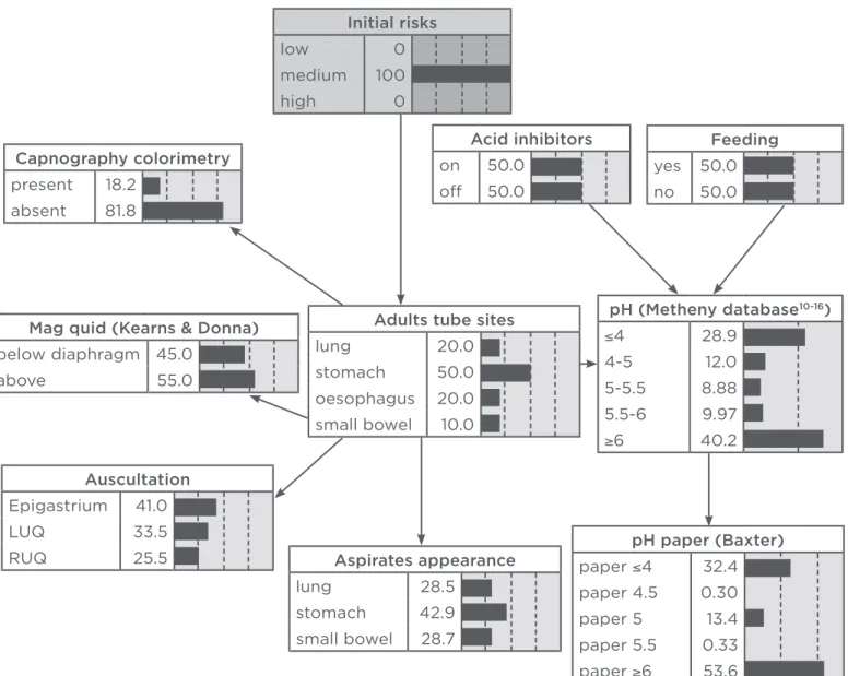
existing or potential bedside tests for locating blindly inserted NG tubes. The BN is shown in
Figure 1. Contained within each bedside test are its indings; next to each inding is the joint (average) probability of observing the inding given the
test validity (Table 1(**)) and the prior probabilities
that the tube is inserted into the lung, stomach, oesophagus, and small bowel, respectively (i.e.
20%, 50%, 20%, and 10%; Figure 1). Note that we
included the discredited auscultation test in our analysis, for two reasons: irstly to provide a check
on the validity of model predictions and secondly, to analyse its potential when used in combination with other tests. For the aspirate pH, we chose 4.0,
5.0, 5.5, and 6.0 as the cut-ofs (indings). Table 1
presents the combined (averaged) sensitivity of each inding of each test. The pH from oesophageal intubation was extrapolated from studies on relux patients, which together demonstrate that the median percentage time with oesophageal pH measured <4.0, is between 0.5-3.1% of recorded
24-hour periods in healthy individuals;19-24 sensitivity
of the pH test above 4 was assumed to be evenly distributed. A lack of high-quality evidence for auscultation test also led us to assume that the loudest sound was equally likely to be heard in epigastrium, left upper quadrant (LUQ), and right upper quadrant (33% in each case) through lung tubes.
Figure 1: The Bayesian Network model for the safe veriication of nasogastric tubes. LUQ/RUQ: left/right upper quadrant.
Initial risks
low 0
medium 100
high 0
Capnography colorimetry
present 18.2 absent 81.8
Mag quid (Kearns & Donna)
below diaphragm 45.0
above 55.0
Auscultation
Epigastrium 41.0
LUQ 33.5
RUQ 25.5 Aspirates appearance
lung 28.5
stomach 42.9 small bowel 28.7
Adults tube sites
lung 20.0
stomach 50.0 oesophagus 20.0 small bowel 10.0
pH paper (Baxter)
paper ≤4 32.4 paper 4.5 0.30 paper 5 13.4 paper 5.5 0.33 paper ≥6 53.6
pH (Metheny database10-16) ≤4 28.9
4-5 12.0
5-5.5 8.88 5.5-6 9.97
≥6 40.2
Acid inhibitors
on 50.0 of 50.0
Feeding
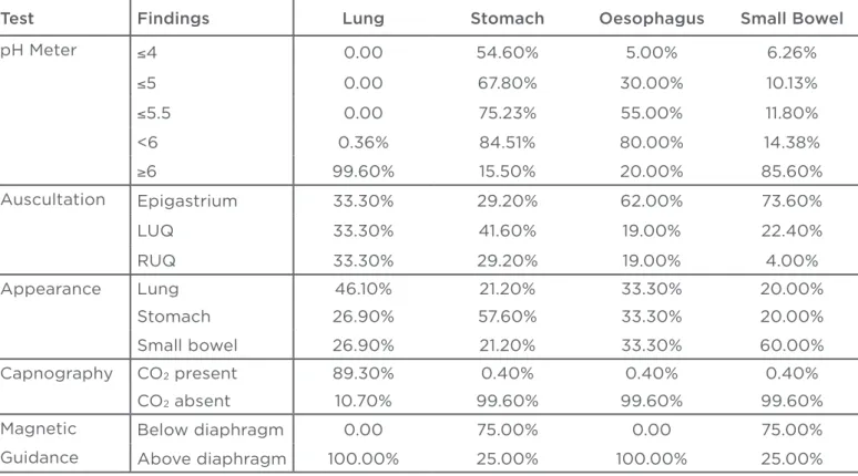
Table 1: Sensitivity of the bedside tests in positioning blindly inserted NG tubes.
Test Findings Lung Stomach Oesophagus Small Bowel
pH Meter ≤4 0.00 54.60% 5.00% 6.26%
≤5 0.00 67.80% 30.00% 10.13%
≤5.5 0.00 75.23% 55.00% 11.80%
<6 0.36% 84.51% 80.00% 14.38%
≥6 99.60% 15.50% 20.00% 85.60%
Auscultation Epigastrium 33.30% 29.20% 62.00% 73.60%
LUQ 33.30% 41.60% 19.00% 22.40%
RUQ 33.30% 29.20% 19.00% 4.00%
Appearance Lung 46.10% 21.20% 33.30% 20.00%
Stomach 26.90% 57.60% 33.30% 20.00%
Small bowel 26.90% 21.20% 33.30% 60.00%
Capnography CO2 present 89.30% 0.40% 0.40% 0.40%
CO2 absent 10.70% 99.60% 99.60% 99.60%
Magnetic Below diaphragm 0.00 75.00% 0.00 75.00%
Guidance Above diaphragm 100.00% 25.00% 100.00% 25.00%
NG: nasogastric; LUQ/RUQ: left/right upper quadrant.
Table 2: The efectiveness of the bedside tests to rule out lung and oesophagus (ininite LRs).
Test Findings LR1 LR2 LR3
pH ≤4 Ininite 10.92 8.72
≤5 Ininite 2.26 6.69
≤5.5 Ininite 1.37 6.38
<6 207.22 1.06 5.88
≥6 0.26 1.29 0.18
Auscultation epigastrium 1.29 0.47 0.40
LUQ 1.01 2.19 1.86
RUQ 0.71 1.54 7.30
Appearance lung 0.52 0.64 1.06
stomach 1.74 1.73 2.88
small bowel 1.08 0.64 0.35
Capnography CO2 present 0.004 1.00 1.00
CO2 absent 9.31 1.00 1.00
Magnetic guidance below diaphragm Ininite Ininite 1.00
above diaphragm 0.44 0.25 1.00
LRs: likelihood ratios; LR1: p(inding|not lung)/p(inding|lung); LR2: p(inding|stomach)/
p(inding|oesophagus); LR3: p(inding|stomach)/p(inding|small bowel); LUQ/RUQ: left/right
Table 2(**) presents the LRs based on test validity
(Table 1) and the prior. A quick scan of Table 2
shows that the best tests to detect lung intubation,
as indicated by an ininite LR1, are pH (5.5 or lower)
or magnetic guidance (below diaphragm), and the best tests to detect oesophageal intubation, as
indicated by an ininite LR2, are magnetic guidance
(below diaphragm), followed by aspirate pH with a cut-of at 4. The latter can reduce the chance of oesophagus placement relative to
stomach placement by nearly 10-fold (LR2=10.92).
In contrast, the chance of oesophagus placement would barely change when a pH of 5.5 or less is
observed (LR2=1.37).
Using the BN model in Netica, a pH of 5.5 or less would predict the probabilities of lung, stomach, oesophagus, and small bowel placements are 0%, 75.5%, 22.1%, and 2.37% respectively, in contrast to the initial 20%, 50%, 20%, and 10%. A pH of 4.0 or less would predict the probabilities of lung, stomach, oesophagus, and small bowel placements are 0%, 94.4%, 3.46%, and 2.16%, respectively. That is, a pH at 4 or less reduces the risk of oesophageal intubation from 20% to 3.46%, i.e. from fairly uncertain to ‘beyond reasonable doubt’.
Auscultation and appearance are not useful on their own as their LRs clustered around 1. The only inding useful in terms of lung and oesophagus intubations is the auscultation test which found
the loudest sound heard in the LUQ (LR2=2.19).
This would halve the chance of oesophageal placement (from 20% to 11.3%) relative to stomach placement (from 50-62.1%). Using capnography
or colourimetry, detecting CO2 would increase the
chance of lung tubes from 20-98% (LR1=0.004).
However, the absence of CO2 cannot be taken as
deinitive evidence that the tube is outside the
lung (LR1 =9.31), and the revised belief of lung
placement is 2.62%.
Feeding, Antacid Medication, and Measurement
Technique Efects on pH Test
Recent feeding, administration of antacid therapy, or using pH paper instead of meter to measure pH, all reduce sensitivity of the pH test. Given a pH of 5.5 or less, receiving antacids would increase the chance of oesophagus placement from 22.1-23.6%, though feeding had little impact. Given the inding of a pH of 4.0 or less, receiving antacids would increase the chance of oesophagus placement from 3.46-4.05% and further to 4.20% if the patient has recently been fed. If Baxter paper is used instead
of pH, the reading of 4 or less would predict the probabilities of stomach, oesophagus, and small bowel placements to be 89.2%, 8.49%, and 2.30%, whereas a reading of 5.5 or less would predict the probabilities of lung, stomach, oesophagus, and small bowel placements to be 0.024%, 78%, 19.6%, and 2.41%.
Impact of Low or High Initial Risks on pH
If a pH of 4 or less was observed, the predicted probabilities of lung, stomach, oesophagus, and small bowel placements were respectively 0%, 98.9%, 1.13%; 0% under low level of initial risks; and 0%, 91.1%, 5.96%, and 2.98% under high level of initial risks. If a pH of 5.5 or less was observed, the chances of lung, stomach, oesophagus, and small bowel placements were 0%, 91.6%, 8.37%, and 0% likely under low level of initial risks and 0%, 63.8%, 33.3%, and 2.86% likely under high level of initial risks.
Assume a worst case scenario where the initial insertions have a high risk of misplacements and the veriication is done by Baxter paper instead of pH meter. A inding of a pH of 5.5 or less would predict lung, oesophagus, and small bowel misplacements to be 0%, 54.6%, and 2.03% respectively, whereas a pH of 4 or less would predict the probabilities of lung, oesophagus, and small bowel misplacements to be 0%, 8.49%, and 2.30%, respectively. That is, if a patient is fed after a pH of 5.5 or less is observed in the worst case scenario, then half of the time the feeding would be in the oesophagus instead of the stomach.
DISCUSSION
Five bedside tests were investigated, i.e. magnetic guidance, aspirate pH (with cut-ofs 4, 5, 5.5, and 6), auscultation, aspirate appearance, and capnography/colourimetry. Consistent with the existing literature and the recommendation of NPSA, neither auscultation nor aspirate appearance can be recommended for use on their own to detect tube misplacements in the lung or oesophagus. It is worth noting that if capnography/colourimetry
is used, the absence of CO2 cannot be taken as
evidence for safe feeding (outside the lung) because such indings are observed in 10.7% of the lung
placements (Table 1). The safest tests are magnetic
sites, whereas a pH test with cut-ofs at 5.5 or lower can rule out lung misplacements. Further lowering the cut-of to 4 or less would minimise oesophagus misplacements. Magnetic guidance is a relatively new technology and has a relatively small, though growing, evidence base (n=243).
Magnetic guidance has been studied in adults and children, particularly in the context of post-pyloric
feeding with retrospective studies.25-27 More recent
prospective studies indicate encouraging accuracy
of the technique28 but there are additional costs
of technical equipment and devices required for
every tube placement29 and the process does not
eliminate the risk of adverse soft tissue injury.30
Further validation studies are needed.
The pH test of tube aspirates is widely used, well-studied, and has an established evidence base (nearly 800 cases in our database). Current practice also recommends the use of aspirate pH, though
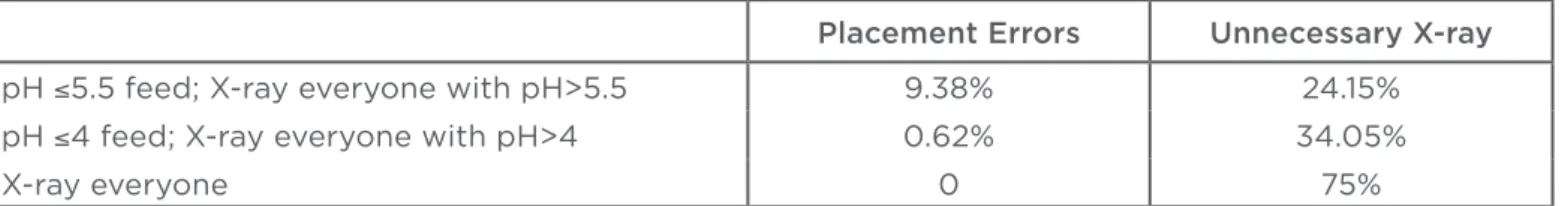
with a cut-of of 5.5. Our analysis shows lowering the pH cut-of from 5.5 to 4.0 can enhance safety in oesophageal intubations. Furthermore, the use of Baxter paper, feeding, medication history of a patient, and potential variations in the risks in the initial insertions of the tube, means a lower pH would provide an extra layer of safety for reducing oesophageal feeding. Lowering the pH threshold would result in more patients with tubes correctly placed in the stomach to be sent for X-rays (unnecessary X-rays). It is therefore a trade-of that needs careful assessment: minimising placement errors (mainly in the oesophagus) versus minimising unnecessary X-rays. Consider three strategies in
Table 3, i.e. X-ray all patients, X-ray only patients with pH higher than 5.5, or X-ray only patients with pH higher than 4. Under the assumption of 50% initial insertion errors, adopting a pH with a cut-of of 4 would reduce placement errors from 9.38% to 0.62% whilst increasing unnecessary X-rays from
24.15-34.05% (Table 3).
Table 3: Outcomes of clinical guidelines.
Placement Errors Unnecessary X-ray
pH ≤5.5 feed; X-ray everyone with pH>5.5 9.38% 24.15%
pH ≤4 feed; X-ray everyone with pH>4 0.62% 34.05%
X-ray everyone 0 75%
One criticism of our recommendation of lowering the pH cut-of is that X-ray facilities are not widely available and therefore lowering the pH may lead to feeding delays and potential harm from lack of
nutrition.6 Another criticism surrounds the liability
of chest radiographs to be misinterpreted. Reducing the pH cut-of used for tube aspirate pH testing may expose patients to a risk of inadvertent feeding if the consequent increase in radiographs to check tube position is associated with an accompanying increase in X-ray misinterpretation. This is debatable as misinterpretation of radiographs afects a cohort of patients with a tube aspirate pH between 4.0 and 5.5. Using the current guideline with a higher pH cut-of (5.5), all of these patients will be fed through the tube regardless of the actual tube site. Given a constant rate of tube misplacement, it is not possible to increase the number of inadvertent feeding errors using a lower pH cut-of, regardless of the risk of X-ray misinterpretation.
In terms of using multiple tests instead of a single test, consider safety needs to rule out lung and oesophagus placements. Magnetic guidance can achieve both ends on its own; the best test to be used with aspirate pH is one that is sensitive to oesophageal misplacement. Auscultation has the potential to halve the chance of oesophagus placement, but the method is subject to interpretation errors and is therefore unreliable.
CONCLUSIONS
Acknowledgements
We are most grateful to members of the clinical consortium (alphabetically: Peter Chow, Annemarie Knight, Richard Leonard, Sandra McLellan, William Oldield, Wendy Slack, Paris Tekkis, Gillian Wheatley) and steering group (alphabetically: Lynne Colagiovanni, Pauline Fellows, Jamil Khair, Gill Lazonby, Alison O’Donnell, Jef Perring, Kate Pickering, Peter Turner) panel of experts for their contribution to the project.
Their participation in decision conferences and feedback on the proposed safety guidelines has been invaluable. We are indebted to the National Patient Safety Agency (alphabetically: Patricia Bain, Kevin Cleary, Frances Healey, Caroline Lecko, John Scarpello, Elaine Stevenson) for coordinating the steering group and providing specialist advice at every stage. We also thank Prof Norma Metheny for generously sharing with us her pH data that proved instrumental to this research, and also Prof Allan Hutchinson and
Mr Hugh Mackenzie for their insightful comments to the previous versions of this paper.
REFERENCES
1. Benya R et al. Flexible nasogastric feeding tube tip malposition immediately after placement. JPEN J Parenter Enteral Nutr. 1990;14(1):108-9.
2. Ellett ML. What is the prevalence of feeding tube placement errors and what are the associated risk factors? Online J Knowl Synth Nurs. 1997;4:5.
3. NHS National Patient Safety Agency, Patient safety alert 05: Reducing the harm caused by misplaced nasogastric feeding tubes. 2005.
4. NHS National Patient Safety Agency. Reducing the harm caused by misplaced nasogastric feeding tubes: interim advice for healthcare staf - February 2005. 5. NPSA. Incidents in contravention to NG tube alert. National Patient Safety Agency. 2008.
6. Lamont T et al. Checking placement of nasogastric feeding tubes in adults (interpretation of x ray images): summary of a safety report from the National Patient Safety Agency. BMJ. 2011;342:d2586. 7. Shachter RD. Evaluating Inluence Diagrams. Oper Res. 1986;34(6):871-82. 8. Pearl J, Probabilistic reasoning in intelligent systems: networks of plausible inference (1988), San Mateo, California: Morgan Kaufmann Publishers.
9. Lauritzen SL, Spiegelhalter DJ. Local computations with probabilities on graphical structures and their application to expert systems. J Roy Stat Soc B. 1988;50(2):157-224.
10. Metheny N et al. Efectiveness of the auscultatory method in predicting feeding tube location. Nurs Res. 1990;39(5):262-7.
11. Metheny N et al. Visual characteristics of aspirates from feeding tubes as a method for predicting tube location. Nurs Res. 1994;43(5):282-7.
12. Metheny N et al. Efectiveness of pH measurements in predicting feeding tube placement: an update. Nurs Res. 1993;42(6):324-31.
13. Metheny N et al. Testing feeding tube placement. Auscultation vs. pH method. Am J Nurs. 1998;98(5):37-42; quiz 42-3. 14. Metheny NA et al. pH testing of feeding-tube aspirates to determine placement. Nutr Clin Pract. 1994;9(5): 185-90.
15. Metheny NA et al. Development of a reliable and valid bedside test for bilirubin and its utility for improving prediction of feeding tube location. Nurs Res.
Footnotes
(*) The conditional probability of a negative inding (e.g. not lung) is the joint probability of the inding and not lung divided by the prior probability of not lung: p(inding|not lung) = (p[inding & stomach]+ p[indings & oesophagus]+p[indings & small bowel])/(p[stomach]+p[oesophagus]+p[small bowel]), where p(inding & tube site) = p(inding|tube site)*p(tube site).
(**) The pH indings were deined diferently in the BN than in Tables 1 and 2. The BN software
(Netica) rendered it impossible to present pH values in terms of cut-ofs, e.g. lower or higher than 5.5.
We therefore expressed the pH as discrete categories, i.e. ≤4, between 4 and 5, between 5 and 5.5,
between 5.5 and 6, and ≥6. In Tables 1 and 2 the probabilities were binary based on the pH cut-of which
is how the test is used in reality. These two methods of expression were consistent. p(pH≤cut-of) is the
sum of the discrete probabilities accumulated until that cut-of and the p(pH>cut-of) = 1-p(pH≤cut-of).
incidents of pneumothorax among 187 tube misplacements with an associated increase in
mortality.31 Techniques to improve the accuracy
of feeding tube insertion such as electromagnetic guidance may prevent both pulmonary tree injury and the consequences of tube malposition. Considering the strength and reliability of available
2000;49(6):302-9.
16. Metheny NA, Titler MG. Assessing placement of feeding tubes. Am J Nurs. 2001;101(5):36-45.
17. Neumann MJ et al. Hold that X-ray: aspirate pH and auscultation prove enteral tube placement. J Clin Gastroenterol. 1995;20(4):293-5.
18. Kearns PJ, Donna C. A controlled comparison of traditional feeding tube veriication methods to a bedside, electromagnetic technique. J Parenter Enteral Nutr. 2001;25(4):210-5.
19. Richter JE. Ambulatory esophageal pH monitoring. Am J Med. 1997;103(5A): 130S-4S.
20. Fass R et al. Age- and gender-related diferences in 24-hour esophageal pH monitoring of normal subjects. Dig Dis Sci. 1993;38(10):1926-8.
21. Freedman J et al. Ambulatory combined pH, bile and manometric monitoring of the oesophagus in asymptomatic healthy volunteers. Clin Physiol Funct Imaging.
2004;24(6):368-73.
22. Shay S et al. Twenty-four hour ambulatory simultaneous impedance and pH monitoring: a multicenter report of normal values from 60 healthy volunteers. Am J Gastroenterol. 2004;99(6):1037-43. 23. Zentilin P et al. Normal values of 24-h ambulatory intraluminal impedance combined with pH-metry in subjects eating a Mediterranean diet. Dig Liver Dis. 2006;38(4):226-32.
24. Fackler WK et al. Ambulatory gastric pH monitoring: proper probe placement and normal values. Aliment Pharmacol Ther. 2001;15(8):1155-62.
25. Koopmann MC et al. A team-based protocol and electromagnetic technology eliminate feeding tube placement complications. Ann Surg. 2011;253(2): 287-302.
26. October TW, Hardart GE. Successful placement of postpyloric enteral tubes using electromagnetic guidance in critically ill children. Pediatric Critical
Care Medicine. 2009;10(2):196-200. 27. Windle EM et al. Implementation of an electromagnetic imaging system to facilitate nasogastric and post-pyloric feeding tube placement in patients with and without critical illness. J Hum Nutr Diet. 2010;23(1):61-8.
28. Taylor S et al. Conirming nasogastric tube position with electromagnetic tracking versus pH or X-ray and tube radio-opacity. Br J Nurs. 2014;23(7):352, 354-8.
29. Krenitsky J. Blind Bedside Placement of Feeding Tubes: Treatment or Threat? Practical Gastroenterology. 2011;35:11. 30. Khasawneh FA et al. Nasopharyngeal perforation by a new electromagnetically visualised enteral feeding tube. BMJ Case Rep. 2013;2013.