Temporary carotid shunt embolism for popliteal artery
Embolia de shunt carotídeo temporário para a artéria poplítea
Leonardo Pires de Sá Nóbrega1,Alcides José Araújo Ribeiro1,Cláudio Eluan Kalume2
Introduction
The occurence of intravascular foreign bodies (FB) is rare. Most cases of embolization occur in the deep ve-nous system and the FB usually migrates to structures such as the superior vena cava, right ventricle, pulmo-nary artery or a combination of such structures1. FBs are
less frequently found in the aorta or any of its branches; however, the higher frequency of percutaneous endovas-cular procedures has caused an increase in the number of cases of intra-arterial FB cases, most frequently stents, catheters and fragments of guide wire2. The displacement
of a temporary shunt to a more distal arterial branch is a rare complication3. This case report describes the
au-thors’ experience with the use of arterial Doppler ultra-sonography to find a segment of nasogastric tube, used as a temporary carotid stunt, that had migrated to the left popliteal artery.
Case report
A male 36-year-old patient, victim of multiple self-inlicted stab wounds, was evaluated and treated by a gen-eral surgery team in the Emergency Room of a secondary care hospital. Penetrating wounds injuries were diagnosed in the abdomen, chest, and let cervical region. Initial care consisted of chest tube drainage, laparotomy and let neck exploration.
Laringeal and let common carotid artery (LCCA) in-juries were found at surgical exploration of the neck.. he general surgeon, ater dissecting the LCCA proximal and distal stumps, placed a temporary carotid shunt made of a 8.5 cm long segment of a #16 nasogastric tube, that was se-cured in place with silk sutures around the proximal and distal carotid stumps. his way, vessel patency was satis-factorily maintained. he laryngeal injury was treated with simple sutures and tracheotomy.
Abstract
he displacement of a shunt to a more distal arterial branch, along a path, is a rare complication. Vascular ultrasound can be presented as an excellent diagnostic modality for identifying the site of embolization of intravascular foreign bodies. he authors report a rare case of temporary shunt migration, implanted in the left common carotid artery, to left popliteal artery. he authors also describe the use of arterial vascular ultrasound to ind the point of embolization, with an exact dermatography, that was essential to an easily surgical approach through a restrict and precise access way.
Keywords: carotid common artery; popliteal artery; ultrasonography doppler color.
Resumo
O deslocamento de um shunt para um ramo arterial mais distal, percorrendo um longo trajeto, é uma complicação rara. A ultrassonograia vascular pode se apresentar como uma excelente modalidade diagnóstica para identiicar o sítio de embolização de corpos estranhos intravasculares. Os autores relatam um caso raro de migração de um shunt temporário, implantado na carótida comum esquerda, para artéria poplítea esquerda. Descrevem, ainda, a utilização do ecocolor doppler arterial que, além de localizar o ponto de embolização, com uma dermatograia exata, foi fundamental para que a abordagem cirúrgica transcorresse com facilidade através de uma via de acesso restrita e precisa.
Palavras-chave: artéria carótida primitiva; artéria poplítea; ultrassonograia doppler em cores.
Study carried out at the Hospital de Base do Distrito Federal (HBDF) – Brasília (DF), Brazil.
1Staf Surgeon at the Angiology and Vascular Surgery Service, HBDF and Clínica de Veias – Brasília (DF), Brazil. 2Chief of the the Angiology and Vascular Surgery Service, HBDF – Brasília (DF), Brazil.
eration, it was concluded that interposition of a grat was not required, as a primary repair was possible by doing an end-to-end anastomosis between the LCCA stumps. However, at the moment of removing the ixation of the carotid shunt, it was inadvertently released into the aortic arch. At the end of carotid repair, the patient was stable, with palpable pulses in all limbs.
In order to identify the site to where the tempo-rary shunt had embolized, abdominal, pelvic and lower limb vascular ultrasound scans were performed, us-ing a Aplio® duplex scanner (Toshiba America Medical Systems, Inc. Tustin, California, USA), which em-ployed multi-frequency linear transducers of 6-11 and 6.2-12.0 MHz. The foreign body (FB) was located in the left superficial femoral artery (SFA). At the exam, an initial assessment using mode B (Figure 1), it was pos-sible to detect not only the FB inside the left SFA, but also the acoustic shadow it cast along its length.. After that, using the power doppler (Figure 2), it was possible to identify blood flowing through the FB, confirming its patency.
As the surgical intervention to remove the FB was possible only hours later, another Doppler ultrasound exam was performed with the objective of marking its exact location and making the procedure easier. The second exam, performed with a 1.9-6.0 MHz multi-frequency convex transducer, showed that the FB had migrated to the left proximal popliteal artery. (Figures 3 and 4).
Thus, with the mark made precisely on the skin of the lower thigh (Figure 5), the surgeons made a relative-ly small incision and, through a medial access, did not have any problem to dissect the proximal third of the left popliteal artery. The FB was removed from the arterial lumen, via transverse arteriotomy (Figures 6, 7 and 8). Fogarty® catheters nº 3 and 4 (Edwards Lifesciences Macchi, São Paulo, SP, Brazil) were used to remove fresh thrombi from the popliteal artery, followed by proximal and distal instillation of a saline solution with heparin. After completion of the procedure, the patient had pal-pable distal pulses in the left lower limb. The patient was transferred back to the General Surgery ward 24 hours later, to continue treatment. He was placed in antico-agulation therapy and had no neurologic sequelae at the time of discharge from the hospital.
Figure 1. Mode B: foreign body into the left supericial femoral artery (note the posterior acoustic shadowing).
Figure 3. Mode B: distal end of the foreign body into the left popliteal artery.
Figure 5. Dermatography showing the foreign body (CE, in the illustra-tion) migration from the left supericial femoral artery (AFSE, in the illus-tration) to the ipsilateral popliteal artery (APOPE, in the illusillus-tration).
Figure 6. Transverse arteriotomy of the left popliteal artery with the nasogastric probe in its lumen (note that the incision was compatible with the preoperative marks on the skin).
Figure 7. Removal of the foreign body from inside the left popliteal artery lumen.
Figure 8. Foreign body: nasogastric probe segment.
Discussion
Intravascular shunts are artificial conduits intro-duced into the lumen of an injured blood vessel to tem-porarily restore flow, until the definitive reconstruction is feasible. It can be regarded as a damage control tech-nique. In this case report, the general surgery team elect-ed to use a 8.5 cm long segment of #16 nasogastric tube. The shunt material selection is a question of individual preference, as any sterile, smooth, rigid synthetic tube of proper caliber can be used as a temporary shunt3.
Temporary intravascular shunts main complica-tions are thrombosis and displacement. The former is more frequent and can be a natural consequence of the procedure, if the temporary shunt is not removed Figure 4. Mode B + power doppler: distal end of the foreign body into
Temporary Shunt
Foreign Body - Left Superficial Femoral Artery
Foreign Body – Left Popliteal Artery
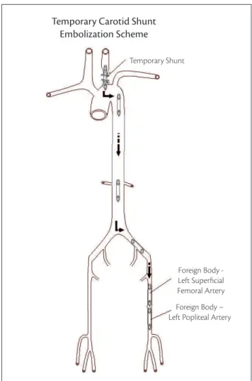
Figure 9. Scheme showing the long path of the temporary shunt.
cal literature3.
Embolization within the vascular system depends on several factors, such as: point of entry, the effects of gravity and patient’s position at the moment of the accident. A FB end location depends upon its size and rigidity, as well as the flow patterns in vessels through it travels (Figure 9). FB migrations to remote locations are rare in the medical literature1.
The clinical picture of arterial FB embolization de-pends on its location and the acute manifestations are well known to any physician: pain, paresthesia, cold-ness, paleness and no pulses distally to the occlusion site. Those findings were not present in this case, as the FB was a tube with a patent lumen, as documented by Doppler ultrasonography, thus rendering the patient asymptomatic4.
As there was no clinical indication of the FB loca-tion, a thorough scanning with arterial vascular ultra-sound was recommended, a method that has already been used in some situations to detect intra-arterial FBs, especially in studies analyzing stents that had migrated from their original sites5-7. In the specialized medical
literature consulted by the authors, no case report was found in which abdominal, pelvic and lower limb scan-ning was used to detect a temporary shunt that had migrated to a more distal artery. Arteriography would be a good option in case the FB could not be found by ecocolor Doppler ultrasonography., Although the FB is radiotransparent, it could have been identified from indirect signs, but involving higher cost and morbidity associated with the procedure1,2.
FBs from the thoracic or abdominal aorta usually migrate to arteries of the lower limbs. The left lower limb is affected three times more often than the right, which might be explained as the result of the left com-mon iliac artery coming out of the central axis of the aorta at a more acute angle (30º) than the right com-mon iliac artery (45º)8,9.
As the FB initially migrated to the LSFA, its location was easily determined by color Doppler ultrasound of lower limbs, which used linear transducers. The identi-fication of flow through the FB, with the power Doppler, confirmed the LSFA patency, justifying palpable pulses in the left dorsalis pedis and posterior tibial arteries.
As there was no possibility of prompt surgical inter-vention, a new Doppler ultrasonogram was extremely
opportune, as it showed the FB had migrated to the popliteal artery. In the second exam, a technical prob-lem occurred during the proximal popliteal artery iden-tification, due to thick thigh musculature, which was overcome with the utilization of a 1.9–6.0 MHz multi-frequency convex transducer. The precise marking of the FB location was essential for an easy surgical approach, without tne need to enlarge the incision. Percutaneous removal of the FB would not not have been possible due to the unavailability of materials that could be used to retrieve it and the FB size and caliber2,3.
References
1. Ulacker R, Lima S, Melichar AC. Intravascular foreign bodies: per-cutaneous retrieval. Radiology. 1986;160(3):731-5.
2. Małecka B, Kutarski A, Zabek A, et al. Percutaneous removal of endocardial implantable cardioverter-deibrillator lead displaced to the right pulmonary artery. Cardiol J. 2010;17(3):293-8.
3. Hirshberg A, Scott BG. Controle de danos no trauma vascular. In: Rich NM, Mattox KL, Hirshberg A, editores. Trauma Vascular. Rio de Janeiro: DiLivros. 2ª ed. 2006. p. 184-5.
4. Iyisoy A, Kursaklioglu H, Celik T, et al. Coronary stent embolization into right common femoral artery - the role of computed tomog-raphy angiogtomog-raphy. Clin Cardiol. 2009 Jul;32(7):E9.
5. Ashar RM, Huettl EA, Halligan R. Percutaneous retrieval of a Wallstent from the pulmonary artery following stent migration from the iliac vein. J Interv Cardiol. 2002;15(2):101-6.
6. Wijesinghe LD, Coughlin PA, Gill K. An unusual cause of femoral embolus. Cardiovasc Surg. 2000;8(4):287-8.
7. Prabhudesai A, Khan MZ. An unusual cause of femoral embolism: angioseal. Ann R Coll Surg Engl. 2000;82(5):355-6.
8. Patel KR, Cortes LE, Semel L, Sharma PV, Clauss RH. Bullet embo-lism. J Cardiovasc Surg (Torino). 1989;30(4):584-90.
9. Burkitt DS, Dhasmana JP, Mortensen NJ, Wisheart JD. ‘Bullet embo-lism’ to the popliteal artery following air rile injury of the thoracic aorta. Br J Surg. 1984;71(1):61.
Correspondence Leonardo Pires de Sá Nóbrega SEPS 715/915 – Conjunto A – Bloco D – Ed. Pacini, salas 317 e 318 – Asa Sul CEP 70390-155 – Brasília (DF), Brazil E-mail: [email protected]
Author´s contributions Conception and design: LPSN Analysis and interpretation: LPSN Data collection: LPSN Writing the article: LPSN Critical revision of the article: AJAR Final approval of the article*: LPSN, AJAR, CEK