Utility of Serum miR-125b as a Diagnostic
and Prognostic Indicator and Its Alliance with
a Panel of Tumor Suppressor Genes in
Epithelial Ovarian Cancer
Mariyam Zuberi1☯, Imran Khan2☯, Rashid Mir3‡, Gauri Gandhi4‡, Prakash Chandra Ray1‡, Alpana Saxena1☯*
1Department of Biochemistry, Maulana Azad Medical College and Associated Hospitals, New Delhi, India, 2Department of Surgery, University of Illinois at Chicago, Chicago, Illinois, United States of America, 3Prince Fahd Bin Sultan Research Chair, Faculty of Applied Medical Sciences, University of Tabuk, Saudi Arabia, Tabuk-71491,4Department of Gynaecology and Obstetrics, Lok Nayak Hospital, New Delhi, India
☯These authors contributed equally to this work.
‡These authors also contributed equally to this work. *[email protected]
Abstract
MicroRNAs (miRNAs) have been found to be dysregulated in epithelial ovarian cancer (EOC) and may function as either tumor suppressor genes (TSGs) or as oncogenes. Hyper-methylation of miRNA silences the tumour suppressive function of a miRNA or hypermethy-lation of a TSG regulating that miRNA (or vice versa) leads to its loss of function. The present study aims to evaluate the impact of aberrant microRNA-125b (miR-125b) expres-sion on various clinicopathological features in epithelial ovarian cancer and its association with anomalous methylation of several TSGs. We enrolled 70 newly diagnosed cases of epithelial ovarian cancer, recorded their clinical history and 70 healthy female volunteers. Serum miR-125b levels were determined by quantitative reverse transcription polymerase chain reaction (qRT-PCR) and the methylation status of various TSGs was investigated by methylation specific PCR. ROC curves were constructed to estimate the diagnostic and prognostic usefulness of miR-125b. The Kaplan—Meier method was applied to compare survival curves. Expression of miR-125b was found to be significantly upregulated (p<0.0001) in comparison with healthy controls. The expression level of miR-125b was found to be significantly associated with FIGO stage, lymph node and distant metastasis. ROC curve for diagnostic potential yielded significant AUC with an equitable sensitivity and specificity. ROC curves for prognosis yielded significant AUCs for histological grade, distal metastasis, lymph node status and survival. The expression of miR-125b also correlated significantly with the hypermethylation of TSGs. Our results indicate that DNA hypermethy-lation may be involved in the inactivation of miR-125b and miR-125b may function as a potential independent biomarker for clinical outcome in EOC.
a11111
OPEN ACCESS
Citation:Zuberi M, Khan I, Mir R, Gandhi G, Ray PC, Saxena A (2016) Utility of Serum miR-125b as a Diagnostic and Prognostic Indicator and Its Alliance with a Panel of Tumor Suppressor Genes in Epithelial Ovarian Cancer. PLoS ONE 11(4): e0153902. doi:10.1371/journal.pone.0153902
Editor:Ratna B. Ray, SAINT LOUIS UNIVERSITY, UNITED STATES
Received:February 4, 2016
Accepted:April 5, 2016
Published:April 19, 2016
Copyright:© 2016 Zuberi et al. This is an open access article distributed under the terms of the Creative Commons Attribution License, which permits unrestricted use, distribution, and reproduction in any medium, provided the original author and source are credited.
Data Availability Statement:All relevant data has been included within the paper and its Supporting Information files.
Funding:The authors received no specific funding for this work.
Introduction
Ovarian cancer is the leading cause of cancer related deaths worldwide. The high rate of mortal-ity may be due to difficulties in diagnosing it at an early stage and lack of effective treatments for patients with an advanced or recurrent disease[1]. Therefore, there is a critical prerequisite for developing prognostic and predictive markers to detect it early and to help optimize treatment.
MicroRNAs (miRNAs) are a class of small non-coding RNA molecules that could induce post-transcriptional silencing of target genes by interacting with their 30-UTR (untranslated
region), thus regulating many important biological processes such as cell development, differ-entiation and apoptosis[2]. Taking into account the important regulatory roles miRNA play in cancer development and progression, several studies have been conducted to identify miRNA as potential biomarkers for cancer diagnosis, prognosis and personalized therapy[3].
Aberrant miRNA expression has been frequently observed in various types of human tumors. Studies suggest that miRNAs may function as either tumor-suppressor genes (TSGs) or oncogenes[4]. Recent studies reported dysregulation of miR-125b in various cancers[5,6]. However, the mechanism by which miR-125b contributes to epithelial ovarian cancer (EOC) has not been documented and is still relatively unclear. High expression of miR-125 had been observed in gliomas and prostate cancer[7,8] while miR-125 levels were seen to decrease in breast and gastric cancer[9,10]. To date, the expression level of miR-125b in epithelial ovarian cancer is not clear yet. As per the putative promoter region, the promoter of miR-125b is embedded in CpG island[11].
Considering the above findings, we tested the expression of miR-125b in serum of epithelial ovarian cancer patients to elucidate its utility as a diagnostic/prognostic marker. We also ana-lysed the methylation pattern in a panel of tumour suppressor genes such as DAPK1, p16, RASSF1A, PTEN, BRCA1 and p14 and explored its association with the aberrant expression of miR-125b.
Materials and Methods
Patient Samples
Blood samples were procured from clinically diagnosed epithelial ovarian cancer patients. The patients (n = 70) were recruited from the Department of Gynaecology and Obstetrics, Lok Nayak Hospital, New Delhi between June 2012 and October 2014. Equal number (n = 70) of female volunteers (age matched) served as controls. Serum was separated by centrifugation. All samples were collected and stored at -70°C and thawed immediately before assay. Complete clinical data and current therapy plan was obtained. The purpose of the sampling was explained to all patients and written informed consent was obtained prior to enrolment. The study was approved by the Institutional Ethics Committee at Maulana Azad Medical College, New Delhi, India.
DNA Extraction
Genomic DNA was isolated from blood specimens of patients and controls by using the Gen-eaid DNA Isolation Kit (Taiwan) in accordance with the manufacturer's instructions. The qual-ity and integrqual-ity of DNA from these tissues were checked by electrophoresis on 1% agarose gel, quantified spectrophotometrically, and then stored at−20°C for further use.
Bisulfite Modification of DNA
cytosines to uracil nucleotides but does not modify methylated cytosine nucleotides. Bisulphite conversion of DNA was carried out at 95°C for 20 minutes during which unmethylated cyto-sines were converted into uracil completely. This was followed by the converted DNA clean-up and storage at -80°C prior to use.
Methylation-specific PCR
Methylation-specific PCR (MSP) is sensitive and specific for methylation of virtually any block of CpG sites within a CpG island. The frequency of CpG sites in CpG islands renders this tech-nique useful and extremely sensitive for such regions. Bisulfite treated DNA was amplified with specific PCR primers (S1 Table) that distinguish methylated and unmethylated DNA. These primers amplify different sized products for DAPK1, p16, RASSF1A, PTEN, BRCA1 and p14 (for methylated and unmethylated sequences) as shown in the supplementary file (S1 Fig). PCR was carried out in a 25μL mixture containing 10μL of Dream Taq Master Mix
(Fer-mentas) containing optimized buffer, 4 mM MgCl2, recombinant Taq DNA polymerase, 0.4 mM of each dNTP, 0.6μM of each primer, and 4.0μL of bisulfite-treated template DNA.
RNA Extraction, polyadenylation and cDNA synthesis
RNA from serum samples was isolated by Trizol method and stored in RNase free tubes at -70°C. The quality and integrity of the RNA was determined by the A260/280ratio. The Poly-A tailing was carried out using Poly A Polymerase enzyme and rATP (Agilent, Cat#600036). Reverse Transcriptase and other necessary reagents for cDNA synthesis were subsequently added to convert the poly(A) tailed miRNAs into cDNA using an oligo-dT adapter primer pro-vided with the kit. The adapter primer has a unique sequence at its 5’end which allows amplifi-cation of cDNAs in real-time qRT-PCR reactions.
miRNA detection by qPCR
Relative quantitative real-time PCR (qPCR) was used to measure the expression of miRNA. qPCR was carried out on a Rotor Gene (Qiagen), using Maxima SYBR Green qPCR mastermix (Fermentas), miR-125b specific forward primer (Qiagen) (Table 1), a common universal reverse primer and snU6 as an endogenous control. 100 ng of the cDNA product amplified in the reverse transcription step above were quantified in a 20μl reaction volume using the
fol-lowing amplification program: DNATaqpolymerase activation at 95°C for 5 min, followed by 40 amplification cycles of denaturation at 95°C for 10 s, annealing at 61°C for 20 s, and elonga-tion at 72°C for 10 s. Lastly, a melting curve was generated by taking fluorescent measurements every 0.5°C for 25 s from 50°C until 95°C to ensure a single PCR product. The real-time PCR experiments were repeated atleast thrice.
The Ct values of housekeeping U6 snRNA and test mir-125b were used to calculate the delta Ct (ΔCt) values between test and reference genes in both normal healthy controls and test patients. Delta delta Ct (ΔΔCt) values between healthy controls and patients were based on dif-ference inΔCt values between the two sets. This was used to calculate the exponential differ-ence based on 2–ΔΔCt. The values were normalized and expressed in terms of fold expression relative to healthy controls.
Table 1. Sequence of mature miR-125b primer used.
Name Chromosomal location Pre-miRNA length Sequence (mature miRNA)
miR-125b Chromosome 11 88 5'UCCCUGAGACCCUAACUUGUGA
Statistical Analysis
The correlations between the methylation levels of tumour suppressor genes and the clinical and pathological parameters were analyzed with Chi-square test or Fisher exact probability analysis. The difference of miR-125b expression levels between epithelial ovarian cancer patients and con-trols was examined by Student'st-test. ROC curves were constructed using XLSTAT software. Survival analysis was performed by using Kaplan—Meier plots and log—rank (Mantel-Cox) test. Data analysis for qPCR was performed by the comparative threshold cycle (Ct) method. Each sample was examined in triplicate and the amounts of the PCR products produced were normal-ized to the internal controls. Gene and miRNA expression levels were obtained by relative quan-tification using the 2(-ΔΔCt)method. Findings greater or lesser than 1 were considered to indicate overexpression or underexpression, respectively. All values were standardized relative to the nor-mal control values, which were represented as a value of 1. The relationship of miR-125b and methylation status of TSGs was examined by Mann-WhitneyUtest. SPSS version 16.0 for Win-dows (SPSS Inc, IL, USA) and GraphPad Prism 6 for WinWin-dows (Version 6.05) were used for sta-tistical analysis. Results were regarded as significant whenpwas<0.05.
Results
Patient characteristics
Clinical-pathological features including age, histological grade and type, tumor size, meno-pausal status, FIGO stage, lymph node status, CA125 levels etc. are shown inTable 2. To eluci-date the expression of miR-125b on the onset of cancer, epithelial ovarian cancer patients were divided into two groups,45 years (57.1%) and>45 years (42.8%). Cases were divided
according to the FIGO staging of EOC and histopathological types.
Distribution of miR-125b expression pattern in cases and controls
Table 3depicts the allocation of miR fold change expression amongst cases and controls. There was significant difference(p<0.0001)observed with serum miR-125b expression in epithelial
ovarian cancer. One-sample t-test was used to evaluate the miRNA expression between patients and healthy controls. The level of miR expression in controls was set to one and then calculated miR expression in matched ovarian carcinoma samples. The serum expression of miR-125b was found to be 5 folds less in the controls than the corresponding epithelial ovarian cancer patient (Table 3).
Expression of miR-125b and its diagnostic potential in epithelial ovarian
cancer
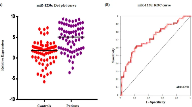
Real time relative quantification analysis showed more than 5 fold increase in serum miR-125b expression among epithelial ovarian cancer patients than healthy controls (Table 3). ROC curve analyses were performed to evaluate the predictive power of miR-125b for malignant ovarian cancer and illustrated inFig 1. Relative expression of the serum miR-125b could dis-criminate patients with malignant ovarian cancer from healthy controls, with a power AUC of 0.728 (95% CI = 0.64–0.81) at a sensitivity of 62.3% and specificity of 77.1% from a cut-off score of 3.76.
miR-125b upregulation in relation to clinicopathological features
Table 2. Association between miR-125b expression and clinicopathological characteristics in epithelial ovarian cancer.
Parameter Number n(%) miR-125b ExpressionHigh (n)(%) p-value
Age (years)
45 40 (57.1) 17 (42.5) 0.13
>45 30 (42.8) 19 (63.3)
Histological grade
Well 25 (35.7) 12 (48.0) 0.04
Moderate 26 (37.1) 18 (69.2)
Poor 19(27.1) 6 (31.5)
Histological type
Mucinous 36(51.4) 18 (50.0) 0.31
Serous 17 (24.2) 11 (64.7)
Papillary 8 (11.4) 2 (25.0)
Others 9 5 (55.5)
Tumor size
<10 cm3 5(7.1) 4 (80.0) 0.22
10–50 cm3 7(10.0) 5(71.4)
50–200 cm3 14(20.0) 9 (64.2)
200–500 cm3 17(24.2) 7 (41.1)
>500 cm3 27(38.5) 11 (40.7)
FIGO stage
I 23(32.8) 23 (100.0) <0.0001
II 10(14.2) 10 (100.0)
III 19(27.1) 2 (10.5)
IV 18(25.7) 1 (5.5)
Distant metastasis
Absent 52(74.2) 35 (67.3) 0.002
Present 18 (25.7) 1 (5.0)
Lymph node status
Absent 51(72.8) 33 (64.7) 0.001
Present 19(27.1) 3 (15.7)
Menopause
Pre 39(55.7) 17 (43.5) 0.21
Post 31(44.2) 19 (61.2)
Nulliparity
Absent 51(44.2) 30 (58.8) 0.07
Present 19(27.1) 6 (31.5)
Serum CA125 (U/ml)
35 9(11.4) 5 (55.5) 0.92
>35 61(88.5) 31 (50.8)
Hemoglobin (gm/dL)
12 59(84.2) 30 (50.8) 0.92
>12 11(15.7) 6 (54.5)
doi:10.1371/journal.pone.0153902.t002
Table 3. Distribution of fold change expression pattern of miR-125b in EOC patients.
miRNA Number of samples (N) Mean fold change expression±SD 95% C.I p-value
miR-125b 70 5.25±1.04 0.845–1.66 <0.0001
showed high expression of miR-125b. Almost 70% patients having moderate grade tumour exhibited increased expression of miR-125b apart from 48% patients with well differentiated tumour and 32% poorly differentiated tumour. Elevated expression of miR-125b was seen in around 65% serous adenocarcinoma patients and about 50% patients with mucinous adenocar-cinoma. When correlated with tumour size, high expression of miR-125b was observed when tumour was at its initial stage i.e.<10cm3. Moreover, all patients in the early stage (stage I and
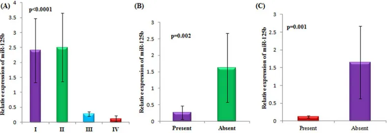
II) showed high expression of miR-125b. Patients having no metastasis ie, involving those patients in the early stage, summed up almost 67% of patients with increased expression of miR-125b as compared to patients (5%) with metastatic disease. Similarly, 65% patients with no lymph node metastasis showed higher expression of miR-125b. Menopausal status, nulli-parity, serum CA125 levels and haemoglobin (Hb) concentration did not associate significantly with the expression of miR-125b.Fig 2demonstrates the significant relationship with respect to stage, lymph node metastasis and distant metastasis status with the expression of miR-125b in epithelial ovarian cancer patients.
miR-125b with respect to stage of epithelial ovarian cancer. A lucid demarcation was observed with respect to the expression of miR-125b and the FIGO stages in epithelial ovarian cancer. All patients in Stage I and stage II presented higher expression of miR-125b while patients in stage III and IV counted merely 10% and 5% of patients with increased expression respectively (Fig 2A).
miR-125b is upregulated in non metastasized tumours. A statistically significant
(p = 0.002)correlation was observed between the metastasis status of epithelial ovarian cancer patients and miR-125b expression. Expression of miR-125b was found to be specifically increased in patients with no metastasis (Fig 2B).
Fig 1. Serum expression of miR-125b.(A) Dot plot showing the relative expression of miR-125b in patients and controls (B) ROC curve for miR-125b exhibiting its diagnostic potential in epithelial ovarian cancer.
miR-125b is upregulated in patients with no lymph node metastasis. As with distant metastasis, patients with no lymph node metastasis showed a statistically significant
(p = 0.001)correlation with the expression of miR-125b (Fig 2C).
Expression of miR-125b and its prognostic potential in epithelial ovarian
cancer
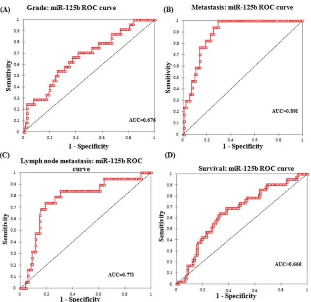
miR-125b was analyzed for its disease progression, prognostic significance and survival out-come in EOC. miR-125b was evaluated for disease progression by the association of its expres-sion with the FIGO stages and various other clinicopathological parameters of epithelial ovarian cancer. The prognostic significance was elucidated by ROC curves. To use ROC curve analysis, the clinical and tumor characteristics were made binary and therefore dichotomized. Stage was dichotomized as early (I+II) or late (III+IV), tumor grade as low (G1+G2) or high (G3), node involvement as no lymph node involvement or any lymph node involvement, dis-tant metastasis as present or absent, survival as death due to epithelial ovarian cancer or other (censored/alive).
The expression of miR-125b and histological grade yielded a significant AUC of 0.676 (95% CI: 0.558–0.793) with a sensitivity of 66.7% and specificity of 62.6% from an optimal cut-off value of 3.91 (Fig 3A) (Table 4). ROC curve for distant metastasis yielded an AUC of 0.891 (95% CI: 0.838–0.944) with a sensitivity and specificity of 94.1% and 70.5% respectively at an optimal cut-off value of 4.34 (Fig 3B). An AUC of 0.773 (95% CI: 0.658–0.887) was obtained in ROC curve with respect to lymph node metastasis and at an optimal cut-off value of 5.85, the sensitivity was 73.7% and specificity was 80.8% (Fig 3C). ROC curve for survival yielded an AUC of 0.660 (95% CI: 0.564–0.756) with a sensitivity of 64.3% and specificity of 67.0% at an optimal cut-off score of 3.91 (Fig 3D).
The survival analysis was done by Kaplan-Meier curve and log-rank test (Fig 4A). There was no significant association found between the miR-125b expression and overall survival of patients with1 fold expression of miR-125b. However, when histological subgroups were estimated for survival separately, it was found that patients with mucinous histotypes had a survival time of 16 months whereas, patients with serous histotypes exhibited a survival time of 9 months (Fig 4B).
Fig 2. Association of serum miR-125b and clinicopathological characteristics in epithelial ovarian cancer patients with respect to A)Stage, (B) Lymph node metastasis and (C)Distant metastasis status.
Expression of miR-125b in association with promoter hypermethylation
of tumour suppressor genes
The relative expression of miR-125b was assessed in relation to the promoter hypermethylation of well known tumour suppressor genes—DAPK1, p16, RASSF1A, PTEN, BRCA1 and p14. Fig 3. Area under curve (AUC) of receiver operating characteristic (ROC) for miR-125b corresponding to (A)Tumor grade (B)Metastasis (C)Lymph node status and (D)Survival of EOC patients.
doi:10.1371/journal.pone.0153902.g003
Table 4. AUC for ROC curve corresponding to the diagnostic value of miR-125b in EOC.
Parameter AUC Standard Error 95% C.I.
Histological grade 0.676 0.060 558–0.793
Distant metastasis 0.891 0.027 0.838–0.944
Lymph node 0.773 0.059 0.658–0.887
Survival 0.660 0.049 0.564–0.756
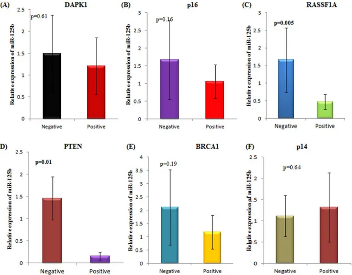
Out of these, RASSF1A and PTEN genes showed a statistically significant correlation between the aberrant hypermethylation of their promoter and the relative expression of miR-125b in epithelial ovarian cancer patients. Patients with RASSF1A promoter hypermethylation demon-strated downregulation of miR-125b expression(p = 0.005)(Fig 5). Similarly, patients with PTEN promoter hypermethylation showed downregulation of miR-125b expression
(p = 0.01). No other tumour suppressor gene exhibited a statistically significant association with miR-125b expression.
Discussion
The development of epithelial ovarian carcinogenesis is a multistep procedure entailing the dysregulation of oncogenes and tumor suppressor genes. Contemporary studies have reported that microRNA-associated transcriptionally regulated gene expression plays a crucial role in the initiation, development and progression of epithelial ovarian cancer, by keeping a regula-tory check on the cell proliferation, cell cycle, apoptosis, and metastasis. miR-125b has been reported to be involved in multiple cancers[12]. miR-125b functions either as an oncogene or tumor suppressor gene. It is upregulated and displays oncogenic potential in some cancers as it triggers cell division, while hampering apoptosis. On the other hand, it is overtly downregu-lated in various other cancers. Our understanding with respect to the contradictory roles of miR-125b in plethora of cancers is still limited. One aspect of our aim was to explore the aber-rant expression of miR-125b in epithelial ovarian cancer in relation to the clinicopathological attributes. The other aspect was to test the expression of miR-125b with reference to the anom-alously methylated TSGs in order to vividly conclude their possible role in either regulating or being regulated by miR-125b.
miR-125b is thought to target the hallmarks of cancer in one way or the other, regulating multiple genes involved in the pathway, leading to the development of the cancer phenotype. Aberrant DNA methylation has a critical outcome on epithelial ovarian cancer initiation and progression. Several investigators have looked for the hypermethylation of the miRNA pro-moter in different cancers as a reason for the inactivation of that particular miRNA and highlighting the putative role of epigenetic disruption in producing the aberrant patterns of expression of miRNAs in cancer cells[13]. Several lines of evidence support the idea that dys-regulation of miRNA can lead to aberrant DNA methylation in cancer and that miRNAs Fig 4. Kaplan Meier survival curve with respect to (A) fold change (B) mucinous and serous histopathological subtypes.
able to regulateDNMTgenes are reportedly downregulated in cancer[14,15]. miRNAs which act as tumour suppressors have been found to be methylated in tumour cells indicating the significance the studying the two forces together- epigenetics and miRNAs[16]. Also, the DNA methylation profile of tumors can be used as a signature to define tumor type, clinical prognosis and treatment response[17,18]. miRNAs transcribed from CpG islands undergo DNA methylation-associated repression with a similar chromatin context to coding genes, such as binding of the transcriptional repressor methyl-CpG-binding domain proteins [19,20]. Such genetic unmasking studies have paved way in identifying the mechanisms responsible for causing the cancer havoc at the molecular level. Based on similar lines, we selected a panel of tumour suppressor genes, which are known to be susceptible in epithelial ovarian cancer to investigate whether their aberrant methylation directly or indirectly affects the expression of miR-125b which in turn leads to the development of cancer. Of the studied tumour suppressor genes, RASSF1A and PTEN genes emerged as likely candidates which might be playing a crucial role in modulating the expression of miR-125b. Poliseno et al. have demonstrated the role of PTEN transcripts in modulating miRNA targeting by Fig 5. Association of miR-125b expression with the promoter hypermethylation of a panel of tumour suppressor genes.(A)DAPK1 (B) p16 (C) RASSF1A (D) PTEN (E) BRCA1 (F) p14. Positive and negative depicts the presence or absence of promoter hypermethylation of the specified tumour suppressor gene respectively.
examining PTENP1 levels[21]. The identification and validation of numerousPTEN -target-ing microRNAs indicates that post-transcriptional regulation plays a central role in deter-mining PTEN abundance in cancer cells[22–25]. Cells are highly sensitive to even slight decrease in PTEN levels, highlighting the importance of microRNA-mediated PTEN regula-tion in cancer[26]. RASSF1A is a tumor suppressor that is a negative regulator in the RAS--MAPK pathway and has reduced expression in various cancers. It has been proposed that RASSF1A participates in cell proliferation and apoptosis by regulating the MAPK pathway and has effects on carcinogenesis[27]. Another study by Banno et al. reported that reduced expression of RASSF1A gene occurs by hypermethylation or suppression of gene expression by miRNAs. Expression of the miRNA itself may be increased or decreased by promoter methylation, based on differences between normal and cancerous cells[28]. Hence our study has extrapolated these results and might give a new direction to examine the expression of miR-125b in a new dimension.
Recent technological advancements have aided the detection of miRNAs at optimum sen-sitivity and specificity. Circulating miRNAs are emerging as promising diagnostic biomarkers for cancer though their usefulness for detecting early neoplasms still remains unclear. miR-125b has been shown to target multiple genes which are more or less relevant in the forma-tion of tumour in different cell types. miR-125b acts as classical oncogene in a variety of tumours where its overexpression may lead to the suppression of a tumour suppressor gene or block its apoptotic machinery. This is further supported by the finding that miR-125b expression levels are correlated with reduced survival rates[29,30]. A recent study by Yamada et al. has also reported an elevated level of miR-125b in colorectal cancer. They identified miR-125b as an early detection biomarker in colorectal cancer owing to the considerable AUC (0.806) in the ROC curve[31]. Giray et al. found an upregulation of miR-125b expres-sion in hepatocellular carcinoma and suggested that it could serve as a novel, non invasive biomarker in this cancer[32]. Jiang and colleagues also showed an upregulation of miR-125b in vivo and in vitro in endometrial carcinoma[33]. In addition, overexpression of miR-125b has been investigated in several cancers such as pancreatic cancer[34], prostate cancer[35], breast cancer[36,37], lung cancer[38], glioblastoma[39], oligodendroglial cancer[7] amongst other cancers. Conversely, miR-125b has been shown to be downregulated in a number of tumours such as bladder cancer[40], esophageal cancer[41], osteosarcoma[42], prostate can-cer[43], liver cancer[44], breast cancer[45], non-small cell lung cancer (NSCLC)[46], breast cancer[47], gastric cancer[48], glioblastoma[49] etc. In ovarian cancer, contradictory studies on miR-125b have made it a necessity to survey its role profoundly. Lee et al. reported a downregulation of miR-125b in ovarian cancer tissue and cell lines compared to healthy con-trols[50]. Luo and colleagues have also recently reported a downregulation of miR-125b in ovarian cancer tissue and cell lines[51]. Gadducci et al. also observed similar results[52]. He et al. suggested a novel therapeutic approach using miR-125b mimic in the treatment of ovar-ian cancer[53]. Guan et al. reported that overexpression of miR-125b resulted in the inhibi-tion of ovarian cancer cell proliferainhibi-tion[54]. On the other hand, Kong and colleagues reported an upregulation of miR-125 in ovarian cancer and suggested that up-regulation of miR-125b expression contributes to cisplatin resistance through suppression of Bak1 expres-sion[55]. Diverse expression of miR-125b was reported in drug-resistant ovarian cancer cell lines by Sorrentino et al[56].
Conclusion
Increasing number of studies have shown that miR-125b plays a crucial role in diverse cellular processes and many diseases especially carcinomas. It is noteworthy that the functions of 125b are controversial in different types of cancers. Hence, the controversial properties of miR-125b in various tumors suggest that miR-miR-125b have distinct functions in cancer pathogenesis and progression, while the underlying mechanisms on different cell context need further vali-dation. The advances that miR-125b holds in clinical applications need a closer look for future treatment of cancer.
Supporting Information
S1 Fig. Electrophoretic band pattern for hypermethylation for a panel of tumor suppressor genes visualized on 2% agarose gel under UV transillumination.
(EPS)
S1 Table. Sequence of primers with annealing temperatures and band size used for MS-PCR.
(RTF)
Acknowledgments
We are grateful to Dr. V Sreenivas for his help and cooperation in the statistical analysis of this manuscript.
Author Contributions
Conceived and designed the experiments: MZ RM AS. Performed the experiments: MZ. Ana-lyzed the data: MZ IK. Contributed reagents/materials/analysis tools: GG PCR AS RM. Wrote the paper: MZ IK RM. Helped in recruiting the subjects: GG PCR.
References
1. Kinose Y, Sawada K, Nakamura K, Kimura T, Kinose Y, Sawada K, et al. The Role of MicroRNAs in Ovarian Cancer, The Role of MicroRNAs in Ovarian Cancer. BioMed Res Int BioMed Res Int. 2014 Sep 10; 2014, 2014:e249393.
2. Catto JWF, Alcaraz A, Bjartell AS, De Vere White R, Evans CP, Fussel S, et al. MicroRNA in prostate, bladder, and kidney cancer: a systematic review. Eur Urol. 2011 May; 59(5):671–81. doi:10.1016/j. eururo.2011.01.044PMID:21296484
3. Heinzelmann J, Unrein A, Wickmann U, Baumgart S, Stapf M, Szendroi A, et al. MicroRNAs with prog-nostic potential for metastasis in clear cell renal cell carcinoma: a comparison of primary tumors and distant metastases. Ann Surg Oncol. 2014 Mar; 21(3):1046–54. doi:10.1245/s10434-013-3361-3
PMID:24242678
4. Ryan BM, Robles AI, Harris CC. Genetic variation in microRNA networks: the implications for cancer research. Nat Rev Cancer. 2010 Jun; 10(6):389–402. doi:10.1038/nrc2867PMID:20495573
5. Vargas Romero P, Cialfi S, Palermo R, De Blasio C, Checquolo S, Bellavia D, et al. The deregulated expression of miR-125b in acute myeloid leukemia is dependent on the transcription factor C/EBPα. Leukemia. 2015 Dec; 29(12):2442–5. doi:10.1038/leu.2015.117PMID:25982911
6. Ribeiro J, Marinho-Dias J, Monteiro P, Loureiro J, Baldaque I, Medeiros R, et al. 34a and miR-125b Expression in HPV Infection and Cervical Cancer Development. BioMed Res Int. 2015; 2015:304584. doi:10.1155/2015/304584PMID:26180794
7. NELSON PT, BALDWIN DA, KLOOSTERMAN WP, KAUPPINEN S, PLASTERK RHA, MOURELA-TOS Z. RAKE and LNA-ISH reveal microRNA expression and localization in archival human brain. RNA. 2006 Feb; 12(2):187–91. PMID:16373485
9. Scott GK, Goga A, Bhaumik D, Berger CE, Sullivan CS, Benz CC. Coordinate suppression of ERBB2 and ERBB3 by enforced expression of micro-RNA miR-125a or miR-125b. J Biol Chem. 2007 Jan 12; 282(2):1479–86. PMID:17110380
10. Nishida N, Mimori K, Fabbri M, Yokobori T, Sudo T, Tanaka F, et al. MicroRNA-125a-5p is an indepen-dent prognostic factor in gastric cancer and inhibits the proliferation of human gastric cancer cells in combination with trastuzumab. Clin Cancer Res Off J Am Assoc Cancer Res. 2011 May 1; 17(9):2725–
33.
11. Chen H, Xu Z. Hypermethylation-Associated Silencing of miR-125a and miR-125b: A Potential Marker in Colorectal Cancer. Dis Markers [Internet]. 2015 [cited 2016 Feb 2];2015. Available from:http://www. ncbi.nlm.nih.gov/pmc/articles/PMC4674596/
12. Banzhaf-Strathmann J, Edbauer D. Good guy or bad guy: the opposing roles of microRNA 125b in can-cer. Cell Commun Signal CCS. 2014; 12:30. doi:10.1186/1478-811X-12-30PMID:24774301
13. Suzuki H, Maruyama R, Yamamoto E, Kai M. DNA methylation and microRNA dysregulation in cancer. Mol Oncol. 2012 Dec; 6(6):567–78. doi:10.1016/j.molonc.2012.07.007PMID:22902148
14. Cummins JM, He Y, Leary RJ, Pagliarini R, Diaz LA, Sjoblom T, et al. The colorectal microRNAome. Proc Natl Acad Sci U S A. 2006 Mar 7; 103(10):3687–92. PMID:16505370
15. Fabbri M, Garzon R, Cimmino A, Liu Z, Zanesi N, Callegari E, et al. MicroRNA-29 family reverts aber-rant methylation in lung cancer by targeting DNA methyltransferases 3A and 3B. Proc Natl Acad Sci U S A. 2007 Oct 2; 104(40):15805–10. PMID:17890317
16. Lujambio A, Esteller M. CpG island hypermethylation of tumor suppressor microRNAs in human can-cer. Cell Cycle Georget Tex. 2007 Jun 15; 6(12):1455–9.
17. Esteller M. Epigenetics in cancer. N Engl J Med. 2008 Mar 13; 358(11):1148–59. doi:10.1056/ NEJMra072067PMID:18337604
18. Rodríguez-Paredes M, Esteller M. Cancer epigenetics reaches mainstream oncology. Nat Med. 2011 Mar; 17(3):330–9. doi:10.1038/nm.2305PMID:21386836
19. Urdinguio RG, Fernandez AF, Lopez-Nieva P, Rossi S, Huertas D, Kulis M, et al. Disrupted microRNA expression caused by Mecp2 loss in a mouse model of Rett syndrome. Epigenetics. 2010 Oct 1; 5 (7):656–63. doi:10.4161/epi.5.7.13055PMID:20716963
20. Lujambio A, Ropero S, Ballestar E, Fraga MF, Cerrato C, Setién F, et al. Genetic unmasking of an epi-genetically silenced microRNA in human cancer cells. Cancer Res. 2007 Feb 15; 67(4):1424–9. PMID:
17308079
21. Poliseno L, Salmena L, Zhang J, Carver B, Haveman WJ, Pandolfi PP. A coding-independent function of gene and pseudogene mRNAs regulates tumour biology. Nature. 2010 Jun 24; 465(7301):1033–8. doi:10.1038/nature09144PMID:20577206
22. Takakura S, Mitsutake N, Nakashima M, Namba H, Saenko VA, Rogounovitch TI, et al. Oncogenic role of miR-17-92 cluster in anaplastic thyroid cancer cells. Cancer Sci. 2008 Jun; 99(6):1147–54. doi:10. 1111/j.1349-7006.2008.00800.xPMID:18429962
23. Kato M, Putta S, Wang M, Yuan H, Lanting L, Nair I, et al. TGF-beta activates Akt kinase through a microRNA-dependent amplifying circuit targeting PTEN. Nat Cell Biol. 2009 Jul; 11(7):881–9. doi:10. 1038/ncb1897PMID:19543271
24. Lewis BP, Shih I -hun., Jones-Rhoades MW, Bartel DP, Burge CB. Prediction of mammalian microRNA targets. Cell. 2003 Dec 26; 115(7):787–98. PMID:14697198
25. Huse JT, Brennan C, Hambardzumyan D, Wee B, Pena J, Rouhanifard SH, et al. The PTEN-regulating microRNA miR-26a is amplified in high-grade glioma and facilitates gliomagenesis in vivo. Genes Dev. 2009 Jun 1; 23(11):1327–37. doi:10.1101/gad.1777409PMID:19487573
26. Alimonti A, Carracedo A, Clohessy JG, Trotman LC, Nardella C, Egia A, et al. Subtle variations in Pten dose determine cancer susceptibility. Nat Genet. 2010 May; 42(5):454–8. doi:10.1038/ng.556PMID:
20400965
27. Pallarés J, Velasco A, Eritja N, Santacana M, Dolcet X, Cuatrecasas M, et al. Promoter hypermethyla-tion and reduced expression of RASSF1A are frequent molecular alterahypermethyla-tions of endometrial carcinoma. Mod Pathol Off J U S Can Acad Pathol Inc. 2008 Jun; 21(6):691–9.
28. Banno K, Kisu I, Yanokura M, Masuda K, Kobayashi Y, Ueki A, et al. Endometrial Cancer and Hyper-methylation: Regulation of DNA and MicroRNA by Epigenetics, Endometrial Cancer and Hypermethy-lation: Regulation of DNA and MicroRNA by Epigenetics. Biochem Res Int Biochem Res Int. 2012 Apr 3; 2012, 2012:e738274.
30. Yuxia M, Zhennan T, Wei Z. Circulating miR-125b is a novel biomarker for screening non-small-cell lung cancer and predicts poor prognosis. J Cancer Res Clin Oncol. 2012 Dec; 138(12):2045–50. doi:
10.1007/s00432-012-1285-0PMID:22806310
31. Yamada A, Horimatsu T, Okugawa Y, Nishida N, Honjo H, Ida H, et al. Serum miR-21, miR-29a, and miR-125b Are Promising Biomarkers for the Early Detection of Colorectal Neoplasia. Clin Cancer Res Off J Am Assoc Cancer Res. 2015 Sep 15; 21(18):4234–42.
32. Giray BG, Emekdas G, Tezcan S, Ulger M, Serin MS, Sezgin O, et al. Profiles of serum microRNAs; miR-125b-5p and miR223-3p serve as novel biomarkers for HBV-positive hepatocellular carcinoma. Mol Biol Rep. 2014 Jul; 41(7):4513–9. doi:10.1007/s11033-014-3322-3PMID:24595450
33. Jiang F, Liu T, He Y, Yan Q, Chen X, Wang H, et al. MiR-125b promotes proliferation and migration of type II endometrial carcinoma cells through targeting TP53INP1 tumor suppressor in vitro and in vivo. BMC Cancer. 2011; 11:425. doi:10.1186/1471-2407-11-425PMID:21970405
34. Bloomston M, Frankel WL, Petrocca F, Volinia S, Alder H, Hagan JP, et al. MicroRNA expression pat-terns to differentiate pancreatic adenocarcinoma from normal pancreas and chronic pancreatitis. JAMA. 2007 May 2; 297(17):1901–8. PMID:17473300
35. Shi X-B, Xue L, Yang J, Ma A-H, Zhao J, Xu M, et al. An androgen-regulated miRNA suppresses Bak1 expression and induces androgen-independent growth of prostate cancer cells. Proc Natl Acad Sci U S A. 2007 Dec 11; 104(50):19983–8. PMID:18056640
36. Vilquin P, Donini CF, Villedieu M, Grisard E, Corbo L, Bachelot T, et al. MicroRNA-125b upregulation confers aromatase inhibitor resistance and is a novel marker of poor prognosis in breast cancer. Breast Cancer Res BCR. 2015; 17:13. doi:10.1186/s13058-015-0515-1PMID:25633049
37. Wang H, Tan G, Dong L, Cheng L, Li K, Wang Z, et al. Circulating MiR-125b as a Marker Predicting Chemoresistance in Breast Cancer. PLoS ONE. 2012 Apr 16; 7(4):e34210. doi:10.1371/journal.pone. 0034210PMID:22523546
38. Wang X, Zhang Y, Fu Y, Zhang J, Yin L, Pu Y, et al. MicroRNA-125b may function as an oncogene in lung cancer cells. Mol Med Rep. 2015 May; 11(5):3880–7. doi:10.3892/mmr.2014.3142PMID:
25573191
39. Wu N, Lin X, Zhao X, Zheng L, Xiao L, Liu J, et al. MiR-125b acts as an oncogene in glioblastoma cells and inhibits cell apoptosis through p53 and p38MAPK-independent pathways. Br J Cancer. 2013 Nov 26; 109(11):2853–63. doi:10.1038/bjc.2013.672PMID:24169356
40. Zhao X, He W, Li J, Huang S, Wan X, Luo H, et al. MiRNA-125b inhibits proliferation and migration by targeting SphK1 in bladder cancer. Am J Transl Res. 2015; 7(11):2346–54. PMID:26807182
41. Zang B, Huang G, Wang X, Zheng S. HPV-16 E6 promotes cell growth of esophageal cancer via down-regulation of miR-125b and activation of Wnt/β-catenin signaling pathway. Int J Clin Exp Pathol. 2015; 8(10):13687–94. PMID:26722596
42. Wang F, Yu D, Liu Z, Wang R, Xu Y, Cui H, et al. MiR-125b Functions as a Tumor Suppressor and Enhances Chemosensitivity to Cisplatin in Osteosarcoma. Technol Cancer Res Treat. 2016 Jan 6; 43. Budd WT, Seashols-Williams SJ, Clark GC, Weaver D, Calvert V, Petricoin E, et al. Dual Action of
miR-125b As a Tumor Suppressor and OncomiR-22 Promotes Prostate Cancer Tumorigenesis. PloS One. 2015; 10(11):e0142373. doi:10.1371/journal.pone.0142373PMID:26544868
44. Zhou J-N, Zeng Q, Wang H-Y, Zhang B, Li S-T, Nan X, et al. MicroRNA-125b attenuates epithelial-mes-enchymal transitions and targets stem-like liver cancer cells through small mothers against decapenta-plegic 2 and 4. Hepatol Baltim Md. 2015 Sep; 62(3):801–15.
45. Xie X, Hu Y, Xu L, Fu Y, Tu J, Zhao H, et al. The role of miR-125b-mitochondria-caspase-3 pathway in doxorubicin resistance and therapy in human breast cancer. Tumour Biol J Int Soc Oncodevelopmental Biol Med. 2015 Aug; 36(9):7185–94.
46. Wang M, Zhu X, Sha Z, Li N, Li D, Chen L. High expression of kinesin light chain-2, a novel target of miR-125b, is associated with poor clinical outcome of elderly non-small-cell lung cancer patients. Br J Cancer. 2015 Mar 3; 112(5):874–82. doi:10.1038/bjc.2015.3PMID:25668010
47. Yang Q, Wang Y, Lu X, Zhao Z, Zhu L, Chen S, et al. MiR-125b regulates epithelial-mesenchymal tran-sition via targeting Sema4C in paclitaxel-resistant breast cancer cells. Oncotarget. 2015 Feb 20; 6 (5):3268–79. PMID:25605244
48. Wu S, Liu F, Xie L, Peng Y, Lv X, Zhu Y, et al. miR-125b Suppresses Proliferation and Invasion by Tar-geting MCL1 in Gastric Cancer, miR-125b Suppresses Proliferation and Invasion by TarTar-geting MCL1 in Gastric Cancer. BioMed Res Int BioMed Res Int. 2015 Oct 4; 2015, 2015:e365273.
49. Wan Y, Sun G, Wang Z, Guo J, Shi L. miR-125b promotes cell proliferation by directly targeting Lin28 in glioblastoma stem cells with low expression levels of miR-125b. Neuroreport. 2014 Mar 26; 25(5):289–
50. Lee M, Kim EJ, Jeon MJ. MicroRNAs 125a and 125b inhibit ovarian cancer cells through post-transcrip-tional inactivation of EIF4EBP1. Oncotarget. 2015 Dec 5;
51. Luo S, Wang J, Ma Y, Yao Z, Pan H. PPARγinhibits ovarian cancer cells proliferation through upregula-tion of miR-125b. Biochem Biophys Res Commun. 2015 Jun 26; 462(2):85–90. doi:10.1016/j.bbrc. 2015.04.023PMID:25944662
52. Gadducci A, Sergiampietri C, Lanfredini N, Guiggi I. Micro-RNAs and ovarian cancer: the state of art and perspectives of clinical research. Gynecol Endocrinol Off J Int Soc Gynecol Endocrinol. 2014 Apr; 30(4):266–71.
53. He J, Jing Y, Li W, Qian X, Xu Q, Li F-S, et al. Roles and mechanism of miR-199a and miR-125b in tumor angiogenesis. PloS One. 2013; 8(2):e56647. doi:10.1371/journal.pone.0056647PMID:
23437196
54. Guan Y, Yao H, Zheng Z, Qiu G, Sun K. MiR-125b targets BCL3 and suppresses ovarian cancer prolif-eration. Int J Cancer J Int Cancer. 2011 May 15; 128(10):2274–83.
55. Kong F, Sun C, Wang Z, Han L, Weng D, Lu Y, et al. miR-125b confers resistance of ovarian cancer cells to cisplatin by targeting pro-apoptotic Bcl-2 antagonist killer 1. J Huazhong Univ Sci Technol Med Sci Hua Zhong Ke Ji Xue Xue Bao Yi Xue Ying Wen Ban Huazhong Keji Daxue Xuebao Yixue Yingde-wen Ban. 2011 Aug; 31(4):543–9.
56. Sorrentino A, Liu C-G, Addario A, Peschle C, Scambia G, Ferlini C. Role of microRNAs in drug-resistant ovarian cancer cells. Gynecol Oncol. 2008 Dec; 111(3):478–86. doi:10.1016/j.ygyno.2008.08.017