Arq Bras Oftalmol 2004;67:451-4
Análise retrospectiva uni-institucional: tratamento de melanomas de coróide com
braquiterapia utilizando cobalto-60
1Professor - Department of Radiation Oncology, Centro de Tratamento Pesquisa Hospital do Câncer – AC Camargo, Radiotherapy Service - Instituto Arnaldo Vieira de Carvalho, São Paulo.
2Professor - Department of Radiation Oncology, Centro de Tratamento Pesquisa Hospital do Câncer – AC Camargo.
3Professor - Department of Onco-opthalmology, Centro de Tratamento Pesquisa Hospital do Câncer – AC Camargo.
4Professor - Department of Onco-opthalmology, Escola Paulista de Medicina e Centro de Tratamento Pesquisa Hospital do Câncer – AC Camargo.
Address to correspondence: Antônio Cássio Assis Pellizzon, Department of Radiation Oncology Centro de Tratamento Pesquisa Hospital do Câncer – AC Camargo - Rua Professor Antonio Prudente, 211 - São Paulo (SP) CEP 01509-900
E-mail: [email protected]
Recebido para publicação em 01.08.2002 Versão revisada recebida em 28.10.2002 Aprovação em 17.12.2003
Nota Editorial: Pela análise deste trabalho e por sua anuência na divulgação desta nota, agradecemos às Dras. Zélia Maria da Silva Corrêa e Maria Teresa B. C. Bonanomi. Antônio Cássio Assis Pellizzon1 João Victor Salvajoli2
Paulo Eduardo Novaes2 Ricardo Fogaroli1 Robson Ferrigno2
Maria Aparecida Conte Maia2 Marta Motono Chojniak3 Clélia Maria Erwene4
INTRODUCTION
The most frequent primary intraocular malignancies are: uveal melanoma in adults and retinoblastoma in children. The natural history of choroidal melanoma (MM-CH) is variable and not fully delineated. The diagnostic challenges are the differentiation of MM-CH from choroidal nevus, choroidal metastasis, choroidal hemangiomas, subchoroidal hematomas and exudative form of age-related macular degeneration(1). The diagnosis
can usually be made by slit lamp biomicroscopy or indirect ophthalmoscopy. The use of other diagnostic methods such as fluorescein angiography, ultrasonography, magnetic resonance imaging and fine needle biopsy can occasionally be used to establish the diagnosis in atypical cases(2).
The management of MM-CH has been the subject of considerable controversy and treatment methods include enucleation and other techniques designed to preserve the eye, such as local resection, plaque radiotherapy, charged particle radiotherapy, laser photocoagulation, and thermotherapy. Eye conservation can be achieved in these patients by several techniques, with external beam charged particle (proton) therapy and episcleral plaque therapy being used most commonly for MM-CH.
The probability of visual preservation and of eye retention with either technique is related to tumor size and location(3). The useful vision is
Single institutional retrospective analysis: treatment of
choroidal melanomas with cobalt-60 brachytherapy
Purpose: To evaluate the outcome of patients with choroidal melanoma treated with conservative therapy with brachytherapy (episcleral Co-60 plaque therapy) at the "Hospital do Cancer" São Paulo, Brazil. Methods: We evaluated 102 patients consecutively treated from January, 1999 to June, 1999. Median age, maximum tumor base diameter and apex size were 55.5 years, 9.75 mm and 5 mm, respectively. Doses at the base of the tumor, including 1 mm of sclera, ranged from 157 to 487 Gy (median 284.5 Gy) and to the apex from 37 to 220 Gy (median 106 Gy). Results: The crude eye preservation rate with conservative therapy alone was 78.5%. Five-year overall actuarial survival rate was 92.2% and eye conservation rate was 78%. Side effects were mostly an uncomplicated retinopathy in 39/102 patients (38.2%); macular degeneration or scarring led to poor central vision in 31/102 patients (30.3%) of cases. Conclusion: Our experience with cobalt-60 plaque brachytherapy achieved a satisfactory rate of local tumor control, despite the oversized base diameters of treated tumors.
ABSTRACT
Arq Bras Oftalmol 2004;67:451-4
452Single institutional retrospective analysis: treatment of choroidal melanomas with cobalt-60 brachytherapy
preserved in eyes with tumors occurring in a favorable loca-tion with respect to the optic disc or macula, but an average of 16.3% of patients treated with radiotherapy subsequently re-quire enucleation due tumor regrowth or uncontrollable neo-vascular glaucoma(4).
Many series have shown that patients with MM-CH were diagnosed too late and in these cases enucleation was the preferred method of treatment(4-5), but the current emphasis is
organ preservation with total or partial visual sparing(6).
At the Hospital A. C. Camargo, we started a conservative treatment policy since January, 1989 using Stallard Cobalt-60 plaques for MM-CH. To date there are 287 patients treated, including MM-CH, retinoblastoma, hemangioma and intrao-cular metastasis.
METHODS
This is a retrospective analysis conducted at the "Centro de Tratamento e Pesquisa – Hospital do Câncer”, São Paulo, Brazil. From January, 1995 to June, 1999 there were 132 pa-tients with intraocular tumors consecutively treated at the Brachytherapy Service and Onco-ophthalmology Department and of these, 108 had MM-CH.
Of the total of patients presenting MM-CH, 102 had ocular confined disease and 6 extraocular extensions and were excluded from the analysis. All patients had a minimum follow-up of 24 months and were evaluated regarding tumor characte-ristics, treatment times, doses to the apex and base of tumor and clinical outcome.
Survival analysis was performed using Kaplan-Meier esti-mates and differences in quantitative variables through chi-square tests(7).
All patients were diagnosed by slit lamp biomicroscopy, indirect ophthalmoscopy and ultrasonography. Pulmonary, bone and liver scan were performed in selected suspicious cases. There was no fine needle biopsy used for diagnosis in this group of patients.
RESULTS
Median age of patients was 55.5 years (range15 to 85). There was a slight predominance of males (54/48) and left-sided (52/50) lesions.
Median tumor base size was 9.75 mm (range 5 to 15 mm) and median apex size was 5 mm (range 2 to 13 mm), determined by ultrasound. There was a predominance of peripheral (72 ca-ses) over central or paracentral (30 caca-ses) lesions.
Patients were admitted to the hospital for an average of 6 days (range 3 to 16) with positioned plaques. Doses at the base of the tumor, including 1 mm of sclera, ranged from 157 to 487 Gy (median 284.5 Gy) and to the apex from 37 to 220 Gy (median 106 Gy) (Table 1). The crude eye preservation rate with conservative therapy alone was 78.5%. Twenty-two (21.5%) patients presented lo-cal uncontrolled disease after a median follow-up of 34 months
Table 1. Characteristics of patients
Variable Mean Median Range
Age (years) 50.4 55.5 15 - 85
Base size (mm) 10.0 9.7 5 - 15
Apex size (mm) 5.8 5.0 2 - 13
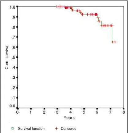
Base dose (Gy) 298.2 284.5 157 - 487 Apex dose (Gy) 113.6 106.0 37 - 220 Stay in hospital (days) 6.7 6.0 3 - 16 (range 19-64), and underwent enucleation. The 5-year overall actuarial survival rate was 92.2% (Figure 1) and eye conserva-tion rate was 78% (Figure 2).
The differences in the size of the tumor base, apex, dose to the apex and treatment days were not statistically significant predictors of clinical outcome in this analysis (Table 2). The dose given to the tumor base was the most important factor to determine a favorable clinical outcome, but without statistical significance (p=0.075).
Central or paracentral tumors showed 50% of the prethera-peutic vision after 2 years, and 80% of the vision was preser-ved in those with peripheral tumors. The main side effects were mostly an uncomplicated retinopathy in 39/102 patients (38.2%); macular degeneration or scarring led to poor central vision in 31/102 patients (30.3% of cases).
After completion of treatment patients were evaluated every 4 months in the first year and every six months thereafter. In the follow-up examinations such as serial photographs, ultrasound, and transillumination, among others, were performed.
Cum survival
Figure 1 - Kaplan-Meier: overall survival analysis Years
Arq Bras Oftalmol 2004;67:451-4
Single institutional retrospective analysis: treatment of choroidal melanomas with cobalt-60 brachytherapy 453
Table 2. Tumor and treatment variables and clinical outcome. Univariate analysis
Status
Variable NED Enucleated p
Base size < 6 1
-(mm) > 6 < 9 18 3
> 9 < 12 28 9 0.804
>12 33 10
Apex size < 5 37 11
(mm) > 5 < 8 30 3
> 8 < 12 10 7 0.892
>12 3 1
Base dose < 200 4
-(Gy) > 200 < 250 18 5 > 250 < 300 27 6
> 300 < 350 22 5 0.075 > 350 < 400 4 2
> 400 5 4
Apex dose < 40 5 3
(Gy) > 40 < 60 16 3
> 60 < 80 15 6 0.179 > 80 < 100 16 2
> 100 28 8
Treatment < 4 7 2
(days) > 4 < 8 53 14 0.989 > 8 < 10 11 2
> 10 9 4
Total 80 22
NED - no evidence of disease in activity
DISCUSSION
Enucleation as a standard treatment for choroidal melano-ma has been questioned since the sixties, because of the loss of sight on that side and the nearly 50% death rate at five years due to metastases(8).
As none of the different treatments, enucleation or brachy-therapy offer a survival advantage, a key factor in choosing among treatment options is their differential impact on the patients’ quality of life.
Of interest is a recent survey reporting that the choice of treatment for uveal melanoma did not seem to be associated with large differences in quality of life when assessed at long-term follow-up(1).
The reduction of the lesion depends on individual charac-teristics of the tumor and must be recorded individually. Pa-tients who are submitted to conservative treatment, if they have a regrowth of the lesions after brachytherapy, to record it with serial photographs, ultrasound, and transillumination is suggested. In a series of 82 MM-CH treated with brachythe-rapy (cobalt-60 and iodine-125), the serial ultrasonic measure-ments showed that no two patients had identical patterns of change. The mean absolute change in tumor height after treat-ment was 1.8 mm at six months, 5.6 mm at 48 months for large tumors, and 0.9 and 1.9 mm for medium sized tumours(9).
A publication evaluating the rate and extent of regression of the first 100 consecutive patients with a MM-CH managed by Cobalt-60 brachytherapy, reported that half of the lesions did not regress rapidly to a flat, depigmented scar but shrank slowly and persisted as a residual mass with approximately 50% of the thickness of the original tumor at 54 months follo-wing brachytherapy(10).
Another report of 62 patients with local relapse, who were matched with an equal number of relapse-free patients in terms of known clinical prognostic factors for both MM-CH specific mortality and local tumor relapse, estimated a 5-year actuarial survival of 87% versus 58% for relapsed patients(11).
In a recent publication no large difference in MM-CH-specific 15-year mortality rate was observed, in patients treated by enucleation versus brachytherapy, after eliminating indivi-dual clinical variables(12).
In our experience, Co-60 plaque brachytherapy has achie-ved a satisfactory rate of tumor local control, despite the over-sized base diameters of treated tumors.
The relative high incidence of presented side effects may be due the used isotope Cobalt-60, a gamma emitter (median energy -1.25 MV) with a relative lack of shielding, with greater amount of intraocular and extraocular tissue being exposed to high doses of radiation when compared to other isotope applicators.
In our analysis we observed a trend toward to a higher dose to the tumor base related to local control, which did not corre-late with a higher incidence of side effects. On the other hand, treatment outcome did not seem to be improved by increasing the apical dose.
Eye preservation rate
Figure 2 - Kaplan-Meier: eye preservation estimates Years
Arq Bras Oftalmol 2004;67:451-4
454Single institutional retrospective analysis: treatment of choroidal melanomas with cobalt-60 brachytherapy
Currently, I-125 and Ru106 are preferred because their lower energy allows easy shielding, leading to better radiation pro-tection for personnel, intra- and extraocular normal tissue(13).
CONCLUSION
Our experience with cobalt-60 plaque brachytherapy achieved a satisfactory rate of local tumor control, despite the oversized base diameters of tumors treated.
RESUMO
Objetivos: Para avaliar o resultado de tratamento pacientes portadores de melanoma de coróide tratados com braquitera-pia (placas episclerais de Co-60) no Hospital do Câncer, São Paulo, Brasil. Métodos: Foram avaliados 108 pacientes trata-dos consecutivamente de janeiro de 1995 a junho de 1999, com idade mediana de 55,5 anos, diâmetro da base do tumor e altura máximos 9,75 mm e 5 mm, respectivamente. As doses na base do tumor, incluindo 1 mm de esclera variaram de 157 a 487 Gy (mediana 284,5 Gy) e para o ápice de 37 até 220 Gy (mediana 106 Gy). Resultados: A taxa de preservação ocular foi 78,5%, com sobrevida atuarial global em cinco anos, e a taxa de con-servação ocular atuarial foram 92,2% e 78%, respectivamente. Os principais efeitos colaterais relacionados ao tratamento foram retinopatia em 39/102 (38,2%) pacientes e degeneração macular, levando a déficit visual central em 31/102 (30,3%) pacientes. Conclusão: Nossa experiência com placas episcle-rais de Co-60 alcançou taxa satisfatória de controle de tumor local, apesar dos grandes diâmetros das bases dos tumores.
Descritores: Neoplasias da coróide; Melanoma; Radioisóto-pos de cobalto/uso terapêutico; Enucleação ocular; Braquite-raia; Dosagem radioterapêutica
REFERENCES
1. Munzenrider JE. Uveal melanomas. Conservation treatment. Hematol Oncol Clin North Am 2001;15:389-402.
2. Shields JA, Shields CL, De Potter P, Singh AD. Diagnosis and treatment of uveal melanoma. Semin Oncol 1996;23:763-7.
3. Shields CL, Shields JA, Gunduz K, Freire JE, Mercado G. Radiation therapy for uveal malignant melanoma. Ophthalmic Surg Lasers 1998;29:397-409. 4. Finger PT. Radiation therapy for choroidal melanoma. Surv Ophthalmol
1997;42:215-32.
5. Starzycka M, Szpakowicz U, Slomska J. Diagnostic problems in intraocular melanoma. Klin Oczna 1999;101:11-7.
6. Hungerford JL. Current trends in the treatment of ocular melanoma by radio-therapy. Clin Experiment Ophthalmol 2003;31:8-13.
7. Kaplan EL, Meier P. Non-parametric estimation from incomplete observa-tions. J Am Stat Assoc 1958;53:457-61.
8. Foss AJ, Lamping DL, Schroter S, Hungerford J. Development and validation of a patient based measure of outcome in ocular melanoma [commented on Br J Ophthalmol 200;84:343-4]. Br J Ophthalmol 2000;84:347-51.
9. Abramson DH, Servodidio CA, McCormick B, Fass D, Zang E. Changes in height of choroidal melanomas after plaque therapy. Br J Ophthalmol 1990; 74:359-62.
10. Cruess AF, Augsburger JJ, Shields JA, Brady LW, Markoe AM, Day JL. Regression of posterior uveal melanomas following cobalt-60 plaque radiothe-rapy. Ophthalmology 1984;91:1716-9.
11. Vrabec TR, Augsburger JJ, Gamel JW, Brady LW, Hernandez C, Woodleigh R. Impact of local tumor relapse on patient survival after cobalt 60 plaque radiotherapy. Ophthalmology 1991;98:984-8.
12. Augsburger JJ, Correa ZM, Freire J, Brady LW. Long-term survival in choroidal and ciliary body melanoma after enucleation versus plaque radiation therapy. Ophthalmology 1998;105:1670-8.