157
ENVIRONMENTAL SURVEY AND QUALITY CONTROL
TESTS OF X-RAY DIAGNOSTIC FACILITY OF A LARGE
NIGERIAN HOSPITAL
1OLUWAFISOYE P.A,2 OLOWOOKERE C.J.3OBED R.I. 4EFUNWOLE H.O and 5AKINPELU J.A
1
College of Science,Engineering and Technology,Osun State University, Osogbo.
2
Department of Physical Science, Ajayi Crowther University, OYO
3
Physics Department, University of Ibadan’ Ibadan.
4
Faculty of Science, Osun State Polytechnic, Iree.
5
Department of Physics with Solar Energy, Bowen University, Iwo
ABSTRACT
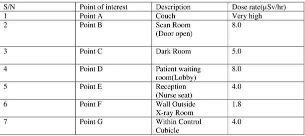
Environmental monitoring and quality control test of X-ray facilities of a large Nigerian hospital was carried out. The results show that the facilities for safety were grossly inadequate and the dose rates of 4.0µSv/hr and 5.0 µSv/hr were recorded at the reception and outside the entrance door respectively. The dose rates at the adjacent ultrasound scan room and waiting lobby are at least a factor of 40 higher than the background dose rate each, indicating higher health risk to the visitors and personnel at the hospital. However, recommendations on how to improve on the safety of the patient and personnel were sent to the management of the X-ray unit of the hospital. Nevertheless, follow-up study indicate improved facilities and safety measures.
INTRODUCTION
Patients and medical personnels receive various doses of ionizing radiation from both naturally occuring and man made sources. The level of doses received depends on the occupation, level of radiation in the environment and where an individual lives. Depending on where an individual lives, most people receive an exposure in the range of 1mSv Rem per year from cosmic radiation from outer space and from naturally occuring isotopes in the ground, air, food and water. Nevertheless, X-ray examinations are common place and contribute by far the largest man made source of ionizing radiation exposure for the population.[1]. More than ever before, in the recent times, there has been a constant increase in the number and frequency of X-ray examinations [2] because of the increase in availability of the X-ray facilities in developing countries. In Nigeria, almost every state owned hospitals has at least an X-ray unit.The University Teaching hospital and Federal medical centres have between two and four X-ray units. The private hospitals (including mission hospitals) have at least an X-ray unit. In some University Teaching hospitals (UTHs) and private hospitals there are Computer Tomography (CT) units.
Meanwhile, these hospitals which spread all over Nigeria contribute certain doses of ionizing radiation to the existing background radiation dose level. Due to the fact that radiation does not respect nationality, experience and professionalism, it is expected that workplace be monitored at a frequency that will ensure safe working conditions [3].
The purpose of a radiation monitoring programme is to identify all sources of radiation exposure within an operation area, to assess the level of radiation exposure of the employee and members of the public so that timely detection of changes in radiation parameters which may lead to increased exposures, and to produce sufficient information for optimization purpose [4].
158
However, in the recent times the Nigerian Nuclear Regulator Authority (NNRA) is saddled with the responsibilities of licencing, training, research and monitoring. Inspite of the presence of the regulatory body in Nigeria, earlier survey reported in another place showed that out of the 22 hospitals in 8 states 9.1% have never used any dose monitoring device and 9.1% have never calibrated their equipment. Also, 81.8% have never calculated the dose to the patient as required by international regulatory bodies [4], while many hospitals have never carried out any quality control (QC) test of their facilities. And inmost cases environmental monitoring has never been carried out. A recent survey carried out in northwestern geopolitical zone of Nigeria, for example, showed that out of 124 institutions using ionizing radiation 253 X-ray installations were found with only 90 sealed radiation sources. Specifically, in Ahmadu Bello University Teaching hospital (ABUTH, Zaria) complex an average of 250 diagnostic X-ray examinations are carried out per day in three hospital facilities located in Zaria, Kaduna and Malunfasi [2]. As a result of these radiation activities, the annual collective doses to the patients, workers and members of the public could be quite significant from such exposures.
Exposure to ionizing radiation is most strongly asociated with leukemia and cancer of the thyroid, breast and lung. An association has been reported at the absorbed dose of less than 0.2Gy [5]. The risk of developing cancer, however, depends to some extent on age at exposure, level of exposure (dose received). Moreover, the risk of carcinogenesis is generally greater for children than the adults and the genetic consequence of doses to the gonad in paediatric patients are also higher than in adults [6]. Additionally, some evidence suggest that lung cancer risk may be most strongly related to exposure of latter age in life. Relationship between radiation exposure and cancer of the salivary gland, stomach, bladder, ovary, colon, central nervous system (CNS) and skin have been reported usually at dose greater than 1Gy [7,8]. It is evident from reports that enviromental and personnel monitoring are still non-existent at most facilities.
However, because of increased radiation risk to children, adults and medical personnel, radiation monitoring programme is essential and required especially in Nigeria with more than 5000 X-ray units. Out of which less than 5% are under regulatory control, thereby posing serious challenges [9].
This paper presents the result of environmental survey and quality control test of immediate surroundings of the X-ray facilities of a large hospital in Nigeria. This monitoring is an attempt at ensuring quality of facilities and environment at our X-ray units. It is also part of ongoing quality assurance programme in Nigeria.
MATERIALS AND METHODS:
The enviromental monitoring in this study was carried out using calibrated radiation monitor device model minirad 1000+, while the factory calibrated non-invasive X-ray test device model 4000 m+ was used for the quality control tests. Questionnaires were also used to elicit information from the most senior personnel of the hospital. The study was carried out at the X-ray unit of the Jon-Ken hospital, Lagos (private hospital).
RESULTS AND DISCUSSIONS:
Table 1 shows the X-ray machine specific data. The data show that the X-ray machine is analogue manufactured more than 26 years ago and installed 22 years ago.
159
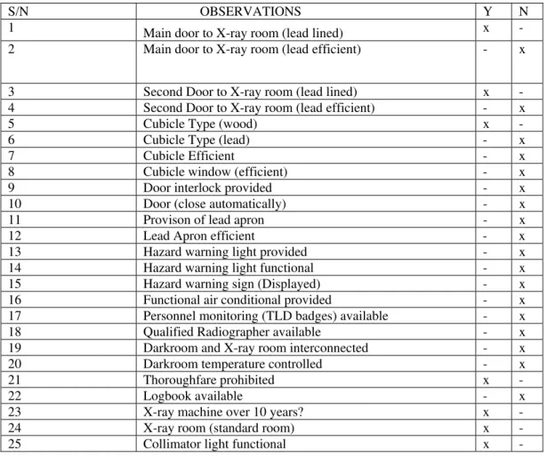
Table 3 shows the general observation in terms of facilities for radiation safety within and around the X-ray machine. The results indicate that the door that leads to the X-ray room was not efficiently lead lined, this inadequacy could have led to the high dose rate at the reception and patient waiting area.The results also show that the cubicle is not efficiently lead lined. Interlock was not provided for the door and the door could not close automatically during the exposure to prevent intruders. It is necessary to note that controlled access to areas where radiation exposure may be taking place is required. It is also evident from table 3 that hazards warning light and personnel monitoring (TLD badges) were not provided. In addition, qualify radiographer was not available and log books for keeping records of radiation protection activities in the unit were not available. The lead apron required to be worn by the radiographer during exposure was visibly missing. It therefore, implies that in the X-ray unit of the hospital, the issue of safety of personnel and patients are not adequately taken into consideration. Apparently the preoccupation of the management and and the personnel was the image quality at the expense of patient health risk. This trend is an indication that the principle of as low as reasonably achievable (ALARA ) principle was not adopted in the hospital.
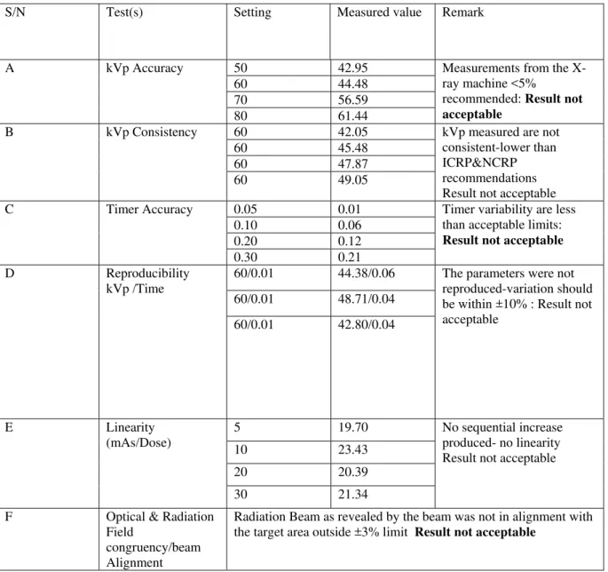
Table 4 is the summary of the quality control test ( test of accuracy, consistency, reproducibility, linearity and congruency). The test for kVp accuracy , kVp consistency and timer accuracy were outside the acceptable limit. Other quality control tests carried out include reproducibility of kVp, linearity of mAs and optical radiation field congruency and were found not acceptable. This inconsistent nature of the kvp could have adverse effect on the image contrast and leads to repeated exposure of the patient. Since the length of exposure affects the total quantity of radiation (mAs) emitted from an X-ray tube, therefore an accurate exposure timer is essential for good radiographic imaging. Non linearity of milliampere seconds (mAs) means that sequential increase in milliampere second does not produce the same sequential increase in exposure. Also, the non alignment within the acceptable limit of the beam with the target area could adversely affect the quality of radiograph produced.
More importantly, qualified radiographer, darkroom operators and medical physicist were not available to carry out the necessary procedures and to look into the safety report of the activities of the hospitals. Available data indicate that, the X-ray centre has never employed the services of Radiation Protection officer and Medical Physicist whose role are very important in diagnostic radiology.
Based on the recommendation of these findings and the follow-up studies, general overhauling of the facilities has since commenced. Recommended safety and radiation monitoring materials have been put in place. Moreover new X-ray machine has been ordered.
In this study we have undertaken the quality control test and environmental monitoring of the facilities of X-ray unit of a large Nigerian hospital. The quality control test obtained fall short of the required standard of International Commission on Radiological Protection (ICRP) and National Council on Radiation Protection (NCRP). In addition, facilities for the safety of both the public and personnel were grossly inadequate.
REFERENCES:
[1]Mahogora W.E and Nyanda A.M. The potential for reduction of radiation doses to patients undergoing some common X-ray examination in Tanzania. Radiation protection Dosimetry 381 – 384 (2000).
[2]Mallam S.P, Akpa M.D, Oladipupo M.D and Sa’id, A. Reappraisal of existing expressions for estimating Radiation output from Diagnostic X-ray machine. Nigerian Journal of physics 16 (2); 2004: 30- [3]Elegba, S.B. Keynote address on Radiation safety officers in diagnostic and interventional Radiology. University of Ibadan Nigeria (2006). Unpublished Keynote address.
[4]Olowookere, C.J Obed, R.I. Adefisoye, P.A. Vincent U.E. Medical Physicist: Missing component of Nigerian Radiological Crew. Journal of Scientific and Industrial Studies,7 (1), 2009: 110-117.
[5]Bhatai, S; Sather, H.N. Pabustan, O.B, Trigg, M.E, Gaynon, P.S and Rabison, L.L. Low incidence of second neoplasm’s among children diagnosed with acute lymphoblast lenkemia after 1983. Blood 99(12): 4257 – 64.
[6]Ware, D.E, Huda, W, Mergo, P.J and Litwiller, A.L. Radiation effective dose to patients undergoing Abdominal C T examination. Radiology 1999; 210: 645 – 650.
160
[8]Licher, M.D; Karagas, M.R, M.H, L.A Spencer S.P, stukel,TA. Greenburg, E.F. Therapentic ionizing radiation and incidence of basal cell carcinomal and squamous cell carcinoma. The New Hamsphire skin cancer study Group. Arch Dematol 136 (80): 1007 – 11; 2000.
[9]Klecnerman , R.A, Boice, J.D, Storm, H.H, Sparen, P, Anderson, A, Pukkala, E. Lynch, C.F, Hankey, B.F, and Flannery, J.T. Second primary cancer after treatment for cervical cancer. An international cancer registries study cancer. 76(3): 442 – 52.
Table 1: X-ray machine specific data Machine name Mediront -4
Age of machine Above 26 years Date of installation 1987
Model 18299 (Budapest, Hungary)
Table 2: Dose Rate measured at different locations
S/N Point of interest Description Dose rate(µSv/hr)
1 Point A Couch Very high
2 Point B Scan Room
(Door open)
8.0
3 Point C Dark Room 5.0
4 Point D Patient waiting
room(Lobby)
8.0
5 Point E Reception
(Nurse seat)
4.0
6 Point F Wall Outside
X-ray Room
1.8
7 Point G Within Control
Cubicle
4.0
161
Table 3: General Observations
S/N OBSERVATIONS Y N
1 Main door to X-ray room (lead lined) x -
2 Main door to X-ray room (lead efficient) - x
3 Second Door to X-ray room (lead lined) x - 4 Second Door to X-ray room (lead efficient) - x
5 Cubicle Type (wood) x -
6 Cubicle Type (lead) - x
7 Cubicle Efficient - x
8 Cubicle window (efficient) - x
9 Door interlock provided - x
10 Door (close automatically) - x
11 Provison of lead apron - x
12 Lead Apron efficient - x
13 Hazard warning light provided - x
14 Hazard warning light functional - x
15 Hazard warning sign (Displayed) - x
16 Functional air conditional provided - x 17 Personnel monitoring (TLD badges) available - x
18 Qualified Radiographer available - x
19 Darkroom and X-ray room interconnected - x
20 Darkroom temperature controlled - x
21 Thoroughfare prohibited x -
22 Logbook available - x
23 X-ray machine over 10 years? x -
24 X-ray room (standard room) x -
25 Collimator light functional x -
162
Table 4: Summary of the quality control tests (QC)
S/N Test(s) Setting Measured value Remark
A kVp Accuracy 50 42.95 Measurements from the X-ray machine <5%
recommended: Result not acceptable
60 44.48 70 56.59 80 61.44
B kVp Consistency 60 42.05 kVp measured are not consistent-lower than ICRP&NCRP recommendations Result not acceptable 60 45.48
60 47.87 60 49.05
C Timer Accuracy 0.05 0.01 Timer variability are less than acceptable limits:
Result not acceptable
0.10 0.06 0.20 0.12 0.30 0.21 D Reproducibility
kVp /Time
60/0.01 44.38/0.06 The parameters were not reproduced-variation should be within ±10% : Result not acceptable
60/0.01 48.71/0.04
60/0.01 42.80/0.04
E Linearity (mAs/Dose)
5 19.70 No sequential increase produced- no linearity Result not acceptable 10 23.43
20 20.39
30 21.34
F Optical & Radiation Field
congruency/beam Alignment