unesp
PROGRAMA DE PÓS-GRADUAÇÃO EM CIÊNCIAS BIOLÓGICAS (BIOLOGIA CELULAR E MOLECULAR)
Alterações morfológicas em glândulas salivares de fêmeas de
carrapatos Amblyomma cajennense Fabricius, 1787,
(Acari:Ixodidae) em diferentes estágios de alimentação
durante sucessivas infestações em coelhos.
PABLO HENRIQUE NUNES
Tese apresentada ao Instituto de Biociências do Campus de Rio Claro, Universidade Estadual Paulista, como parte dos requisitos para obtenção do título de Doutor em Ciências Biológicas (Biologia Celular e Molecular).
unesp
PROGRAMA DE PÓS-GRADUAÇÃO EM CIÊNCIAS BIOLÓGICAS (BIOLOGIA CELULAR E MOLECULAR)
Alterações morfológicas em glândulas salivares de fêmeas de
carrapatos Amblyomma cajennense Fabricius, 1787,
(Acari:Ixodidae) em diferentes estágios de alimentação
durante sucessivas infestações em coelhos
PABLO HENRIQUE NUNES
Tese apresentada ao Instituto de Biociências do Campus de Rio Claro, Universidade Estadual Paulista, como parte dos requisitos para obtenção do título de Doutor em Ciências Biológicas (Biologia Celular e Molecular).
Rio Claro
Estado de São Paulo - Brasil
Agosto- 2009
Orientadora: Profa Dra. Maria Izabel Camargo Mathias
! "
#
$ !
% & $
'
( % & ) * +
" !
,- ,
./01% 1 2
) 3 % &4 $ % / 3 ) 4
5
0 6 0
2 1 * 3
. ) !
4 8 )4 1 9 & :
4
Sumário
Resumo... 6
Abstract ... 8
1. Introdução ... 10
2. Objetivos ... 16
3. Material e Métodos ... 18
3.1. Material ... 18
3.1.1. Carrapatos ... 18
3.1.2. Hospedeiros ... 18
3.1.3. Construção da Câmara de Alimentação dos Carrapatos para Alocação dos
Casais de A. cajennense ... 18
3.1.4. Fixação da Câmara de Alimentação no Hospedeiro ... 19
3.1.5. Deposição dos Casais de Amblyomma cajennense na Câmara de
Alimentação ... 19
3.2. Métodos ... 20
3.2.1. Histologia ... 20
3.2.2. Histoquímica ... 21
4. Resultados ... 24
4.1. Capítulo 1: Necessary feeding time for the engorgement of females of
Amblyomma cajennense Fabricius, 1787 (Acari:Ixodidae) in naive and resistant
rabbits. ... 24
4.2. Capítulo 2: Morphological changes in the salivary glands of Amblyomma
cajennense females (Acari: Ixodidae) in different feeding stages on rabbits at first
infestation ... 38
4.3. Capítulo 3: Secretion dynamics of the salivary gland on semi and engorged tick
females Amblyomma cajennense Fabricius, 1787 (Acari: Ixodidae) during the
second infestation in rabbit. ... 40
4.4. Capítulo 4: Secretory process of salivary glands of Amblyomma cajennense
Fabricius, 1787 (Acari: Ixodidae) female ticks fed on resistant rabbits. ... 61
5. Discussão Geral ... 90
6. Conclusões ... 98
Pablo Henrique Nunes
Resumo
Pablo Henrique Nunes
Abstract
Pablo Henrique Nunes
1. Introdução
O carrapato adulto da espécie Amblyomma cajennense é conhecido popularmente como carrapato-estrela, carrapato do cavalo ou rodoleiro e suas formas imaturas por micuins. São hematófagos obrigatórios e necessitam de repasto sangüíneo em três hospedeiros para completar seu ciclo de vida (FLECHTMANN, 1985). Esses carrapatos apresentam grande importância sanitária, uma vez que podem transmitir, entre outras doenças, a febre maculosa, também conhecida como febre das Montanhas Rochosas, febre do carrapato, febre negra ou doença azul, causada pela bactéria Rickettsia rickettsii. O carrapato A. cajennense encontra-se amplamente distribuído no continente americano, desde o sudoeste dos Estados Unidos, América Central até América do Sul, com exceção do Chile e do Uruguai (COOLEY e KOHLS, 1944).
As fêmeas dos carrapatos A. cajennense, depois de fecundadas e ingurgitadas, desprendem-se do hospedeiro e caem no solo para realizar a postura (em torno de 5.000 a 8.000 ovos) antes de morrerem. Após aproximadamente 12 dias, elas iniciam a oviposição, que dura em média 25 dias, na qual depositam os ovos, dos quais aproximadamente 95% são viáveis. As larvas produzidas (micuins) permanecerão em jejum por um período de até seis meses até encontrarem o seu primeiro hospedeiro. As larvas alojadas nas gramíneas e nos arbustos esperarão a passagem dos hospedeiros, e após os encontrarem e deles sugarem o sangue por cerca de três a seis dias, os ectoparasitas desprendem-se e caem no solo, onde ocorrerá a muda (18 a 26 dias). Dessa muda, surgirão as ninfas, que subirão e descerão diariamente das folhas e dos ramos das plantas, à procura de um novo hospedeiro. No estágio de ninfa, o ectoparasita pode permanecer em jejum por um período estimado de um ano ou mais. Depois de encontrado o segundo hospedeiro, as ninfas se fixarão aí e se alimentarão por aproximadamente cinco a sete dias. Depois de completamente ingurgitadas, se soltarão e cairão no solo, onde realizarão a segunda muda em local protegido (FLECHTMANN, 1985).
Pablo Henrique Nunes
Após esse período, as fêmeas desprender-se-ão do hospedeiro e, no solo, iniciarão uma nova geração (FLECHTMANN, 1985).
Os carrapatos em geral, quando vão se alimentar, primeiramente caminham sobre a pele do hospedeiro, tocando-a com a extremidade dos palpos maxilares, onde se encontram estruturas sensoriais. Assim que encontrado o ponto adequado, eles se prendem firmemente à pele e a penetram utilizando suas partes bucais altamente especializadas, que por meio de ganchos, forçam o hipostômio contra a pele, penetrando-a lentamente e funcionando como órgão de fixação durante todo o repasto sangüíneo.
As glândulas salivares presentes em A. cajennense, assim como nos ixodídeos em geral, são consideradas órgãos vitais para o sucesso biológico desse grupo, pois apresentam grande diversidade de funções que estão envolvidas com a produção de diferentes componentes. Sendo assim, atribuem-se às glândulas salivares as funções de produção de substâncias ligadas à fixação e à alimentação dos parasitas (SCHUMAKER; SERRA FREIRE, 1991).
Pablo Henrique Nunes
na região bucal dos machos e das fêmeas, para absorção da água atmosférica, o que contribui para a hidratação do animal nos períodos de não parasitismo e de seca (GREGSON, 1960, 1967).
As glândulas salivares são órgãos que estão presentes tanto nos machos como nas fêmeas, localizadas ao longo da porção ventral, nas regiões antero-laterais da cavidade corpórea e desembocando na cavidade oral (OLIVIERI; SERRA-FREIRE, 1992; TILL, 1961; WALKER et al., 1985). São estruturas pares e de coloração esbranquiçada (SCHUMAKER; SERRA FREIRE, 1991; SONENSHINE, 1991), sendo constituídas por uma porção secretora e uma excretora, porém desprovidas de reservatório para armazenamento. A porção secretora é formada por diferentes tipos de ácinos (I, II, III e IV), sendo o IV exclusivo dos machos.
Segundo Sonenshine (1991) e Olivieri & Serra-Freire (1992) os ácinos I são aqueles denominados agranulares e os II, III e IV, granulares, devido à presença de grande quantidade de grânulos de secreção no citoplasma de suas células. A porção excretora é composta por um sistema de ductos ramificados, havendo um ducto principal ou excretor comum, tubo central longo e de maior calibre, que se abre na porção anterior do carrapato para eliminar a secreção na sua cavidade bucal. Do ducto excretor comum, partem os intermediários, de menor calibre, que ainda se subdividem ao longo da glândula, terminando em pequenos canalículos, que coletam a secreção produzida diretamente por cada ácino (ducto acinar) (TILL, 1961; WALKER et al, 1985; FAWCETT et al, 1986; SCHUMAKER; SERRA FREIRE, 1991; BALASHOV, 1979).
As glândulas salivares dos carrapatos têm como característica apresentar um ciclo secretor bem definido, marcado por uma fase de desenvolvimento e outra de involução. Esse ciclo secretor é determinado pelo estado fisiológico em que o carrapato se encontra, momento caracterizado como jejum ou alimentado.
Estudos realizados por Tatchell (1967), durante a alimentação de Boophilus microplus, mostraram que o tecido glandular sofre rápida transformação estrutural e funcional. Os ácinos I, encontrados em indivíduos de ambos os sexos, sofrem poucas mudanças estruturais, por exemplo, no diâmetro. Os ácinos II aumentam de tamanho, bem como aumentam sua atividade secretora, sendo os ácinos dominantes, nas fêmeas, na produção de secreção no final do estágio alimentar (WALKER et al., 1985). Os ácinos III sofrem rápida transformação estrutural e funcional (SAUER; HAIR, 1986). Os ácinos IV apresentam mudanças significativas, tanto que em carrapatos em jejum são denominados de indiferenciados.
pré-Pablo Henrique Nunes
secretora, quando as células glandulares estão pouco ativas. A fixação dos animais no hospedeiro seria o estímulo para as glândulas salivares se desenvolverem, processo esse que não ocorreria por completo até que o carrapato iniciasse sua alimentação (WALKER et al., 1985). Durante a alimentação dos carrapatos sabe-se que as glândulas salivares encontram-se em alta atividade secretora. Na terceira fase, que teria início após a completa alimentação das fêmeas (desprendimento destas do hospedeiro), as glândulas entrariam em degeneração. Após a oviposição no solo, a degeneração completa do órgão ocorreria (WALKER et al., 1985; TILL, 1961).
Ainda sobre a degeneração das glândulas salivares em carrapatos, sabe-se que o tecido glandular de fêmeas ingurgitadas perde quase toda sua capacidade secretora, e então as células sofrem autólise e os ácinos degeneram. Já a interrupção na alimentação, sem que a fêmea tenha atingido o seu “peso crítico”, resulta na perda parcial da atividade das glândulas salivares, situação que é rapidamente reestruturada quando os carrapatos voltam a se alimentar. A literatura tem relatado que a degeneração das glândulas salivares seria controlada por alguns fatores tais como os hormônios identificados como ecdisteróides (ecdisona e 20-hidroxiecdisona) (KAUFMAN, 1986), bem como por substâncias conhecidas como fator de macho/ingurgitamento (WEISS & KAUFMAN, 2004) que seriam introduzidas juntamente com o espermatóforo durante a cópula. Segundo a literatura, o fator de macho somado a sinais neurais seria o elemento chave para o inicio da produção de ecdisteróides, explicação dada para a perda da atividade secretora das glândulas salivares em fêmeas ingurgitadas fecundadas, mas não para fêmeas ingurgitadas virgens e machos alimentados (SONENSHINE, 1991). Após a completa degeneração das glândulas salivares, permanece apenas o sistema de ductos, bem como uma massa de tecido glandular (TILL, 1961).
Pablo Henrique Nunes
salivares e de outros tecidos dos carrapatos (WIKEL, 1981; JITTAPALAPONG et al., 2000/2008).
Pablo Henrique Nunes
2. Objetivos
Diante das informações expostas e considerando que a redução no peso atingido pelas fêmeas e o aumento do tempo de alimentação dos carrapatos em hospedeiros resistentes são reflexos de alterações que ocorrem nas suas glândulas salivares, estruturas responsáveis pela fixação e alimentação dos mesmos, o objetivo geral deste trabalho foi o de analisar e comparar morfo-histoquimicamente as glândulas salivares de fêmeas de Amblyomma
cajennense nas seguintes situações:
1. Fêmeas em jejum
2. Fêmeas semi-ingurgitadas e ingurgitadas fixadas em coelhos naive (primeira infestação).
3. Fêmeas semi-ingurgitadas e ingurgitadas fixadas em coelhos em situação de segunda infestação.
Pablo Henrique Nunes
3. Material e Métodos
3.1. Material
3.1.1. Carrapatos
Para a realização deste trabalho foram utilizadas fêmeas de Amblyomma cajennense em jejum, semi-ingurgitadas, ingurgitadas e com dois e cinco dias após o ingurgitamento. Para tanto, cento e oitenta casais de carrapatos adultos em jejum, cedidos pelo Prof. Dr. Gervásio Henrique Bechara do Departamento de Patologia Veterinária da UNESP campus de Jaboticabal (SP), a partir de colônia mantida em laboratório em condições controladas (29 oC, 80% de umidade e fotoperíodo de 12 horas) em estufa BOD, foram depositados nos hospedeiros(coelhos) para se alimentar, segundo o procedimento descrito abaixo:
3.1.2. Hospedeiros
Foram utilizados nas infestações seis coelhos adultos (New Zealand White) sadios, pesando cerca de 1kg, procedentes do Biotério Central da UNESP, campus de Botucatu, que foram alimentados com ração apropriada para a espécie e recebendo água ad libitum.
3.1.3. Construção da Câmara de Alimentação dos Carrapatos para Alocação dos Casais de A. cajennense (BECHARA et al., 1995)
Pablo Henrique Nunes
3.1.4. Fixação da Câmara de Alimentação no Hospedeiro (BECHARA et al., 1995)
Os coelhos tiveram uma área da região dorsal tosada, a qual recebeu uma camada de cola Britânica (cola especial e atóxica). Da mesma forma, a região revestida com tecido de algodão da câmara de alimentação (acima descrita) recebeu uma camada desta cola. Em seguida, a câmara foi fixada na pele do coelho. Esta fixação foi reforçada com esparadrapo, que cobriu parte da câmara e da região tosada dos hospedeiros (Figs. 1 C-G).
A câmara de alimentação já fixada permaneceu por 24 horas destampada para eliminação do odor da cola, para então serem depositados os carrapatos.
3.1.5. Deposição dos Casais de Amblyomma cajennense na Câmara de Alimentação (BECHARA et al., 1995)
Depois de decorridas 24 horas da fixação da câmara, os casais de carrapatos foram colocados no interior das câmaras de alimentação (Figura 1 H).
A primeira observação realizou-se 12 horas após a deposição dos casais (tempo necessário para eles se acomodarem), e a partir daí foram realizadas a cada 3 horas, para acompanhar a fixação dos carrapatos no hospedeiro, até que todas as fêmeas estivessem fixadas.
Foram utilizados 6 coelhos e em cada um foram realizadas 3 infestações com intervalos de 30 dias entre elas. Em cada infestação foram fixadas 2 câmaras alimentadoras/coelho e em cada câmara foram depositados 5 casais de A. cajennense.
Pablo Henrique Nunes
dependeria, dentre outros fatores da cópula com o macho pelo macho e, portanto, seria inadequado realizar o controle pelo tempo de fixação. Os pesos das fêmeas ingurgitadas foram obtidos em três infestações pilotos realizadas em três coelhos. Foram analisadas ao todo 90 fêmeas (10/coelho/infestação). O peso médio das fêmeas ingurgitadas, bem como o peso das fêmeas semi-ingurgitadas utilizadas nas análises encontram-se na tabela abaixo.
INFESTAÇÃO MAIOR PESO
(INGURGITADAS)
MENOR PESO (INGURGITADAS)
PESO UTILIZADO SEMI-INGURGITADAS
1 0.676 0.362 entre 0.036 e 0.067
2 0.442 0.182 entre 0.018 e 0.044
3 0.441 0.167 entre 0.016 e 0.040
PESO (EM GRAMAS) DAS FÊMEAS SEMI INGURGITADAS (OBTIDOS EM INFESTAÇÕES PILOTO)
Assim que completados os períodos de alimentação as glândulas salivares das fêmeas de A. cajennense foram retiradas e encaminhadas para processamento.
3.2. Métodos
3.2.1. Histologia
3.2.1.1. Coloração pela Hematoxilina de Harris-Eosina Aquosa (HE) (JUNQUEIRA, 1983).
As fêmeas de A. cajennense foram colocadas em congelador (5 minutos) para anestesia por choque térmico, nos laboratórios de Histologia do Departamento de Biologia da UNESP campus de Rio Claro-SP Brasil. Na seqüência, foram retiradas suas glândulas salivares em solução salina (7.5 g de NaCl + 2.38 g de Na2HPO4 + 2.72 g de KH2PO4 + 1000 mL de água destilada) e na sequencia fixadas em paraformaldeído 4%.
Pablo Henrique Nunes
com eosina por 10 minutos e novamente lavadas com água para retirar o excesso de corante. Após a secagem procedeu-se a montagem das lâminas histológicas com bálsamo do Canadá as quais foram posteriormente observadas e fotografadas em microscópio MOTIC BA 300.
3.2.2. Histoquímica
3.2.2.1. Reação de PAS (Ácido Periódico- Schiff) com Contra-Coloração pelo Verde de Metila (McManus 1946)
As glândulas salivares foram fixadas em mistura de Bouin aquoso, desidratadas em concentrações crescentes de álcool, transferidas para resina de embebição, incluídas e seccionadas. A embebição e a inclusão foram efetuadas em resina Leica. As secções, com espessura de 3 µm, foram recolhidas em lâminas de vidro, reidratadas em água destilada por 1 minuto e transferidas para solução de ácido periódico por 10 minutos. Novamente foram lavadas em água destilada por 1 minuto. Na seqüência foram colocadas, por 1 hora, no reagente de Schiff e posteriormente lavadas, por 30 minutos, em água corrente. O material foi contra-corado, por 20 segundos com verde metila, lavado, seco e montado com Bálsamo do Canadá para observação e documentação em fotomicroscópio MOTIC BA 300.
Pablo Henrique Nunes
4. Resultados
Os resultados estão apresentados sob a forma de capítulos cada um com um artigo publicado ou submetido a periódico especializado na área.
Desta forma, a tese é composta por quatro capítulos apresentados pelos seguintes artigos:
4.1. Capítulo 1: Necessary feeding time for the engorgement of females of Amblyomma
cajennense Fabricius, 1787 (Acari:Ixodidae) in naive and resistant rabbits.
Authors: Nunes P.H., Bechara G.H., Camargo Mathias M.I.
Periódico: Tick and Tick-borne Diseases (Elsevier)
Pablo Henrique Nunes
Necessary feeding time for the engorgement of females of Amblyomma cajennense Fabricius, 1787 (Acari:Ixodidae) in naive and resistant rabbits.
Authors: Nunes P.H.(1), Bechara G.H.(2), Camargo Mathias M.I.(3)
1 – Instituto de Biociências - UNESP campus de Rio Claro Av. 24 A nº 1515 Bela Vista, Rio Claro, São Paulo- Brazil CEP. 13506-900
3 - Departamento de Patologia Veterinária - UNESP campus de Jaboticabal Via de acesso Professor Paulo Donato Castellane, s/n, Rural
CEP. 14884-900 - Jaboticabal, São Paulo- Brasil
2 - Corresponding author
Instituto de Biociências - UNESP campus de Rio Claro Av. 24 A nº 1515 Bela Vista, Rio Claro, São Paulo- Brazil CEP. 13506-900,
Pablo Henrique Nunes
RESUMO
Pablo Henrique Nunes
ABSTRACT
In tick-host relationships the inoculation of tick saliva can induce the acquisition of resistance by the host as demonstrated by alterations in several alimentary and reproductive biological parameters of the ectoparasite. This study was aimed to analyse the feeding time necessary to reach full engorgement as well the engorgement weight of Amblyomma
cajennense tick females fed on either naïve or reinfested rabbits. The results herein obtained
revealed that rabbits acquire resistance against the A. cajennense tick from the second infestation on as demonstrated by increase in the female feeding time to full engorgement and decrease in its engorgement weight with possible reduction in both egg mass weight and larval hatch.
Pablo Henrique Nunes
INTRODUCTION
The adult ticks of Amblyomma cajennense species is popularly known as star tick or horse tick and their immature forms by “micuins” (species of small ticks) or “vermelhinhos” (little red ticks). They are hematophagous and need blood meal in three hosts to complete its life cycle (Flechtmann, 1985). These ticks have great health importance once they can transmit among other diseases the spotted fever, also known as Rocky Mountain fever or tick fever, zoonosis which in Brazil is caused by the bacterium Rickettsia rickettsii (Labruna et al., 2004).
Several studies have been carried out to examine the resistance development to several tick species by different host species after successive infestations (McGowan et al., 1980; Rechav, 1992; Mukai et al., 2002a,b; Castagnolli et al., 2003; Hlatshwayo et al., 2004a,b), since the understanding of the host defense mechanism can help solving in the alternative control of these ectoparasites, which have become increasingly resistant to chemical acaricides. Moreover, the excessive use of these products can cause serious damage to the environment besides its high application cost (Utech et al., 1978; Willadsen and Jongejan, 1999).
Some studies show that the host resistance development to ticks varies according to the tick species and the considered host species (Castagnolli et al., 2003; Heller-Haupt et al., 1996; Mukai et al., 2002a,b; Hlatshwayo et al., 2004a,b, Szabó, 1995).
The resistance degree evaluation acquired by the host has been made through the analysis of biological parameters related to feeding and egg laying by females. Among these parameters feeding and pre-oviposition period can be highlighted, the final weight gained by females and conversion rate of food into eggs (Bechara et al., 1995; Castagnolli et al., 2003).
Pablo Henrique Nunes
MATERIAL AND METHODS
Ticks
Adult males (180) and females (180) of the species A. cajennense ticks from the colony of the Department of Veterinary Pathology, FCAV-UNESP, from Jaboticabal (SP) campus had been used and maintained in laboratory under controlled conditions (29oC, 80% humidity and photoperiod of 12 hours) in Fanem BOD.
Hosts
Six New Zealand White rabbits had been used in each infestation, adult and healthy females, weighing about 1kg, from the Central Vivarium UNESP, Botucatu campus, SP, Brazil fed with appropriate diet for the species and receiving water “ad libitum”, having an interval between infestations of 30 days.
Each host had a dorsal region area shaved, where two feeding chambers specially designed for this purpose (Bechara et al., 1995) had been fixed with non toxic glue. After 24 hours of setting the camera, five A. cajennense couples were released inside each chamber.
The first observation was made 12 hours after the couple placement and started the following observations had been made every three hours to monitoring the tick attachment on the rabbits skin, until all the females had been fixed. Two semi-engorged females had been removed from each chamber from all the rabbits in all infestations to perform further analysis, so that the engorged female number in each chamber had been three.
Pablo Henrique Nunes
RESULTS:
The results showed that the A. cajennense female engorgement weight had been lower (p <0.05) in the second and third infestations in relation to the first one (Figure 1). However, the posteriori tests revealed that between the second and third infestations, no difference in weight (p> 0.05) between the second and the third engorged females (Table 1) had happened.
Current effect: F(2, 81)=62,315, p=0,0000
1 2 3
Infestation 0,1 0,2 0,3 0,4 0,5 0,6 0,7 W e ig h t (g )
Figure 1: Engorgement weight of A. cajennense females fed in rabbits on three successive infestations at intervals of 30 days. Results demonstrated in average + standard deviation.
Infestation 1 2
2 0.000108
3 0.000108 0.894616
Table 1: p-value of posteriori Tukey´s test, comparing engorgement weight of A. cajennense females, in three successive infestations in rabbits.
Pablo Henrique Nunes
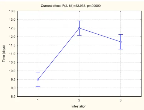
Current effect: F(2, 81)=52,933, p=,00000
1 2 3
Infestation 8,5 9,0 9,5 10,0 10,5 11,0 11,5 12,0 12,5 13,0 13,5 T im e ( d a y s )
Figure 2: Necessary feed time for the A. cajennense females to ingurgitate in three successive infestations in rabbits. Results demonstrated in average + standard deviation.
Infestation 1 2
2 0.000108
3 0.000108 0.017319
Pablo Henrique Nunes
DISCUSSION
The ticks are ectoparasites that can have as hosts: natural mammals, birds, reptiles or amphibians. Among the mammals, the rodents are the preferred ones (Lopes, 1998). In laboratory, several species of ticks feed on rabbits and due to it, they have been widely used in studies of their life cycle. However, what had already been observed is that rabbits can develop resistance against ticks, which cause changes in various biological parameters as in feeding process (decrease in tick number that can be fixed, weight reduction of engorged females and a longer time necessary for the engorgement), in the reproduction process (reduction in egg mass and diminishing in egg viability), depending on tick species considered (Brown, 1986).
Thus, the obtained data in this study with Amblyomma cajennense females showed that when they have been fixed in rabbits previously infested, unlike those fixed in naïve rabbits, feeding difficulty has been shown, such parameter measured by evaluating the engorged female weight and the time that they had been fixed in the host till the complete engorgement. The rabbits subjected to successive infestations showed resistance to ticks, demonstrated by a longer time required for engorgement and lower weight reached by engorged females. These data corroborate those of McGowan et al. (1980) for A. maculatum females fixed in rabbits immunized with tick extracts of the same species and those of Latif et al. (1988) for A. variegatum females fixed in re-infested rabbits. Using the hypersensitivity skin test induced by the extract of A. cajennense adult, Hlatshwayo et al. (2004a) showed that naive rabbits develop a kind of immediate reaction while pre-sensitized animals show besides the immediate response a late-type hypersensitivity, which could be related to the resistance acquisition to these ticks.
In general, the resistance against tick development varies from one host to another. In dogs, the engorgement weight of A. cajennense nymphs does not change significantly even after three successive infestations (Mukai et al., 2002). In horses, A. cajennense nymphs show a trend, although not significant, in reducing the engorgement weight after three infestations, while donkeys show significant resistance acquisition against A. cajennense in all the stages of life (Castagnolli et al., 2003). Furthermore, in goats, the nymphs weight decreased significantly right after the first infestation (Monteiro and Bechara, 2008).
Pablo Henrique Nunes
adult rabbits of the A. cajennense star tick after a single infestation. However, other biological parameters of the ticks should be investigated to confirm the effectiveness of such resistance.
ACKNOWLEDGMENTS:
Pablo Henrique Nunes
REFERENCES
BECHARA, G.H.; SZABÓ, M.P.J.; FERREIRA, B.R.; GARCIA, M.V., 1995. Rhipicephalus
sanguineus tick in Brazil: feeding and reproductive aspects under laboratorial conditions.
Brazilian Journal of Veterinary Parasitology, v.4, n.2, p.61-66.
BROWN, S.J., 1986. Rabbit-acquired resistance to Amblyomma americanum and western blot analysis of salivary gland-derived antigens. Host regulated developmental mechanisms in vector arthropods : proceedings of the Vero Beach Symposium, Vero Beach, Florida. Borovsky, D.Spielman, A. (eds.).- Vero Beach, FL (USA): University of Forida-IFAS. Florida Medical Entomology Laboratory. pp. 108-113.
CASTAGNOLLI, K.C.; FIGUEIREDO, L.B.; SANTANA, D.A.; DE CASTRO, M.B.; ROMANO, M.A.; SZABÓ, M.P.J., 2003. Acquired resistance of horses to Amblyomma
cajennense (Fabricius, 1787) ticks. Veterinary Parasitology, Amsterdam, v.117, p.271- 283.
FLECHTMANN, H. W. C., 1985. Ácaros de Importância Médico-Veterinária. Ed. Nobel, 3ª edição, 192p.
Pablo Henrique Nunes
HLATSHWAYO, M.; SZABÓ, M.P.J.; BECHARA, G. H.; MBATI, P.A., 2004b. Cross-reactivity between antigens from Amblyomma cajennense and A. hebraeum (Acari:Ixodidae). Journal of the South African Veterinary Association, 75: 40-42.
HELLER-HAUPT A., KAGARUKI L.K., VARMA M.G.R., 1996. Resistance and cross-resistance in rabbits to adults of three species of African ticks (Acari: Ixodidae). Experimental and Applied Acarology (20) 3: 155 – 165.
LABRUNA, M.B., 2004. Carta Acarológica. Revista Brasileira de Parasitologia Veterinária, v.13, suplemento 1, p199 – 204.
LATIF, A.A., NEWSON, R. M. and DHADIALLA, T. S., 1988. Feeding performance of Amblyomma variegatum (Acarina: Ixodidae) fed repeatedly on rabbits. Experimental and Applied Acarology. V.5, 1-2 p. 83-92.
LOPES, C.M.L.; LEITE, R.C.; LABRUNA, M.B.; OLIVEIRA, P.R.; BORGES, L.M.F.; RODRIGUES, Z.B.; CARVALHO, H.A.; FREITAS, C.M.V.; VIEIRA Jr., C.R., 1998. Host specificity of Amblyomma cajennense (Fabricius, 1787) (Acari: Ixodidae) with comments on the drop-off rhythm. Mem Inst Oswaldo Cruz, v.93, p.347-351.
MCGOWAN, M.J.; HOMER, J.T.; O'DELL, G.V.; MCNEW, R.W.; BARKER, R.W., 1980. Performance of ticks fed on rabbits inoculated with extracts derived from homogenized ticks
Amblyomma maculatum Kock. The Journal of Parasitology, 66: 42-48.
Pablo Henrique Nunes
MUKAI, L. S., NETTO, A. C., SZABÓ, M. P. J., BECHARA, G. H., 2002a. Development of resistance in dogs to nymphs of Amblyomma cajennense ticks (Acari:Ixodidae). Annals of the New York Academy of Sciences, v.969, p.180 - 183.
MUKAI, L. S., NETTO, A. C., SZABÓ, M. P. J., BECHARA, G. H., 2002b. Hypersensitivity induced in dogs by nymphal extract of Amblyomma cajennense ticks (Acari:Ixodidae). Annals of the New York Academy of Sciences, v.969, p.184 – 186.
RECHAV, Y., 1992. Naturally acquired resistance to ticks - a global view. Ins. Sci. Appl. 13, 495–504.
UTECH, K.B.W.; WHARTON, R.H. AND KERR, J.D., 1978. Resistance to Boophilus
microplus (Canestrini) in different breeds of cattle. Australian Journal of Agricultural
Research, 29: 885 – 859.
Pablo Henrique Nunes
4.2. Capítulo 2: Morphological changes in the salivary glands of Amblyomma cajennense females (Acari: Ixodidae) in different feeding stages on rabbits at first infestation
Autores: Pablo Henrique Nunes, Gervásio Henique Bechara e Maria Izabel Camargo Mathias
Periódico: Experimental and Applied Acarology (2008) 45:199–209
Situação: Publicado
Morphological changes in the salivary glands of Amblyomma cajennense females
(Fabricius, 1787) (Acari:Ixodidae) in different feeding stages on rabbits at first
infestation.
P.H. Nunes (1), G.H. Bechara (2), M.I. Camargo Mathias (3)
1 – Instituto de Biociências - UNESP campus de Rio Claro Av. 24 A nº 1515 Bela Vista, Rio Claro, São Paulo- Brazil CEP. 13506-900
3 - Departamento de Patologia Veterinária - UNESP campus de Jaboticabal Via de acesso Professor Paulo Donato Castellane, s/n, Rural
CEP. 14884-900 - Jaboticabal, São Paulo- Brasil
2 - Corresponding author
Instituto de Biociências - UNESP campus de Rio Claro Av. 24 A nº 1515 Bela Vista, Rio Claro, São Paulo- Brazil CEP. 13506-900,
Pablo Henrique Nunes
Resumo
Pablo Henrique Nunes
4.3. Capítulo 3: Secretion dynamics of the salivary gland on semi and engorged tick females Amblyomma cajennense Fabricius, 1787 (Acari: Ixodidae) during the second infestation in rabbit.
Authors: Nunes P.H., Bechara G.H., Camargo Mathias M.I.
Autores: Pablo Henrique Nunes, Gervásio Henique Bechara e Maria Izabel Camargo Mathias
Periódico: Experimental Parasitology
Pablo Henrique Nunes
Secretion dynamics of the salivary gland on semi and engorged tick females Amblyomma
cajennense (Fabricius, 1787) (Acari: Ixodidae) during the second infestation in rabbit.
Authors: Nunes P.H.(1), Bechara G.H.(2), Camargo Mathias M.I.(3)
1 – Instituto de Biociências - UNESP campus de Rio Claro Av. 24 A nº 1515 Bela Vista, Rio Claro, São Paulo- Brazil CEP. 13506-900
3 - Departamento de Patologia Veterinária - UNESP campus de Jaboticabal Via de acesso Professor Paulo Donato Castellane, s/n, Rural
CEP. 14884-900 - Jaboticabal, São Paulo- Brasil
2 - Corresponding author
Instituto de Biociências - UNESP campus de Rio Claro Av. 24 A nº 1515 Bela Vista, Rio Claro, São Paulo- Brazil CEP. 13506-900,
Pablo Henrique Nunes
RESUMO:
Pablo Henrique Nunes
ABSTRACT:
This study aimed to examine morpho-histochemically, the secretion dynamics of the female salivary glands on Amblyomma cajennense in semi and engorged stages, having as hosts rabbits previously infected. The acini I presented no changes compared to the females fixed on naive rabbits. In acini II the cells c1 there was more intense secretion release during the blood consumption, the c2 remained active until the food period ending, but without morphological changes when compared to the females fixed on naive rabbits. The other acini II cells presented the same development and degeneration pattern observed in females fixed on naive rabbits. In acini III the cells d remained active throughout the feeding process and an increased activity had been observed in engorged females, showing a necessity of increasing the glandular activity in female fixed on re-infested rabbits; such cells presented activity only at the beginning of the food process.
Pablo Henrique Nunes
INTRODUCTION
The salivary glands of ticks in Amblyomma cajennense as in Ixodidae are in general vital organs for the biological success of this group, presenting function in the production of various components including substances related to the attachment and feeding process of them (Schumaker, Serra Freire, 1991 ).
Functionally the salivary glands have a secretory cycle also defined by specific physiological and feeding states where the ticks are, the latter characterized as unfed or in feeding process, which is marked by one activity phase and another by degeneration (Sauer and Hair, 1986).
According to literature data, ticks on stage before the food (unfed) have the salivary glands in pre-secretory phase, as the gland cells, are somewhat active. The fixation process in the host would be the stimulus for the glands to be activated and to develop, which would not happen completely until the tick start its diet (Walker et al., 1985). Thus, during the tick feeding process the salivary glands would be in fully secretory activity. After the complete feeding (in females) and the detachment from the host, the glands would come into degeneration, a process which would be completed after oviposition, remaining only the duct system besides an amorphous mass of residual tissue (Walker et al., 1985 , Till, 1961), once at this moment the salivary glands would not be necessary for the biology of ectoparasites and the female would use the food consumed in the process of vitellogenesis (Coons et al., 1989; Tarnowski and Coons, 1989).
Hosts which are either infested by ticks or artificially immunized with extracts of salivary glands, develop resistance to ectoparasites, causing decrease in engorged female weight, in egg-laying rates by adults and low egg viability (Hlatshwayo et al., 2004). The rabbits would be examples of resistance development against ticks after one infestation, which would also depend on the tick species (Brown, 1986). The resistance is shown among other factors by changes in time that the ectoparasite takes to feed and the weight gained by engorged females (Castagnolli et al., 2003), which are directly controlled by the salivary glands.
Pablo Henrique Nunes
laboratory, to the feeding stages of semi and fully engorged, on second infestation condition, having rabbits as hosts.
MATERIAL AND METHODS
1 - MATERIAL
Ticks
To achieve the various steps, 60 females of Amblyomma cajennense semi, engorged and with two and five day’s post-engorgement had been used. Thus, 60 unfed couples, assigned by Prof. Dr. Gervásio Henrique Bechara, UNESP (Sao Paulo State University) Department of Veterinary Pathology Jaboticabal Campus (SP-Brazil) and obtained from the colony maintained in the laboratory under controlled conditions (29 ºC, 80% humidity and photoperiod of 12 hours) in the oven chamber BOD, had been placed in the host (rabbits) to be fed.
Hosts
Six healthy adult rabbits (New Zealand White) from UNESP, Botucatu Campus (SP, Brazil), weighing each one about 1kg (New Zealand White), had been used in the infestations. They had been fed with appropriate diet for the species and received water “ad libitum”.
Pablo Henrique Nunes
3 - METHODS
Histology
Haematoxylin of Harris-Eosin Aqueous (HE) (JUNQUEIRA, 1983).
In this study, the females had been placed in freezer (5 minutes) for anesthesia by thermal shock in the Histology Laboratory, UNESP (Sao Paulo State University) Department of Biology Rio Claro Campus –SP, Brazil. Moreover, the salivary glands had been removed in saline solution (7.5 g NaCl + 2.38 g of Na2HPO4 + 2.72 g of KH2PO4 + 1000 mL of distilled water) and fixed in 4% paraformaldehyde.
Then, the material had been dehydrated in increasing alcohol concentrations and transferred to imbibitions resin where it remained for 24 hours. The inclusion was in historesin Leica (Hidrixietilmetacrilato) and the blocks containing the material had been sectioned in microtome (3 µm / section). The sections had been taken on glass slides, rehydrated in distilled water for 1 minute, stained for 10 minutes in Harris hematoxylin and kept in water for 10 minutes to react. Thus, they had been washed in water, stained with eosin for 10 minutes and again washed with water to remove the dye excess. After drying the histological slides were covered with Canada balsam and they were subsequently observed and photographed in photomicroscope MOTIC BA 300.
Histochemistry
PAS (periodic acid-Schiff) reaction with counter-staining by the Methyl green (McManus, 1946).
Pablo Henrique Nunes
solution for 10 minutes. They had been washed again in distilled water for 1 minute. In sequence they had been placed for 1 hour in Schiff's reagent and then washed for 30 minutes in running water. The material had been counter-stained for 20 seconds with methyl green, washed, dried and mounted with Canada balsam for observation on MOTIC BA 300 light microscope and subsequent photographic documentation.
This study was approved by the Ethics Committee in Animal Research Protocol under No 44/2008 - EAEC - Faculty of Veterinary Medicine and Animal Science - UNESP, Botucatu, SP - Brasil.
RESULTS
Semi-engorged females
Acinus TYPE I
The acini I which has been directly connected to the common excretory duct (Figs. 1A, E) consists of a large central cell showing a large and rounded nucleus and smaller peripheral cells and with well marked homogeneous nucleus by hematoxylin (Figs . 1A, E).
The lumen of these acini, in this stage, is quite wide. It can be observed in some of their acini the pyknotic nuclei presence with irregular shape (Fig. 1A).
Acinus II
These acini connect to the intermediary ducts through an acinar valve (Fig. 1G). It had been observed in these acini three types of secretory cells a, b and c, the latter ones subdivided into c1, c2, c3 and c4.
The a cells are small and apparently have already eliminated all the cytoplasmic content (Fig. 1B). Their nuclei are rounded and two nucleoli which in most of the time have been very evident (Fig. 1B).
Pablo Henrique Nunes
The c cells are the ones which show the most amount of secretion granules in their cytoplasm (Figs. 1B, FI). The c1 show cytoplasm filled with secretion granules strongly PAS positive (Figs. 1F-G). These cells nuclei are large and rounded (Figs. 1B, F). The c2 cells are small and the cytoplasm is PAS negative. These cells show little cytoplasmic contents suggesting they might have already released all the secretion (Fig. 1F). The nuclei are rounded and show homogeneous staining (Fig. 1F). The c3 cells have cytoplasm filled with secretion granules strongly stained by both techniques (Figs. 1B, FI). The secretion granules in the cytoplasm of these cells have different sizes, however they are larger than those of cells c1 (Figs. 1B, FI). The nuclei of these cells is large and rounded (Figs. 1F, G, I). The c4 cells show fine granulation and negative PAS (Fig. 1H).
Acinus III
In this acini only is possible to differentiate the d cells (Fig. 1C-D, JK), once the others do not have secretion besides being very similar among them. The d cells are located next to the acinar duct and show secretion granules strongly stained by eosin (Figs. 1C-D), therefore they are negative PAS (Figs. 1J-K). The nuclei of these cells are rounded and present clear nucleoli (Figs. 1J-K).
Engorged females
Acinus I
The engorged female acini I are similar to those of semi-engorged with a large central cell with rounded nucleus and evident nucleoli (Figs. 2A, K) and the peripheral cells are smaller. However, the central cell shows now cytoplasm slightly stained by eosin, different from those found in semi-engorged female acini I (Figs 2A, K).
Acinus II
In engorged female acini II the cells a, b, c1, c2, c3 and c4 have also been distinguished.
Pablo Henrique Nunes
have large amounts of secretion granules in the cytoplasm (Figs. 2B-C, E, H), however some of them have very vacuolated cytoplasm (Figs. 2D, H). The c2 cells of engorged females, as the ones semi engorged are small and show negative cytoplasm to the PAS (Figs. 2B, H). The c3 cells show cytoplasm filled with secretion granules (Figs. 2C, E, and HJ). The secretion granules in the cytoplasm of these cells are larger than those in cells c1 (Fig. 2C, J). The c4 cells have cytoplasm with fine granulation and slightly stained by hematoxylin (Figs. 2D-E) but negative PAS (Fig. 2I).
Acinus III
In the acini III the only cells which were identified had been the d cells (Figs. 2F-G). These cells are bigger when compared to those found in semi-engorged females due to the secretion accumulation (Figs. 2F-G). The secretion granules in the cytoplasm of these cells are large and show strongly stained by eosin (Figs. 2F-G). The other acini cells do not show granulation in the cytoplasm, what makes difficult their identification, however, the hypertrophy of them causes the lumen of the acini becomes significantly reduced (Figs. 2F-G).
Females with 2 days post engorgement
Acinus I
In the females with 2 days post engorgement, the acini I show very similar to those found in engorged females (Fig. 2K). The central cytoplasm of the cell is weakly stained by eosin (Fig. 2K), different from that observed in semi-engorged females (Fig. 1).
Acinus II
The acini II cells of females with 2 days post engorgement have irregular shape, very vacuolated and pyknotic nuclei (Figs. 2L, M, O). For this reason it is not possible to identify the most of the other cell types except the type c3.
Pablo Henrique Nunes
As the cells in acini II, the acini III cells show irregular shape and pyknotic nuclei (Figs. 2N, P). These cells show fairly vacuolated cytoplasm and without secretion, which prevents that the identification of each cell could be made.
Females with 5 days post engorgement
Pablo Henrique Nunes
DISCUSSION
This study showed the results from the female salivary glands of Amblyomma
cajennense fixed in re-infested rabbits with the occurrence of small morphological differences
in these organs when compared to that observed in females of these ectoparasites fixed in naive rabbits.
The acini I of the female salivary glands studied showed no significant changes during the feeding period, corroborating Nunes et al. (2008) for females of A. cajennense fixed in naïve rabbits. This probably had been occurred due to the fact that the acini type I would not develop essential functions in the feeding process. Several authors in other studies reported that the salivary glands acini type I from several other species of Ixodidae, act in its osmotic control, as well as in the water capitation, either when it was outside the host or during the feeding (Rudolph & Knulle 1974/78; Sauer and Hair 1986; Grigorieva and Amosova 2009).
This study set clearly that in general the acini II of females fixed in re-infested rabbits are morphologically similar to those found in naive females fixed in rabbits described by Nunes et al. (2008). The same types of cells, i.e. a, b, c1, c2, c3 and c4 had been found here, where the a cells either in the semi-engorged engorged females showed or in the engorged practically did not present granule secretion in the cytoplasm, such result could be explained by the fact that the cells would be active mainly in the beginning of the food process. Some authors suggested that the cells would be involved mainly in the formation of the cement cone (Nunes et al. 2008, Nunes et al. 2006, Binnington 1978), structure of extreme importance for setting the ticks to the hosts and therefore, after ectoparasites attachment on hosts, these cells would lose their function, once after fixed on the host the females only loose themselves after the engorgement. The salivary glands studied here had been obtained from females in semi and engorged feeding stages which had already been fixed in the host.
Pablo Henrique Nunes
salivary glands b cells of female fixed in re-infested rabbits had been found active in both semi and engorged ones.
Regarding c1 cells, they presented a development during the whole blood consumption process and remained active until the end of it and only after that start showing the first degeneration signs. In the engorged females, the c1 cells showed cytoplasmic spaces absent of secretion (vacuolated) suggesting that almost all the secretion produced had been released until the end of the food process. These data differ from those found by Nunes et al. (2008) for females fixed in naive rabbits. In these engorged females’ c1 cells the cytoplasm had still been shown filled with secretion. Walker and Fletcher (1989) also observed a significant increase in size and amount of secretion in the salivary glands c1 cells of female R.
appendiculatus semi engorged fixed in resistant rabbits. The data therefore suggested a
greater consumption of secretion produced by the c1 cells when the ticks had been fed on resistant hosts.
The c2 cells analyzed here showed reduced size in relation to c subtypes and little secretion in the cytoplasm, either in half females or in engorged ones, suggesting that they probably do not answer to the host resistance with the secretion increase. However, the fact that these cells remain still active in the engorged females might be an evidence that they would still be necessary in engorged females, unlike the females fixed in naive rabbits where c2 cells had not been observed in engorged females (NUNES et al. 2008). According to Walker and Fletcher (1989) there would not be significant difference in the c2 cell structure of
R. appendiculatus female fixed in resistant rabbits when compared with those determined in
naive rabbits. Although in A. cajennense evident structural changes in c2 cells had not been observed; data obtained here have suggested an extension in activity time of these cells when the females had been fed on re-infested rabbits.
Pablo Henrique Nunes
This study when examined the acini III, showed secretion only in d cells of semi and engorged females. Furthermore, it was shown that the d cells might show significant increase either in its size or in the amount of cytoplasmic secretion granules when compared the semi with the engorged ones. These data differ from those found by Nunes et al. (2008) for females of A.cajennense fixed in naïve rabbits. According to the authors the d cells would present little secretion in semi-engorged females and little or no secretion in engorged females, suggesting therefore a response of the d cells in the resistance development by the host. These data differed from those described by Walker and Fletcher (1989) for females of R.
appendiculatus fixed in resistant rabbits, which reported that all acini III cells would play the
osmoregulation role after the attachment of the ticks on the hosts. However, the results obtained here show that the d cells still retain their secretory activity until the feeding period had been finished, while the others acini III cells would play osmoregulation activity, because they showed an increase in its cytoplasmic volume and lack of secretion responsible for the decrease in the acini lumen diameter which became extremely low, corroborating findings of other authors (Binnington, 1978; Kaufman and Sauer, 1983, Walker et al., 1985; Fawcet et al., 1986; L'Amoreaux and Coons, 1986 and Sonenshine, 1991).
Once the A. cajennense females finish the engorgement process, two days after the salivary glands began to show the occurrence of degeneration and, during five days after the engorgement; only the glandular ducts, using the cellular folds (cytoplasmic mass besides many apoptotic bodies), could be observed; making impossible to identify the cell types.
ACKNOWLEDGMENTS:
Pablo Henrique Nunes
REFERENCES
BINNINGTON, K.C. (1978). Sequential changes in salivary gland structure during attachment and feeding of the cattle tick Boophilus microplus. International Journal on Parasitology, 8: 97-115.
BROWN, S.J., 1986. Rabbit-acquired resistance to Amblyomma americanum and western blot analysis of salivary gland-derived antigens. Host regulated developmental mechanisms in vector arthropods : proceedings of the Vero Beach symposium, Vero Beach, Florida. Borovsky, D.Spielman, A. (eds.).- Vero Beach, FL (USA): University of Forida-IFAS. Florida Medical Entomology Laboratory. pp. 108-113.
CASTAGNOLLI, K.C.; FIGUEIREDO, L.B.; SANTANA, D.A.; DE CASTRO, M.B.; ROMANO, M.A.; SZABÓ, M.P.J., 2003. Acquired resistance of horses to Amblyomma
cajennense (Fabricius, 1787) ticks. Veterinary Parasitology, Amsterdam, v.117, p.271- 283,
2003.
COONS, L.B.; LAMOREAUX, W.J.; ROSELL-DAVIS, R.; TARNOWSKI, B.I., 1989. Onset of vitellogenin production and vitellogenesis, and their relationship to changes in the midgut epithelium and oocytes in the tick Dermacentor variabilis. Experimental and Applied Acarology 6, 291–305.
Pablo Henrique Nunes
FAWCETT, D.W.; BINNINGTON, K.; VOIGT, W.P., 1986. The cell biology of the ixodid tick salivary gland. In: sauer JR, Hair JA (eds) Morphology, physiology, and behavioral biology of ticks. Ellis Horwood Ltd, Chichester, Great Britain, pp 23–45.
GRIGORIEVA, L.A. AND AMOSOVA, L.I., 2008. Morphological changes of salivary glands os female ixodid ticks of subfamilies Ixidinae and Amblyomminae (Acari: Ixodidae) during feeding and their significance. Journal of Biochemestry and Physiology 44 (6), pp.662 – 635.
HLATSHWAYO, M.; SZABÓ, M.P.; BECHARA, G.H.; MBATI, P.A., 2004. Cross-reactivity between antigens from Amblyomma cajennense and A. hebraeum (Acari:Ixodidae). J. S. Afr. Vet. Assoc. 75(1) pp. 40-2.
JUNQUEIRA, L.C.U. & JUNQUEIRA, L.M.M. S., 1983. Técnicas básicas de citologia e histologia. Livraria Editora Santos. pp.48-81.
KAUFMAN, R.; SAUER, J.R., 1983. Ion and water balance in feeding ticks; mechanisms of tick excretion. In: Obenchain FD, Galun RL (eds) Physiology of ticks. Pergamon Press Ltd, New York, pp 213–44.
Pablo Henrique Nunes
NUNES, E.T.; CAMARGO MATHIAS, M.I.; BECHARA, G.H., 2006. Structural and cytochemical changes in the salivary glands of the Riphicephalus (Boophilus) microplus (Canestrini, 1887) (Acari: Ixodidae) tick female during feeding. Veterinary Parasitology 140, 114-123.
NUNES, P.H.; CAMARGO MATHIAS, M.I.; BECHARA, G.H., 2008. Morphological changes in the salivary glands of Amblyomma cajennense females (Fabricius, 1787) (Acari:Ixodidae) in different feeding stages on rabbits at first infestation. Experimental and Applied Acarology 45, pp. 199–209.
RUDOLPH, D. & KNULLE, W., 1974. Site and mechanism of water vapour uptake from the atmosphere in ixodid ticks. Nature 149: 84 – 85.
RUDOLPH, D. & KNULLE, W., 1978. Uptake of water vapour from air: process, site, and mechanism in ticks. In: Comparative physiology: water, ions and fluid mechanics (K. Schimdt-Nielsen, L. Bolio & S.H. P. Maddrell, eds.), Cambridge University Press. pp. 97 – 113.
SAUER, J.R.; HAIR, J.A., 1986. Morphology, physiology, and behavioral biology of ticks. Ellis Horwood, 509p.
Pablo Henrique Nunes
SONENSHINE, D.E., 1991. Biology of Ticks. Oxford University Press, New York, USA.
TARNOWSKI, B.I.; COONS, L.B., 1989. Ultrastructure of the midgut and blood meal digestion in the adult tick Dermacentor variabilis. Experimental and Applied Acarology 6, 263–289.
TILL, W.M., 1961. A contribution to the anatomy and histology of the brown ear tick
Rhipicephalus appendiculatus Neumann. Memoirs of Entomological Society of South Africa
6:1–124.
WALKER, A.R.; FLETCHER, J.D.; GILL, H.S., 1985. Structural and histochemical changes in the salivary glands of Rhipicephalus appendiculatus during feeding. International Journal on Parasitology, pp. 81-100.
Pablo Henrique Nunes
4.4. Capítulo 4: Secretory process of salivary glands of Amblyomma cajennense Fabricius, 1787 (Acari: Ixodidae) female ticks fed on resistant rabbits.
Autores: Pablo Henrique Nunes, Gervásio Henique Bechara e Maria Izabel Camargo Mathias
Periódico: Veterinary Parasitology
Pablo Henrique Nunes
RESUMO
Pablo Henrique Nunes
Secretory process of salivary glands of Amblyomma cajennense Fabricius, 1787, (Acari: Ixodidae) female ticks fed on resistant rabbits.
Authors: Nunes P.H.(1), Bechara G.H.(2), Camargo Mathias M.I.(3)
1 – Instituto de Biociências - UNESP campus de Rio Claro
Av. 24 A nº 1515 Bela Vista, Rio Claro, São Paulo- Brazil CEP. 13506-900
2 - Departamento de Patologia Veterinária - UNESP campus de Jaboticabal
Via de acesso Professor Paulo Donato Castellane, s/n, Rural CEP. 14884-900 - Jaboticabal, São Paulo- Brasil
3 - Corresponding author
Dra. Maria Izabel Camargo Mathias
Instituto de Biociências - UNESP campus de Rio Claro Av. 24 A nº 1515 Bela Vista, Rio Claro, São Paulo- Brazil CEP. 13506-900,
Pablo Henrique Nunes
Abstract
The ticks in general have great economic and health importance since infested animals have the milk and meat production reduced, and furthermore, causing high costs for the producers in its control. While feeding, the ticks can transmit a large amount of pathogens, including Rickettsia rickettsii responsible for the "spotted fever" or "mountains fever." It is known that animals infested by ticks or artificially immunized with their own salivary gland extracts, develop resistance resulting among other factors in an engorged female weight decrease, in the egg-laying by adults, in the low egg viability and in some cases the transmission capacity of pathogens reduction. This study aimed to examine morpho-histochemically the female salivary glands of Amblyomma cajennense semi and engorged, fed on resistant rabbits. The acini I had no changes compared with those of females fed on naive rabbits. The c cells of acini II showed signs of early degeneration, which may result in feed efficiency decrease. In acini III d cells the activity time had been longer; such occurrence had been associated with the time of female fixation which increased whose females had been fed on resistant hosts.
Pablo Henrique Nunes
Introduction
Adult ticks Amblyomma cajennense species are popularly known as star tick, horse tick or “rodoleiro” and their immature forms by “micuins”. To complete their life cycle these ectoparasites require blood meals from three hosts (Flechtmann, 1985). Such tick species has sanitary importance; once it can transmitte several diseases like spotted fever, also known as "Mountains fever," "tick fever", "black fever" or "blue disease" caused by the bacterium
Rickettsia rickettsii.
The salivary glands, in A. cajennense, as well as in other ixodid ticks considered vital organs to their biological success, since presenting a great diversity of functions relating primarily to the various components production which allow the ectoparasite attachment and feeding on their hosts (Schumaker and Serra-Freire , 1991, Szabó, 1995).
Some studies, which had already been made by other authors, showed that the host resistance development to ticks would get variation according to tick species and hosts (Heller-Haupt et al., 1996; Castagnolli et al., 2003).
Pablo Henrique Nunes
Scholars Heller-Haupt et al. (1996) concluded that A. hebraeum tick species could induce rabbits to the resistance with only one infestation such resistance also spread to other species such as A. variegatum and Rhipicephalus appendiculatus. According to Brown (1988), A. americanum tick species have already induced resistance in rabbits in the first infestation. Other studies show that inoculated rabbits with salivary gland extracts of A.
cajennense would develop hypersensitivity (Hlatshwayo, 2004) and this development would
be related to the resistance level acquired by the hosts (Willadsen et al., 1978).
Studies that evaluate the resistance degree acquired by the host have been performed through the biological parameter analysis related to feeding and egg laying by ectoparasites females. Among these factors, the relationship between female weight and their egg mass weight, as well the final weight reached by females and the whole feeding time (Castagnolli et al., 2003).
Pablo Henrique Nunes
Material and Methods
1- Material
Ticks
To achieve the various stages of this work 60 Amblyomma cajennense semi, and engorged females had been used with two and five days after engorgement. Thus, 60 unfed couples, given by Prof. Dr. Gervásio Henrique Bechara, UNESP Department of Veterinary Pathology, Jabotical (SP) Brazil campus and obtained from the colony maintained in the laboratory under controlled conditions (29 oC, 80% humidity and photoperiod of 12 hours) in BOD oven chamber, had been deposited in the host (rabbits) to be fed.
Hosts
In the infestations, six healthy adult rabbits (New Zealand White) had been used, weighing about 1 kg each, from the UNESP Central vivarium, Botucatu campus (SP, Brazil), fed with appropriate diet for the species and receiving “ad libitum” water.
Pablo Henrique Nunes
2. Methods
Histology
Staining by Hematoxylin-Eosin Aqueous Harris (HE) (JUNQUEIRA, 1983).
Amblyomma. cajennense females had been placed in freezers (5 minutes) for thermal
shock anesthesia, in the Histology Laboratory of Biology Department, UNESP Rio Claro-SP Brazil. In sequence, their salivary glands had been removed in saline (7.5 g NaCl + 2.38 g Na2HPO4 + 2.72 g KH2PO4 + 1000 mL of distilled water) and set in 4% paraformaldehyde.
Then the material was dehydrated in increasing alcohol concentrations and transferred to resin, remaining for 24 hours. Inclusion was in historesin Leica (Hidrixietilmetacrilato) and blocks containing the material were sectioned in microtome (3µm/section). The sections were collected on glass slides, rehydrated in distilled water for 1 minute, stained for 10 minutes in Harris hematoxylin and kept in water for 10 minutes to react. Moreover they were washed in water, stained with eosin for 10 minutes and washed again with water to remove the dye excess. After histological slides had been set with Canada balsam, afterwards they had been observed and photographed in a MOTIC BA 300 photomicroscope.
Histochemistry
PAS reaction (periodic acid-Schiff) Counter-staining by the Methyl Green (McManus
1946)
Pablo Henrique Nunes
rehydrated in distilled water for 1 minute and transferred to periodic acid solution for 10 minutes. They were washed again in distilled water for 1 minute. In the sequence they were placed for 1 hour in Schiff's reagent and then washed for 30 minutes in running water. The material was counter-stained for 20 seconds with methyl green, washed, dried and mounted with Canada balsam for observation and documentation in MOTIC BA 300 photomicroscope.
Pablo Henrique Nunes
Results
Semi-Engorged Females
Acini I
The type I acini is presented intact, with rounded shape, where its central cell is observed larger than the other peripheral ones and minor as well as their respective nuclei (Fig. 1A).
Acini II
The type II acini are the cells a, b, c1, c2, c3 and c4 (Figs. 1C-D, FI). Cells b, c1, c2 and c3 have a large amount of secretion in the cytoplasm (Figs. 1C, F, H-I). Some cells of acini have large vacuolated areas, mainly the cells c1 and c4 (Figs. 1G-I). The nuclei of cells c in general have, mostly dispersed chromatin and heterogeneous staining (Figs. 1C-D, FG). An irregular shape in the nuclei (Figs. 1F, G) had been often observed.
Acini III
The type III acini have intact cells and enlarged lumen (Figs. 1B, E, J). The only cells with activity characteristic are the cells d due to large amount of secretion present in the cytoplasm (Figs. 1B, E, J). The nuclei of cells d is slightly rounded with granular chromatin (Fig. 1E).
Engorged Females
Pablo Henrique Nunes
The type I acini in engorged females have similar characteristics to those found in semi-engorged females where the large central cells had been observed and with rounded nuclei. However, unlike the ones found in semi-engorged females, it was observed in these nuclei that the chromatin presents condensed and located peripherally, close to the nuclear envelope (Fig. 2A).
Acini II
In the engorged female acini II, only the cells c (c1, c2, c3, c4) (Figs. 2B-G, I) had been observed. The c1 and c3 have a large amount of cytoplasmic secretion granules (Figs. 2B, DF). In all cell subtypes has also been observed the presence of large vacuoles in the cytoplasm (Figs. 2B-C, E, G, I). Another cell feature of the engorged female acini II is the presence of highly irregular cell boundaries, where in many situations the cell begins to retract and separate from adjacent cells (Figs. 2B-F, I), as observed in semi-engorged females, the acini II nuclei cells of those engorged females exhibit condensed chromatin and with granular aspect (Figs. 2B-F). In some cases it is observed that the nuclear envelope is broken (Figs. 2B-C).
Acini III