Tolerance in Transgenic Tobacco by Reducing Oxidative
Stress and Improving Photosynthesis Machinery
Narendra Tuteja1*, Mst. Sufara Akhter Banu1, Kazi Md. Kamrul Huda1, Sarvajeet Singh Gill2, Parul Jain1,
Xuan Hoi Pham1¤, Renu Tuteja1
1International Centre for Genetic Engineering and Biotechnology, Aruna Asaf Ali Marg, New Delhi, India,2Stress Physiology and Molecular Biology Lab, Centre for Biotechnology, MD University, Rohtak, India
Abstract
Background:The DEAD-box helicases are required mostly in all aspects of RNA and DNA metabolism and they play a significant role in various abiotic stresses, including salinity. Thep68is an important member of the DEAD-box proteins family and, in animal system, it is involved in RNA metabolism including pre-RNA processing and splicing. In plant system, it has not been well characterized. Here we report the cloning and characterization ofp68from pea (Pisum sativum) and its novel function in salinity stress tolerance in plant.
Results:The pea p68 protein self-interacts and is localized in the cytosol as well as the surrounding of cell nucleus. The transcript of peap68is upregulated in response to high salinity stress in pea. Overexpression ofp68driven by constitutive cauliflower mosaic virus-35S promoter in tobacco transgenic plants confers enhanced tolerances to salinity stress by improving the growth, photosynthesis and antioxidant machinery. Under stress treatment, peap68overexpressing tobacco accumulated higher K+and lower Na+level than the wild-type plants. Reactive oxygen species (ROS) accumulation was remarkably regulated by the overexpression of peap68under salinity stress conditions, as shown from TBARS content, electrolyte leakage, hydrogen peroxide accumulation and 8-OHdG content and antioxidant enzyme activities.
Conclusions:To the best of our knowledge this is the first direct report, which provides the novel function of peap68 helicase in salinity stress tolerance. The results suggest that p68 can also be exploited for engineering abiotic stress tolerance in crop plants of economic importance.
Citation:Tuteja N, Banu MSA, Huda KMK, Gill SS, Jain P, et al. (2014) Pea p68, a DEAD-Box Helicase, Provides Salinity Stress Tolerance in Transgenic Tobacco by Reducing Oxidative Stress and Improving Photosynthesis Machinery. PLoS ONE 9(5): e98287. doi:10.1371/journal.pone.0098287
Editor:Jin-Song Zhang, Institute of Genetics and Developmental Biology, Chinese Academy of Sciences, China
ReceivedMarch 20, 2014;AcceptedApril 30, 2014;PublishedMay 30, 2014
Copyright:ß2014 Tuteja et al. This is an open-access article distributed under the terms of the Creative Commons Attribution License, which permits unrestricted use, distribution, and reproduction in any medium, provided the original author and source are credited.
Data Availability:The authors confirm that all data underlying the findings are fully available without restriction. All data are included in the manuscript.
Funding:Work on RNA/DNA metabolism and plant abiotic stress tolerance in N.T.’s laboratory is partially supported by Department of Biotechnology (DBT), Government of India and Department of Science and Technology (DST), Government of India. S.S.G. acknowledges the receipt of research grants from CSIR, New Delhi for helicase work. The funders had no role in study design, data collection and analysis, decision to publish, or preparation of the manuscript.
Competing Interests:The authors have declared that no competing interests exist. * E-mail: [email protected]
¤ Current address: Department of Plant Molecular Pathology and Abiotic Stress, The Institute of Agricultural Genetics, Hanoi, Vietnam
Introduction
The DEAD-box families of proteins are helicases conserved from bacteria to humans and are involved in a variety of nucleic acid metabolic processes such as replication, repair, recombina-tion, transcriprecombina-tion, pre-mRNA processing, RNA degradarecombina-tion, RNA export, ribosome assembly and translation [1–3]. At the sequence level, helicases have been classified into five superfamilies (SF1–SF5). The largest of these groups are SF1 and SF2. Most of these contain nine conserved helicase motifs, Q, I, Ia, Ib, II, III, IV, V and VI. All the helicases exhibit nucleic acid dependent ATPase activity which provides energy for the helicase action [3,4].
The p68 is a prototype member of DEAD-box family and is one of the best characterized helicases and it plays a very important role in cell/organ development and participates in a variety of
biological processes including pre-mRNA and pre-rRNA process-ing [5–6], rearrangement of RNA secondary structures [6], RNA splicing [7–8] and gene transcription. In human,p68is a RNA-binding protein endowed with an ATP-dependent RNA helicase, RNA-dependent ATPase and RNA-protein complex remodeling activities. In human malaria parasitePlasmodium falciparum,p68 has also been reported as a dual helicase and its helicase and ATPase activities are stimulated after phosphorylation with protein kinase C [9–10].
The DEAD-box RNA helicases are becoming a subject of attention as they play a significant role during development and stress responses in plants [11–14]. Each DEAD box RNA helicase is thought to be differentially regulated during development and in response to environmental stresses in plants [2,13]. Arabidopsis
overexpression of pea DNA helicase (PDH45) was shown to confer salt tolerance in tobacco and rice [12,14]. A rice DEAD-box RNA helicase (OsBIRH1) has been reported to function in defense responses against pathogen and oxidative stresses [16]. OsSUV3, a member of DEAD-box RNA helicase, has been shown to be involved in salinity stress tolerance recently [17]. Splicing factors are also known to affect the alternative splicing of many genes which lead to alter the gene expression through splicing factor networks and thereby involving in plant stress tolerance [18]. The plant homologue of animal splicing factor p68 has not been characterized in detail till date. The transcript ofArabibopsis thaliana
p68 DEAD-box RNA helicase (AtDRH1) was reported to be
accumulated at a high level and almost equally in every part of the
Arabidopsis plant [19]. Studies on MA16 (maize RNA-binding
protein) and ZmDRH1 (Z. mays DEAD-box RNA helicase 1)
revealed that these proteins might be part of a ribonucleoproteins complex involved in ribosomal RNA (rRNA) metabolism [20]. However, the precise role of DEAD-box RNA helicase, especially the role ofp68in abiotic stress tolerance, has not been reported so far. Here, we report detailed characterization of p68 from pea (Pisum sativum). Our results show that pea p68 self-interacts and the overexpression of pea p68 enhances the salinity stress tolerance in tobacco by controlling the generation of stress-induced reactive oxygen species (ROS) through modulating antioxidative defence machinery thereby protecting the photosynthesis and yield.
Materials and Methods
Construction of Plasmid for Tobacco Transformation Thepea p68gene was isolated by screening of pea cDNA library using a heterologous probe fromArabidopsis thaliana.The complete
ORF ofp68was cloned into pGEMT easy vector and sequenced
(Accession number: AF271892.1). The insert (1.8 kb) was release from pGEMT-p68by digesting with EcoRI and BamHI restriction enzymes and ligated into the MCS of pRT101. The
CaMV35S-p68-polyA cassette generated in pRT101 was then cut with Hind III and ligated to the MCS of pCAMBIA-1301 binary vector containing GUS as the reporter and hygromycin
phosphotrans-ferase as the selection marker gene. The pCAMBIA1301-p68
clone was then transformed intoAgrobacteriumLBA4404 strain for generation of transgenic tobacco using standard protocol [21]. Positive transgenic lines were confirmed by PCR using gene
specific primers (FP: 59- GAATTC
ATGTCGTATGTTCCTC-CACAC-3/EcoRI site bold) and (RP: 59- GGATCC
CCAT-TACCTACAAACATGACTGAT-3/BamHI site bold). The
transgenic tobacco plants were analyzed in two independent experiments with three technical replicates.
Transcript Analysis and Yeast-two-hybrid Assay
To analyze the expression of peap68transcripts, northern blots analysis was performed as described earlier [11]. The
pGBKT7-p68 and pGADT7-p68 were generated by inserting a PCR
fragment encoding the complete ORF of p68. The empty yeast
strain AH109, pGADT7, pGBKT7, pGADT7/pGBKT7,
pGADT7/pGBKT7-p68, pGADT7-p68/pGBKT7 and
pGBKT7-p68/pGADT7-p68 were transformed separately on
yeast cells. Yeast cells carrying all the plasmids were selected on the synthetic medium lacking Leu and Trp (SD-Leu-Trp-). The yeast cells were then streaked on SD medium [(Leu), (Trp), (His)] containing 30 mM 3-AT (3-Amino-1, 2, 4-triazole) to determine the expression of HIS3 nutritional reporter. Theb-galactosidase
expression of the fusion proteins encoded by pGBKT7-p68,
pGADT7-p68constructs was assayed by colony filter lift assays as per manufacturer instruction (Clontech).
In vivoLocalization by Immunofluorescence Staining and Confocal Microscopy
Polyclonal antibody against pea p68 was raised in rabbit as per the standard procedure. For in vivo localization, exponentially growing tobacco BY2 [11] suspension cells were fixed in 4% formaldehyde, permeabilized by cellulase and layered onto poly-L-lysine-coated cover slips. The cells were immunostained with p68-specific primary rabbit antibody in 1:2000 dilutions and Alexafluor 488-labeled goat antirabbit secondary antibody (Molecular Probes, Eugene, OR, USA) in 1:1000 dilutions. Counter staining of the cells with DAPI (4/, 6-diamidino-2-phenylindole), confocal laser scanning microscopy and image processing was carried out in a method described earlier [11].
Southern and Western Blots Analysis
Genomic DNA was isolated by CTAB method from PCR positivep68tobacco transgenic lines and WT. For southern blots analysis,,20mg of genomic DNA was digested with HindIII and resolved on agarose gel. The DNA was transferred to nylon membrane (Hybond N, Amersham Pharmacia, http://www. gelifesciences.com/), and hybridized with radiolabelledp68cDNA as described previously [12]. For western blot analysis, the total soluble proteins were isolated from the unstressed tissue samples of transgenic lines as well as WT plants and separated on 12%
SDS-PAGE. Western blot analysis was performed by using anti-p68
(1:5,000 dilutions) as primary and anti-rabbit (alkaline phosphatase conjugated antirabbit antibody-Sigma) as secondary antibody (1:12,500 dilutions). The blot was developed as per manufacturer’s protocol (Sigma, USA).
Histochemical GUS Staining and Morphological Characterization of Transgenic Plants
The seedlings of peap68overexpressing tobacco transgenic lines and WT were vacuum infiltrated for 10 m and histochemicalGUS
staining was performed by a method described earlier [22]. For
in vitropollen germination, mature pollen was isolated aseptically and cultured on pollen germination media (SMM: 0.3 M sucrose,
1.6 mM H3BO3, 3 mM Ca(NO3)2 4H2O, 0.8 mM
MgSO4.7H2O, 1 mM KNO3) supplemented with 100 and
200 mM NaCl and incubated at 26uC in the dark. The
germination status of the pollens was monitored and scored during a 7 d experimentation period. The sensitivity of seed germination to NaCl was assayed on MS agar plates saturated with 200 mM NaCl and incubated at 26uC under cool-white light for germination. Leaf disks assays, measurement of growth characteristics like shoot length, root length, leaf area and plant dry weight, measurement of tolerance index, determination of the total chlorophyll content and yield characteristics of transgenic and wild-type (WT) plants were performed as described earlier [14].
Measurement of Photosynthetic Characteristics and Photosystem II Activity (Fv/Fm)
The photosynthetic characteristics like net photosynthetic rate (PN), stomatal conductance (gs), intercellular CO2 concentration
(Ci) and chlorophyll fluorescence (Fv/Fm) of transgenic lines and WT plants were recorded on the fully expanded leaves using infra–
red gas analyzer (Li–6400, Li–COR, Lincoln, NE, USA) between 11:00 and 12:00 h. The conditions during the measurement were photosynthetically active radiation (PAR) 94568mmol m–2s–1, relative humidity 7566%, temperature 2862uC and an ambient CO2 concentration of 350mmol mol–1. The chlorophyll
also determined on the same leaves used for photosynthetic measurements after dark adaptation for 30 min.
Measurement of Ion Content
The Na+and K+ion content was measured as described earlier [23]. Salinity treated (0, 100 or 200 mM NaCl) leaves of the transgenic lines and WT plants were collected and rinsed with deionised water thoroughly. The fresh weight was determined for each sample. After drying (70uC for 48 h), dry weight was also measured. The samples are then subjected to an overnight digestion with HNO3/H2O2. The materials were picked in 2 M
HCl, and Na+
and K+
ion content was analyzed by using simultaneous inductively coupled plasma emission spectrometry (ICP trace analyzer, Labtam, Braeside, Australia).
Measurement of Oxidative Stress, Enzymatic Antioxidants (SOD, CAT, APX, GR) and Non-enzymatic Antioxidants (AsA and GSH) inp68Transgenic Lines and WT
Oxidative stress was detected by measuring thiobarbituric acid reactive substances (TBARS), hydrogen peroxide (H2O2) content,
electrolyte leakage and oxidative DNA damage (8-OHdG) in the leaves ofp68overexpressing transgenic lines and WT plants by a previously described method [14]. The salinity stressed (0, 100 or 200 mM NaCl) and fully expanded leaves from transgenic and WT plants were used for the measurement of enzymatic antioxidants (superoxide dismutase, catalase, ascorbate peroxidase, glutathione reductase) and non-enzymatic antioxidants (ascorbate and glutathione). Leaf samples (transgenic lines and WT) were homogenized with an extraction buffer containing 100 mM
potassium phosphate buffer (pH 7.0), 0.5% Triton X–100 and
1% polyvinylpyrrolidone (PVP) using pre-chilled mortar and pestle. The homogenate was centrifuged at 15,0006g for 20 min
at 4uC. The supernatant obtained after centrifugation was used for enzyme assays. Measurement of enzymatic antioxidants (SOD, CAT, APX, and GR) and non-enzymatic antioxidants (AsA and GSH) was performed as described previously [14]. All the measurements were carried out 3 weeks after initiating the NaCl treatment.
Transgenic Plants and Salinity Stress Tolerance
For the assay of sensitivity to salinity stress, 14 d-old seedlings of WT and the transgenic plants (grown on vermiculite pots) were transferred to nutrient solutions containing 200 mM NaCl. After 2 d, growth status of the transgenic lines was observed. In another experiment, 40 plants of each line and WT were grown in vermiculite pots and watered for 10 d and then 200 mM NaCl solution was irrigated for every 3 d interval up to 12 d. After treatments, morphological changes were observed. In order to investigate the effect of salinity stress, 35-day-old plants were also subjected to salt stress for 28 d and growth was observed till maturity and photographs were taken.
Statistical Analysis
All the treatments were performed in three independent trials with consistent results. The results from only one representative
experiment are shown, expressed as means6 standard errors.
Analysis of one-way variance (ANOVA) was performed on the data using SPSS (12.0 Inc., USA) to determine the least significant difference (LSD) for the significant data to identify the differences in the mean among the treatments. The means were separated by Duncan’s multiple range tests (DMRT). The graphs were prepared using Sigmaplot Ver. 11. Different letters indicate significant difference atP,0.05.
Results
Isolation and Sequence Analysis of Pea p68 cDNA The pea cDNA library was screened using a 1.9 kb cDNA fragment of p68 from Arabidopsis thaliana (kindly provided by Tetsuo Meshi of Kyoto University, Japan) as a probe. This resulted in the isolation of a positive clone of peap68(Figure S1A). The sequence analysis shows that the open reading frame (ORF)
of peap68cDNA (1.8 kb) encodes a protein of 622 amino acid
residues, with a calculated molecular mass of 67.65 kDa and a pI of 6.46. It also exhibits all the known canonical helicase motifs (Q, I, Ia, Ib, II-VI) (Figure S1B). Phylogenetic analysis identified the closest orthologs of peap68asArabidopsis thalianaandSaccharomyces cerevisiae p68followed byOryza sativa p68(Figure S1C).
Tissue Specific Distribution and Regulation of Peap68 Transcript in Response to Abiotic Stresses
To investigate the expression pattern of the transcript level of peap68in different parts of pea plant, the total RNA was isolated from root, leaf, tendril and flower and was subjected to northern blot analysis using 1.8 kb peap68 cDNA as the probe. For the internal control, 18S ribosome probe was used to show the equal loading. A single transcript of the expected size for pea p68
mRNAs was present in all the tissues (Figure 1A). Further, to analyze the expression of peap68under salinity stress, 7-day old pea seedlings were treated with 200 mM NaCl for 24 h and used for northern blot analysis. The results show that the pea p68
transcript was dramatically up-regulated (,4.8 fold) in response to
salinity stress (Figure 1B).
Peap68Interacts with Itself in the Yeast Two-hybrid System
To verify the self-interaction, we co-expressed BD-pea p68 with a construct encoding the full ORF of peap68fused to the GAL4 activation domain (AD-Pea p68). Yeast cells carrying both the
plasmids of BD-pea p68+AD-Pea p68 were able to grow on 3DO
media [Figure 1C (ii)]. All the clones including negative and positive control transformed in yeast cells were grown in YPD medium [Figure 1C (iii)]. Yeast cell carrying the plasmids of
BD-pea p68+AD-Pea p68 and Gb-pGADT7+Gb-pGBKT7 exhibited
b-galactosidase activity, indicating the expression of reporter genes [Figure 1C (iv)]. These results confirm that the full-length pea p68 is able to self-interact in the yeast two-hybrid system.
In vivoLocalization of p68
The in vivolocalization of p68 was also analyzed by immuno-fluorescence labeling and observed under confocal microscopy. Immunofluorescent labeling of tobacco BY2 cells with anti-pea p68 antibodies showed p68 protein exclusively localized in the cytoplasm as well as in the surrounding of cell nucleus (Figure 2).
Overexpression ofp68and Molecular Analysis of Transgenic Tobacco Lines
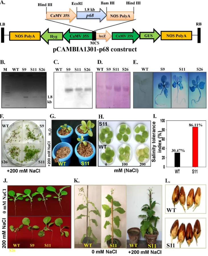
To establish the functional significance of the p68 gene, the complete ORF of the gene was cloned in Hind III site of
pCAMBIA1301 (Figure 3A). Peap68-pCAMBIA-1301 construct
was overexpressed in tobacco plants by using Agrobacterium
-mediated transformation. The integration of the transgene was confirmed by PCR (Figure 3B). The stable integration was also confirmed by Southern blot analysis (Figure 3C). Western blot detected,68 kDa band in transgenic lines (Figure 3D). The GUS
germination efficiency of the seeds was also tested. The seeds of
pea p68 transgenic lines and WT were grown on MS media
containing 200 mM NaCl (Figure 3F). In NaCl containing
medium, the seeds of pea p68 transgenic plants started to
germinate at 3 d with germination efficiency of more than 70% (data not shown). In contrast, the germination of WT seeds was observed at 9 d on the NaCl-containing medium. The highest germination rates of WT seeds were 35.6% on the
NaCl-containing MS medium (data not shown), which was lower than those of peap68transgenic seeds, suggesting the susceptibility of WT plants to 200 mM NaCl. The seedlings ofp68transgenic S11 and WT were transferred to pots supplemented with 200 mM NaCl, WT plants died after sometime whereas S11 survived and resumed the growth (Figure 3G). Salinity stress tolerance was also tested by leaf disk senescence assay. Leaf disks ofp68transgenic line S11 and WT were floated separately on 0 (H2O only), 100 or Figure 1. Expression analysis and self-interaction assay for peap68.(A). Transcript analysis of pea p68 by Northern blot analysis (R: root, S: shoot, T: tendril and F: flower tissue respectively). (B) Transcript level in response 200 mM NaCl stress respectively (UT: untreated samples). About 30mg of total was separated by electrophoresis, blotted and hybridized with the32P-labeled ORF of pea p68 cDNA (1.8 kb). For equal loading of RNA
200 mM NaCl for 72 h. The salinity induced damage was reflected in the degree of bleaching observed in the leaf tissue after 72 h. The leaves of WT plants were bleached, whereas, the leaf disks of S11 retained chlorophyll (Figure 3H). The measure-ment of salinity stress tolerance index of the 200 mM NaCl treated
p68transgenic line S11 and WT plants was made using the data of plant dry weight and it was noted that the tolerance potential of
p68transgenic line S11 was 86.11%, whereas, it was only 30.47% in WT plants (Figure 3I). The performance ofp68transgenic lines in the presence or absence of salt was similar to the WT plants, which revealed the potential of p68 in salinity stress tolerance (Figure 3J).
Thep68transgenic S11 line and WT plants were phenotypically similar when grown in absence of NaCl. To assess the effect of high salt (200 mM NaCl) on growth, morphology and
develop-ment of p68overexpressing and WT plants, the three-week old
seedlings were grown in the presence of continuous salt (200 mM NaCl). In the presence of NaCl some stress induced symptoms appeared on the transgenic plants but it still grew normally and set viable seeds, whereas the WT plants could not survive under continuous salinity stress (Figure 3K). In response to stress, thep68
transgenic set healthy pods similar to H2O grown WT plants
(Figure 3L). The results show that the p68 overexpressing
transgenic lines have better ability to tolerate salinity stress. p68 overexpressing transgenic lines were further analysed for physio-logical and biochemical parameters to understand the mechanism of salinity stress tolerance inp68overexpressing transgenic lines. Segregation Ratio and Seedling Survival of Transgenic and WT Plants Under Salinity Stress
To determine whether the salinity tolerance imparted byp68is functionally and genetically stable, the homozygous T2 progeny
was analyzed. Seeds from the T0 plants, when plated onto
hygromycin containing medium, segregated in 3:1 ratio (Table
S1). The percent seedlings survival of p68 overexpressing
transgenic lines and WT were also observed and it was found that 200 mM NaCl did not affect the seedlings survival and there
was no difference when compared with H2O grown WT plants
(Table S1).
Effect of Salinity on Germination of Pollens and Seeds of Peap68Transgenic Tobacco Plants
To compare the germination efficiency, first pollens of WT and
p68 transgenic lines were sown on pollen germination medium
(PGM) alone or on medium supplemented with either 100 or 200 mM NaCl. Without addition of NaCl, the germination of the pollen of pea p68 transgenic was similar to that of WT pollen (Figure S2). The germination of the WT pollen was repressed in the medium containing 100 mM NaCl while no germination was observed in 200 mM NaCl (Figure S2 B, C).
Effect of Salinity on Growth Performance and Yield Transgenic and WT Plants
Significant growth reduction was noted for the WT plants in
response to 200 mM NaCl treatment whilep68transgenic lines
resist the adverse effects of stress by maintaining vigorous growth. Growth performance measured in terms of shoot length, root length, leaf area and plant dry weight remained almost similar in
p68overexpressing transgenic lines and WT plants under 0 mM
NaCl (Figure S3). High concentration of salt (200 mM NaCl) significantly reduced the shoot length, root length, leaf area and plant dry weight of WT plants by 51.70, 57.31, 64.10 and 61.96%, in comparison to 0 mM NaCl. However, the decrease was 16.67, 19.55, 18.82 and 17.35% in the case of S9; 14.50, 18.04, 18.22 and 13.31% in the case of S11 and 18.85, 18.01, 16.92 and 15.62% in the case of S26 in comparison to their respective controls (Figure S3). Under salinity stress, transgenic lines maintained yield contributing parameters including time required for flowering, number of pods per plant, seed number per pod and seed weight per pod and therefore set normal seeds (Table S1).
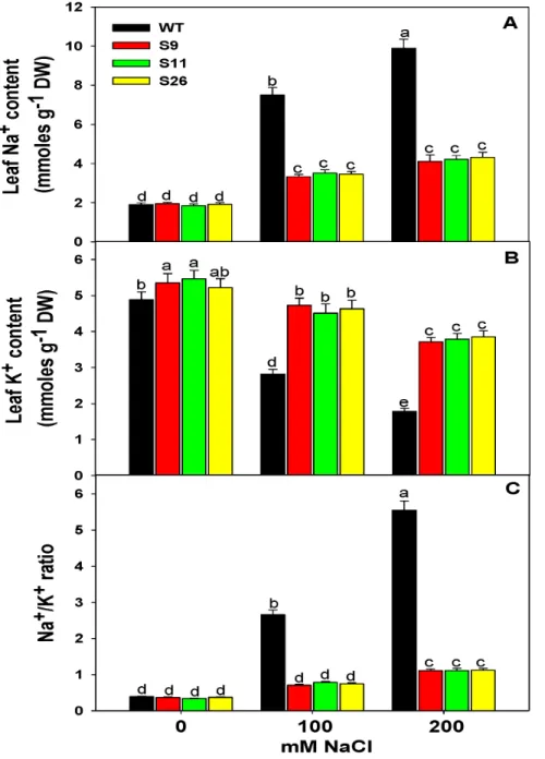
Ion (Na+and K+) Accumulation in Transgenic and WT
Plants under Salinity Stress
To observe Na+and K+accumulation, WT andp68transgenic plants were exposed to salt stress. No significant difference was observed in the accumulation of Na+
in between WT and p68
overexpressing transgenic lines without NaCl treatment. However, with the increasing salt concentration (100 or 200 mM NaCl), the
accumulation of Na+
was significantly increased in WT plants whilep68transformed plants accumulated less Na+
(Figure 4A). The pattern of K+
accumulation was similar in the leaves ofp68
Figure 2.In vivolocalization of the p68 in tobacco BY2 cells.The tobacco BY2 cells were fixed, permeabilized and immunostained with primary antibodies against p68 followed by Alexafluor 488-labeled secondary antibody and then counterstained with DAPI. A single confocal image is shown. (A) Image of cell stained with DAPI (blue). (B) Immunofluorescently stained cell (green). Anti-p68 labeling is restricted to the nucleus and cytosol. (C) Superimposed image of cell.
overexpressing transgenic lines and WT in response to 0 mM NaCl treatment (Figure 4B) but in response to salinity stress,p68
overexpressing transgenic lines retained more K+
compared to the WT plants (Figure 4B). Furthermore, p68 overexpressing trans-genic lines showed lower Na+
/K+
ratio in comparison to WT plants (Figure 4C), which reflect the potential of transgenic lines to tolerate salinity stress.
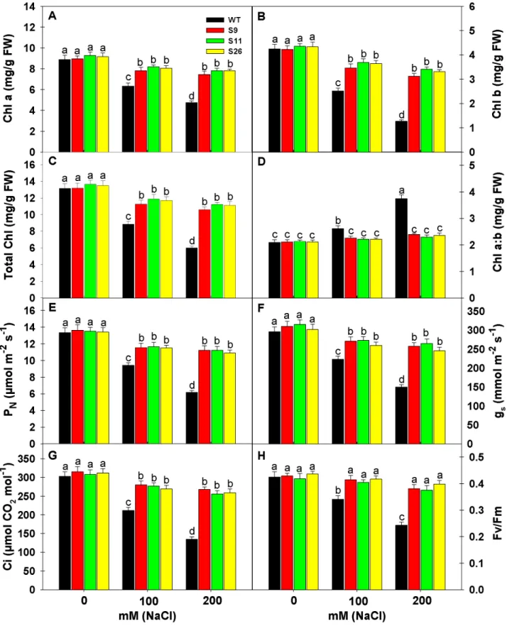
Effect of Salinity on Chlorophyll Content, Photosynthesis and Chlorophyll Fluorescence of Transgenic and WT Plants
To access the effect of salinity on Chlorophyll (Chl), Chl a, Chl
b, total Chl and Chl a: b was measured in p68 overexpressing
transgenic lines and WT plants (Figure 5A–D). Salinity stress (100 or 200 mM NaCl) significantly reduced the Chla, Chlb and total seedlings were transferred to without or with NaCl in nutrient solution (0 and 200 mM NaCl). (K) WT andp68overexpressing plant (S11) in soil pots supplied without or with 200 mM NaCl solution. Note that the WT plant could not sustain growth under salinity stress. (L) Phenotypic comparison of pods grown in water and stress condition of WT and transgenic line (S11).
doi:10.1371/journal.pone.0098287.g003
Figure 4. Analysis of ion content in transgenic and WT tobacco plants.(A) Na+
content in the leaves of transgenic and WT plants. (B) K+
content in the leaves of transgenic and WT plants. (C) Na+
/K+
ratio in the leaves of transgenic and WT plants. The leaves were exposed to 0, 100 or 200 mM NaCl for 3 weeks of salinity treatment. Values are mean6SE (n = 3). Different letters on the top of bars indicate significant differences at P,
0.05 level as determined by Duncan’s multiple range test (DMRT). The results are representative of similar results obtained from two independent experiments.
Chl in transgenic lines and WT plants but the extent of reduction was higher in WT thanp68overexpressing transgenic lines. The reduction in Chl a, Chl b and total Chl under 200 mM NaCl in WT was 46.56, 70.12 and 54.18% in comparison to 0 mM NaCl, whereas, the reduction was 16.65, 26.24 and 19.73% in the case of S9; 15.71, 21.61 and 17.59% in the case of S11 and 14.83, 23.73 and 17.69% in the case of S26 in comparison to their respective controls (Figure 5A–C). Under salinity stress the Chl content remained significantly higher in transgenic than WT plants. In WT plants the Chl a:b ratio followed the reverse pattern as of Chl content and significantly increased with the increasing salt concentration, whereas, no significant change was noted in the case of transgenic lines (Figure 5D). It reflects that Chl b was severely affected by salinity stress than Chl a under increasing salt concentration in WT plants than transgenic lines which led to significant increase in Chla: b ratio.
Salinity stress affects the photosynthetic functions at various levels, therefore, the photosynthetic parameters like net photosyn-thetic rate (PN), stomatal conductance (gs) and internal CO2(Ci)
were measured inp68overexpressing transgenic lines (S9, S11 & S26) and WT plants under different salinity levels (0, 100 or 200 mM NaCl) (Figure 5E–G). High level of salinity (200 mM NaCl) significantly reduced the photosynthetic parameters but the extent of reduction was several folds higher in WT plants than transgenic lines. It is interesting to note that p68overexpressing transgenic lines maintained higher photosynthesis than WT even under 0 mM NaCl. The reduction in PN, gs andCiof WT plants
was 53.75, 49.32 and 55.44% under 200 mM NaCl in comparison to their controls, whereas, the decrease in PN, gs andCiof S9, S11
and S26 was 17.67, 16.77, 14.92%; 16.94, 15.87, 16.88% and 18.85, 18.54, 16.99%, respectively in comparison to their controls. Chlorophyll fluorescence measurement is one of the most commonly used parameters to study the ecophysiology of plants under salinity stress. To understand the response of salt stress, we measured maximal efficiency of PSII photochemistry (Fv/Fm) in
p68 overexpressing transgenic lines and WT plants (Figure 5H). High salinity stress (100 or 200 mM NaCl) significantly reduced the Fv/Fm, whereas, it remained unaltered in transgenic lines. Statistically insignificant change in Fv/Fm inp68 overexpressing transgenic lines reflects that PSII complex did not suffer damage under NaCl stress.
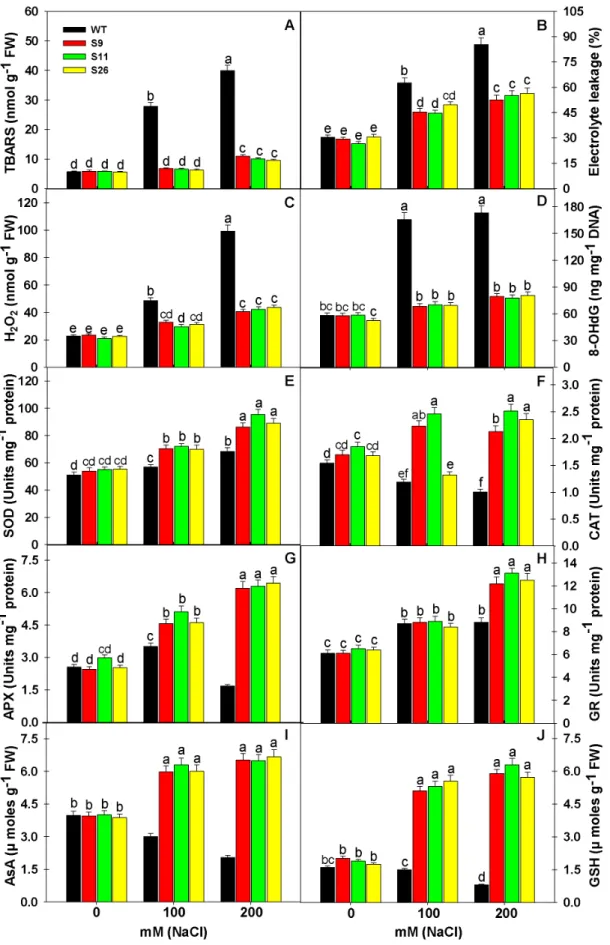
Less Oxidative Stress inp68Transgenic Lines than WT under Salinity Stress
Abiotic stresses including salinity cause overproduction of ROS, which leads to oxidative stress in plants. Therefore, the indicators of oxidative stress such as lipid peroxidation (TBARS content), H2O2 content, electrolyte leakage and oxidative DNA damage
were studied inp68overexpressing transgenic lines and WT plants (Figure 6A–D). High concentration of salt (100 or 200 mM NaCl) significantly increased the extent of oxidative damage and it was significantly higher in WT as compared to p68 transgenic lines. The response of oxidative stress parameters reflects non-significant
difference in the values of TBARS content, H2O2 content,
electrolyte leakage and oxidative DNA damage (8-OHdG) betweenp68overexpressing transgenic lines and WT plants under
no salt i.e. 0 mM NaCl. The increase in TBARS, H2O2,
electrolyte leakage and 8-OHdG under 200 mM NaCl in WT was 600.00, 337.44, 181.03 and 197.59% in comparison to 0 mM NaCl, whereas, the increase was 86.61, 72.76, 79.53 and 37.67% in the case of S9; 74.48, 102.39, 107.08 and 32.48% in the case of S11 and 70.20, 93.78, 84.48 and 53.62% in the case of S26 in comparison to their respective controls (Figure 6A–D). Overall, it is noted that WT plants suffered maximum oxidative damage
reflected in terms of peroxidation of lipids, nucleic acid and electrolyte leakage, whereas, p68 overexpressing transgenic lines experienced less oxidative stress, therefore less oxidative damage.
Overexpression ofp68Enhances ROS Scavenging Capacity inp68Transgenic Lines
Salinity stress is known to cause ROS induced oxidative damage in plant cells. Therefore, we analyzed the response of enzymatic (SOD, CAT, APX and GR) and non-enzymatic antioxidants (AsA and GSH) inp68overexpressing transgenic lines and WT plants under salinity stress. Antioxidant defense machinery protects the plant cells from ROS induced oxidative damage. SOD constitutes the primary step of cellular defense, where SOD dismutates O2Nto
H2O2and O2. The increase in SOD activity was noted in bothp68
transgenic lines and WT plants but activity of SOD was several folds higher as compared to WT plants. Significant increases in
SOD activity in p68 overexpressing transgenic lines under
200 mM NaCl was 59.92, 73.13, 61.12%, whereas, it was just 33.92% in WT in comparison to their respective controls
(Figure 6E). The H2O2 generated by the action of SOD is
restricted through the action of CAT and APX, which reduces H2O2to water. In the present study, reverse to SOD activity, the
CAT and APX activity decreased significantly in WT plants but
CAT and APX activity showed significant increase in p68
overexpressing transgenic lines under 200 mM NaCl (Figure 6F– G). The decrease in CAT and APX activity was 34.80 and 34.12% in WT plants under 200 mM NaCl. The activity of CAT and
APX increased in p68 overexpressing transgenic lines under
200 mM NaCl by 25.29, 153.06%; 35.68, 111.07% and 39.88, 154.19%, respectively, in comparison to their respective controls (Figure 6F–G). GR catalyzes the NADPH-dependent reduction of oxidized GSSG to the reduced GSH. Here, GR activity showed an increasing trend under salinity stress in bothp68overexpressing transgenic lines and WT plants but maximum significant increase
was seen in p68 transgenic lines (Figure 6H). Increase in GR
activity in p68 overexpressing transgenic lines under 200 mM
NaCl was 100.00, 101.54 and 95.31%, respectively, in comparison to their respective controls, whereas, it was just 44.26% in WT plants. Overall, the upregulation of ROS scavenging antioxidant enzymes inp68overexpressing transgenic lines protected the plant cells from the damaging effect of ROS generated by salinity stress. AsA is the only antioxidant buffer in the apoplast and a key antioxidant that reacts with superoxide and hydroxyl radicals, whereas, GSH is a major non-enzymatic scavenger of ROS. The efficient coordination of AsA and GSH in AsA-GSH cycle can protect the plants from salinity induced oxidative damage. Under control condition (0 mM NaCl), ASC and GSH contents were
same in control and transformed plants. However, p68
overex-pression enhances the pool of AsA and GSH in the transgenic lines. The decrease in AsA and GSH content was 48.74 and 50.00% in WT plants under 200 mM NaCl. The content of AsA and GSH increased inp68overexpressing transgenic lines under 200 mM NaCl by 65.32, 193.53%; 61.84, 233.33% and 72.35, 226.86%, respectively, in comparison to their respective controls (Figure 6F–G).
Discussion
Figure 5. Effects of salinity stress on chlorophyll content, photosynthesis and chlorophyll fluorescence of transgenic and WT tobacco plants. (A) Chlorophyll a content. (B) Chlorophyll b content. (C) Total Chlorophyll content. (D) Ratio of Chlorophyll a and b. (E) Measurement of Net photosynthetic rate (PN). (F) Measurement of stomatal conductance (gs). (G) Measurement of internal CO2concentration [(Ci)].
(H) Measurement of Chlorophyll fluorescence (Fv/Fm). Data’s were recorded from WT andp68overexpressing transgenic tobacco lines after 3 weeks exposure to 0, 100 or 200 mM NaCl treatment. Each value represents mean of three replicates6SE. Means were compared using ANOVA. Data followed by the same letters are not significantly different at P,0.05 as determined by least significant difference (LSD) test.a, b, cindicate significant differences at P,0.05 level as determined by Duncan’s multiple range test (DMRT).
Figure 6. Overexpression of pea p68showed less oxidative damage by modulating the ROS machinery under salinity stress.
[8,11–12,14,17,24]. In this study, a novel DEAD-box helicase gene (p68) was isolated from pea plant which is specifically upregulated in response to salinity. The transcript of peap68 is also accumulated at a high level and almost equally in every part (roots, leaves, tendrils and flowers) of the pea plant. This result is consistent with the earlier report of transcript analysis ofAtDRH1
gene expression inA. thaliana[19]. Therefore, this gene could be a potential candidate for developing stress-tolerant transgenic plants. The pea p68 protein contains all conserved domains that are characteristic of the DEAD-box proteins including ‘Q’ and ‘GG’ motifs [25]. The ability of p68 to interact with itself indicated that the oligomerization of p68 may be essential for its proper functioning. The p68 is exclusively localized to the cytoplasm and also seems to surround the nucleus of cell. Similar results were reported for another pea helicase (PDH45) [26]. Previously nuclear localization of some DEAD-box helicases (eg. mammalian
eIF4AIII and A. thaliana UAP56) showed the involvement in nonsense-mediated mRNA decay, mRNA splicing and export [27–28]. RNA splicing process involves the association and dissociation of the pre-mRNA with snRNAs, which may be facilitated by RNA helicases. Previous report in animal system has shown that p68 and p72 RNA helicases are the crucial factors required for efficient RNA splicing [29–30]. Both p68 and p72 interact with the U1 small nuclear ribonucleoprotein that recognizes the 59 splice site [31]. The p68 protein, devoid of RNA helicase or ATPase activity also inhibited the dissociation of U1 from 59splice site, and downregulation of DDX5 resulted in the accumulation of unspliced RNA [29]. The role of plant p68 in splicing has not been yet reported. However, there may be possibility that the p68 is sustaining the splicing activity during the stress condition and therefore allowing the p68 overexpressing tobacco plant to survive under the salinity stress condition.
Previously a number of studies demonstrated that DEAD-box RNA helicases are involved in regulating many stages of plant development processes including plant morphogenesis, embryo-genesis, pollen tube guidance, floral meristems, flowering, plastids and seed development [32–34]. In plant the first report of stress induced helicase gene came by cDNA microarray analysis of 1300
Arabidopsisgenes where the authors reported a DEAD-box helicase gene (accession number AB050574) as a cold stress-inducible gene suggesting a new role of helicases in stress signaling [35]. Later, many plant DEAD-box helicases were identified and found to be activated in response to changing environmental conditions [2,13– 14,36]. Evidence is accumulating that transcript of PDH45 and PDH47 was found to be induced in response to high salt, dehydration and cold stresses [11,13–14,17,26]. In barley, a salt-responsive transcript HVD1 is induced under salt stress, cold stress, and ABA treatment [37]. AvDH1 is another DEAD-box helicase gene from the halophyte dogbane plant that also strongly upregulated in response to salinity and low temperature [24]. Under normal growth conditions relatively high level of basal expression of the peap68gene in different plant parts implies its function in growth and/or development processes. Under salt
treatment, a single species of pea p68 mRNA was detected
abundantly and constitutively in the tissues examined. This indicated that basic activity of cells might be regulated by pea
p68under salt stress.
Genome-wide expression analysis of many DEAD-box helicase genes have been identified and suggested that these genes might be stress regulated [38]. Overexpression analysis in different DEAD-box helicases has been shown to provide multiple abiotic stress tolerance in crop plants by regulating different signalling pathways
[11–12,17]. For example, overexpression ofPDH45andOsSUV3
gene provided salinity stress tolerance in tobacco and rice respectively [12,17]. LOS4 and RCF1 mutant analysis in
Arabidopsis was found to play an important role in response to cold and heat stress [8,15]. Our study showed that overexpression of peap68provides salinity stress tolerance in tobacco.
The reduction in leaf chlorophyll content under abiotic stress has been attributed to the destruction of chlorophyll pigments in various crop plants [3,39–40]. We observed that stress-induced chlorophyll loss was enhanced in WT plant while transgenic lines retained more chlorophyll. This finding has strong correlation with the previous studies in other DEAD-box helicases [12,41–42].
Hence it indicated that overexpression of pea p68 could have
positive effects on the growth and photosynthetic metabolism process. Under stress condition, maintenance of the Na+
, K+ levels
and Na+
/K+
ratio are important indicators for plant stress
tolerance [43–45]. The pea p68 overexpressing tobacco plants
accumulated less Na+and more K+as compared to the WT plants.
Higher K+ content implies delayed leaf senescence in the
transgenic lines. Previously it was reported that lower cytosolic K+
content controlled endonuclease and caspase-like proteases activity in the cells causing leaf senescence under stress conditions [40,46]. Transgenic tobacco plants also extruded more Na+
from cells and, as a result, low Na+
content was detected which indicated that overexpression of peap68enhances stress tolerance
in transgenic plants. In wheat increased K+ uptake and Na+
extrusion was reported in response to salinity stress [47]. The pea p68 overexpressing transgenic plants are also capable of absorbing
more water and diluting the Na+
content. Earlier transgenic approaches with transporter proteins and DESD-box helicase (PDH45) showed that lower Na+
/K+
ratio helps plants to respond to salinity stress tolerance [14,45,48]. We found lower Na+/K+ ratio in transgenic lines which suggested overexpression of peap68
might restrict the entry of Na+
ions into the cells thereby protecting photosynthetic machinery from abiotic stresses.
Stress also leads to the rapid production of ROS including H2O2in plant tissues that ultimately cause damages to the cell
membrane and other cellular components such as plasma membrane, mitochondria and chloroplasts [14,40]. Hence, to avoid any stress-induced injuries plant needs to develop efficient mechanism to remove excess ROS from cells. Enzymatic ROS-scavenging and non-enzymatic antioxidants system are such mechanisms in the plant cells that prevent ROS induced oxidative damage [49–51]. In the present study, stress-induced high H2O2
accumulation was noted more in WT plants in comparison to the transgenic lines. Therefore, we speculated that transgenic plants
are capable of removing excess H2O2 from the cells, hence
preventing cellular damages. Catalase, ascorbate and peroxidase are the major enzymes that are known to be involved in scavenging of cellular production of H2O2[52–53]. Interestingly
in this study the activity of these enzymes increased in transgenic lines in response to stress treatment. This indicated that overexpressing lines could readily scavenge H2O2 either
decom-Measurement of the activities of superoxide dismutase (E), Catalase (F), ascorbate peroxidase (G) guaiacol peroxidase (H) ascorbate content (I) and glutathione content (J) in the leaves of WT and p68 overexpressing transgenic tobacco exposed to 0, 100 or 200 mM NaCl after 3 weeks of salinity treatment. Values are mean6SE (n = 3). Different letters on the top of bars indicate significant differences at P,0.05 level as determined by Duncan’s multiple range test (DMRT). The results are representative of similar results obtained from two independent experiments.
posing it through increased activity of catalase or by ascorbate through the ascorbate/glutathione cycle. Previously, a number of overexpression studies have shown an increased activity of catalase, ascorbate and peroxidase in response to abiotic stress treatment [14,54–56].
The involvement of DEAD-box helicases in various metabolic processes in plant cells might have general implications. The present study provides new insights into the novel function of p68 DEAD-box protein in conferring salinity stress tolerance in transgenic tobacco plants without affecting yield. The
overexpres-sion of stress-induced DEAD-box p68 can also provide a good
example of the exploitation of factors of RNA metabolism pathways including splicing factor for enhanced agricultural production of economically important crops under stress condi-tions.
Supporting Information
Figure S1 Cloning and sequence analysis ofp68.
(TIF)
Figure S2 Percent pollen germination of WT and transgenic lines under salinity stress.
(TIF)
Figure S3 Effects of salinity stress on growth parameter of transgenic and WT tobacco plants.
(TIF)
Table S1 Comparison of segregation ratio, plant seed-lings survival and various yield parameters of the WT and transgenic plants.
(DOCX)
Acknowledgments
We thank Mr Kazi Mostaque Ahmed for helpful corrections.
Author Contributions
Conceived and designed the experiments: NT RT. Performed the experiments: MSAB KMKH PJ SSG XHP. Analyzed the data: NT MSAB KMKH SSG. Contributed reagents/materials/analysis tools: NT. Wrote the paper: NT MSAB KMKH RT.
References
1. Tanner NK, Linder P (2001) DExD/H box RNA helicases: from generic motors to specific dissociation functions. Mol Cell 8: 251–262.
2. Owttrim GW (2006) RNA helicases and abiotic stress. Nucleic Acids Res 34: 3220–3230.
3. Tuteja N, Gill SS, Tuteja R (2012) Helicases in Improving Abiotic Stress Tolerance in Crop Plants. In: Tuteja N, Gill SS, Tiburcio AF, Tuteja R (eds) Improving crop resistance to abiotic stress. Wiley-VCH Verlag GmbH and Co, KGaA, Germany, 433–445.
4. Tuteja N (2000) Plant cell and viral helicases: essential enzymes for nucleic acid transactions. Cri Rev Plant Sci 19: 449–478.
5. Bates GJ, Nicol SM, Wilson BJ, Jacobs AM, Bourdon JC, et al. (2005) The DEAD box protein p68: a novel transcriptional coactivator of the p53 tumour suppressor. EMBO J 24: 543–553.
6. Fuller-Pace FV (2006) DExD/H box RNA helicases: multifunctional proteins with important roles in transcriptional regulation. Nucleic Acids Res 34: 4206– 4215.
7. Ishizuka A, Siomi MC, Siomi H (2002) A Drosophila fragile X protein interacts with components of RNAi and ribosomal proteins. Genes Dev 16: 2497–2508. 8. Guan Q, Wu J, Zhang Y, Jiang C, Liu R, et al. (2013) A DEAD box RNA helicase is critical for pre-mRNA splicing, cold-responsive gene regulation, and cold tolerance inArabidopsis. Plant Cell 25: 342–356.
9. Pradhan A, Chauhan VS, Tuteja R (2005a) A novel ‘DEAD box’ DNA helicase from Plasmodium falciparum is homologous to p68. Mol Biochem Parasitol 140: 55–60.
10. Pradhan A, Chauhan VS, Tuteja R (2005b) Plasmodium falciparum DNA helicase 60 is a schizont stage specific, bipolar and dual helicase stimulated by PKC phosphorylation. Mol Biochem Parasitol 144: 133–141.
11. Vashisht A, Pradhan A, Tuteja R, Tuteja N (2005) Cold and salinity stress-induced pea bipolar pea DNA helicase 47 is involved in protein synthesis and stimulated by phosphorylation with protein kinase C. Plant J 44: 76–87. 12. Sanan-Mishra N, Pham XH, Sopory SK, Tuteja N (2005) Pea DNA helicase 45
overexpression in tobacco confers high salinity tolerance without affecting yield. Proc. Natl Acad. Sci. USA 102: 509–514.
13. Vashisht A, Tuteja N (2006) Stress responsive DEAD box helicases: a new pathway to engineer plant stress tolerance. J Photochem Photobiol 84: 150–160. 14. Gill SS, Tajrishi M, Madan M, Tuteja N (2013) A DESD-box helicase functions in salinity stress tolerance by improving photosynthesis and antioxidant machinery in rice (Oryza sativa L. cv. PB1). Plant Mol Biol 82: 1–22. 15. Gong Z, Dong CH, Lee H, Zhu J, Xiong L, et al. (2005) A DEAD box RNA
helicase is essential for mRNA export and important for development and stress responses inArabidopsis. Plant Cell 17: 256–267.
16. Li D, Zhang H, Wang X, Song F (2008) OsBIRH1, a DEAD box RNA helicase with functions in modulating defence responses against pathogen infection and oxidative stress. J Exp Bot 59: 2133–2146.
17. Tuteja N, Sahoo RK, Garg B, Tuteja R (2013) OsSUV3 dual helicase functions in salinity stress tolerance by maintaining photosynthesis and antioxidant machinery in rice (Oryza sativaL. cv. IR64). Plant J 76: 115–127.
18. Staiger D, Brown JW (2013) Alternative splicing at the intersection of biological timing, development, and stress responses. Plant Cell 25: 3640–3656. 19. Okanami M, Meshi T, Iwabuchi M (1998) Characterization of a DEAD box
ATPase/RNA helicase protein ofArabidopsis thaliana. Nucleic Acids Res 26: 2638–2643.
20. Gendra E, Moreno A, Alba MM, Pages M (2004) Interaction of the plant glycine-rich RNA-binding protein MA16 with a novel nucleolar DEAD box RNA helicase protein from Zea mays. Plant J 38: 875–886.
21. Horsch RB, Fry JE, Hoffmann NL, Eichholtz D, Rogers SG, et al. (1985) A Simple and General Method for Transferring Genes into Plants. Science 227: 1229–1231.
22. Jefferson RA, Kavanagh TA, Bevan MW (1987) GUS fusionsb-glucuronidase as a sensitive and versatile gene fusion marker in higher plants. EMBO J 6: 3901– 3907.
23. Munns R, Wallace PA, Teakle NL, Colmer TD (2010) Measuring soluble ion concentrations (Na+
, K+ , Cl
-) in salt-treated plants. In: Sunkar R (ed-) Methods in molecular biology, vol 639. Plant stress tolerance: methods and protocols. Humana Press. Springer, New York, 371–382.
24. Liu HH, Liu J, Fan SL, Song MZ, Han XL, et al. (2008) Molecular cloning and characterization of a salinity stress-induced gene encoding DEAD-box helicase from the halophyte Apocynum venetum. J Exp Bot 59: 633–644.
25. Tanner NK, Cordin O, Banroques J, Doe’re M, Linder P (2003) The Q motif: a newly identified motif in DEAD box helicases may regulate ATP binding and hydrolysis. Mol Cell 11: 127–138.
26. Pham XH, Reddy MK, Ehtesham NZ, Matta B, Tuteja N (2000) A DNA helicase fromPisum sativumis homologous to translation initiation factor and stimulates topoisomerase I activity. Plant J 24: 219–229.
27. Ferraiuolo MA, Lee C, Ler LW, Hsu JL, Costa-Mattioli M, et al. (2004) A nuclear translation-like factor eIF4AIII is recruited to the mRNA during splicing and functions in nonsense-mediated decay. Proc. Natl Acad. Sci. USA 101: 4118–23.
28. Kammel C, Thomaier M, Sørensen BB, Schubert T, La¨ngst G, et al. (2013) ArabidopsisDEAD-Box RNA Helicase UAP56 Interacts with Both RNA and DNA as well as with mRNA Export Factors. PLoS ONE 8: e60644. 29. Lin C, Yang L, Yang JJ, Huang Y, Liu ZR (2005) ATPase/helicase activities of
p68 RNA helicase are required for pre-mRNA splicing but not for assembly of the spliceosome. Mol. Cell Biol 25: 7484–7493.
30. Janknecht R (2010) Multi-talented DEAD-box proteins and potential tumor promoters: p68 RNA helicase (DDX5) and its paralog, p72 RNA helicase (DDX17). American J Transl Res 2: 223–234.
31. Liu ZR (2002) p68 RNA helicase is an essential human splicing factor that acts at the U1 snRNA- 59splice site duplex. Mol Cell Biol 22: 5443–5450. 32. Shimizu KK, Ito T, Ishiguro S, Okada K (2008) MAA3 (MAGATAMA3)
helicase gene is required for female gametophyte development and pollen tube guidance inArabidopsis thaliana. Plant Cell Physiol 49: 1478–1483.
33. Ohtani M, Demura T, Sugiyama M (2013)Arabidopsisroot initiation defective1, a DEAH-box RNA helicase involved in pre-mRNA splicing, is essential for plant development. Plant Cell 25: 2056–2069.
34. Kanai M, Hayashi M, Kondo M, Nishimura M (2013) The plastidic DEAD-box RNA helicase 22, HS3, is essential for plastid functions both in seed development and in seedling growth. Plant Cell Physiol 54: 1431–1440. 35. Seki M, Narusaka M, Abe H, Kasuga M, Yamaguchi-Shinozaki K, et al. (2001)
Monitoring the expression pattern of 1300Arabidopsisgenes under drought and cold stresses by using a full-length cDNA microarray. Plant Cell 13: 61–72. 36. Mahajan S, Tuteja N (2005) Cold, salinity and drought stresses: an overview.
Arch Biochem Biophys 444: 139–158.
38. Kant P, Kant S, Gordon M, Shaked R, Barak S (2007) STRS1 and STRS2, two DEAD-box RNA helicases that attenuateArabidopsisresponses to multiple abiotic stresses. Plant Physiol 145: 814–830.
39. Zhang ZH, Liu Q, Song HX, Rong XM, Ismail AM (2012) Responses of different rice (Oryza sativaL.) genotypes to salt stress and relation to carbohydrate metabolism and chlorophyll content. Afr J Agric Res 7: 19–27.
40. Huda KM, Banu MS, Garg B, Tula S, Tuteja R, et al. (2013) OsACA6, a P-type IIB Ca2+
ATPase promotes salinity and drought stress tolerance in tobacco by ROS scavenging and enhancing the expression of stress-responsive genes. Plant J 76: 997–1015.
41. Dang HQ, Tran NQ, Gill SS, Tuteja R, Tuteja N (2011) A single subunit MCM6 from pea promotes salinity stress tolerance without affecting yield. Plant Mol Biol 76: 19–34.
42. Sahoo RK, Gill SS, Tuteja N (2012) Pea DNA helicase 45 promotes salinity stress tolerance in IR64 rice with improved yield. Plant Signal Behav 7: 1037– 1041.
43. Cuin TA, Betts SA, Chalmandrier R, Shabala S (20080 A root’s ability to retain K+
correlates with salt tolerance in wheat. J Exp Bot 59: 2697–2706. 44. Gao Z, He X, Zhao B, Zhou C, Liang Y, et al. (2010) Overexpressing a putative
aquaporin gene from wheat, TaNIP, enhances salt tolerance in transgenic Arabidopsis. Plant Cell Physiol 51: 767–775.
45. Hill CB, Jha D, Bacic A, Tester M, Roessner U (2012) Characterization of ion contents and metabolic responses to salt stress of differentArabidopsisAtHKT1;1 genotypes and their parental strains. Mol Plant 6: 350–368.
46. Shabala S (2009) Salinity and programmed cell death: unraveling mechanisms for ion specific signalling. J Exp Bot 60: 709–712.
47. Munns R, James RA, Lauchli A (2006) Approaches to increasing the salt tolerance of wheat and other cereals. J Ex Bot 57: 1025–1043.
48. Rajagopal D, Agarwal P, Tyagi W, Singla-Pareek SL, Reddy MK, et al. (2007) Pennisetum glaucum Na+
/H+
antiporter confers high level of salinity tolerance in transgenicBrassica juncea. Mol. Breed 19: 137–151.
49. Gill SS, Tuteja N (2010) Reactive oxygen species and antioxidant machinery in abiotic stress tolerance in crop plants. Plant Physiol Biochem 48: 909–939. 50. Gill SS, Singh LP, Gill R, Tuteja N (2012) Generation and scavenging of
reactive oxygen species in plants under stress. In: Tuteja N, Gill SS, Tiburcio AF, Tuteja R (eds) Improving crop resistance to abiotic stress. Wiley-VCH Verlag GmbH and Co. KGaA, Germany, 49–70.
51. Bhattacharjee S (2012) The language of reactive oxygen species signaling in plants. J Bot. doi:10.1155/2012/985298.
52. Willekens H, Langebartels C, Tire C, Van Montagu M, Inze D, et al. (1994) Differential expression of catalase genes inNicotiana plumbaginifolia(L.). Proc. Natl Acad. Sci. USA 91: 10450–10454.
53. Noctor G, Foyer CH (1998) Ascorbate glutathione: Keeping active oxygen under control. Annu Rev Plant Physiol Plant Mol Biol 49: 249–279. 54. Jiang M, Zhang J (2002) Water stress-induced abscisic acid accumulation
triggers the increased generation of reactive oxygen species and upregulates the activities of antioxidant enzymes in maize leaves. J Exp Bot 53: 2401–2410. 55. Luna CM, Pastori GM, Driscoll S, Groten K, Bernard S, et al. (2004) Drought
controls on H2O2 accumulation, catalase (CAT) activity and CAT gene
expression in wheat. J Exp Bot 56: 417–423.