ARTICLE
Neuropsychological performance in patients
with subcortical stroke
Perfil neuropsicológico em pacientes com lesões vasculares subcorticais
Silviane Pinheiro Campos de Andrade1, Sônia Maria Dozzi Brucki2, Orlando Francisco Amodeo Bueno3,
José Ibiapina Siqueira Neto4
Vascular cognitive impairment (VCI) has superseded vas-cular dementia and multi-infarct dementia as the concept to be used in cognitive decline secondary to cerebrovascular disease1.
he prevalence of dementia increases with age, and the prevalence of vascular dementia in epidemiological stud-ies varstud-ies between 22 and 26.28%2,3. An epidemiologic study
in Brazil showed a prevalence of 9.3% for vascular dementia (VaD) among demented participants aged 64 years and older4.
Studies in tertiary outpatient clinics report a prevalence of vas-cular dementia between 24.9 and 32.25%5,6, and of 36.9% in a
sample with presenile dementia7.
VCI is a risk factor for the development of dementia. Wentzel8
observed that half of the subjects with vascular cognitive impair-ment with no deimpair-mentia (VCIND) developed deimpair-mentia over a ive-year period. he current indings suggest that poor mental lexibility and verbal retrieval in the context of preserved func-tion in other domains may characterize the prodromal stage of VaD9. Cognitive impairment with vascular disease often occurs
in domains of selective or divided attention, executive function ( frontal lobe function) and processing speed10.
Chui11 compared groups with dementia secondary to
vas-cular disease presenting ischemic subcortical lesions (VADIS)
1 Psychologist, Federal University of São Paulo (UNIFESP), São Paulo SP, Brazil; 2 MD, PhD, UNIFESP, São Paulo SP, Brazil, Santa Marcelina Hospital, São Paulo SP, Brazil; 3 Psychologist, UNIFESP, São Paulo SP, Brazil;
4 MD, Federal University of Ceará (UFC), Fortaleza CE, Brazil.
Correspondence: Sonia Maria Dozzi Brucki; Rio Grande 180/61; 04018-000 São Paulo SP - Brasil; E-mail: [email protected]
Support: This work was supported by a grant provided by the Fundação de Amparo à Pesquisa do Estado de São Paulo - FAPESP (04/11436-5) and Associação Fundo de Incentivo à Psicofarmacologia - AFIP.
Conflict of interest: There is no conflict of interest to declare.
Received 03 November 2010; Received in final form 21 November 2011; Accepted 28 November 2011
ABSTRACT
Vascular cognitive impairment (VCI) is characterized by cognitive compromise predominantly of executive dysfunction. Objectives: To assess cognitive functions in VCI, focusing on executive functions, to observe functional losses in relation to activities of daily living (ADLs) and to detect early symptoms prior to the onset of dementia. Methods: We evaluated healthy subjects matched for gender, education and age to patients with diagnosis of subcortical vascular disease who had a stroke classified into three groups: 1) vascular lesions and no impairment; 2) vascular cognitive impairment with no dementia (VCIND); 3) vascular dementia (VaD). Results and discussion: The performance on neu-ropsychological tests differed among groups, worsening with increased impairment level. The probable VaD group demonstrated impaired performance in memory, processing speed and verbal production, while the VCIND group showed attention deficits. Conclusion: Impairment in executive functions and difficulties in ADLs allow us to differentiate levels of impairment in groups of subcortical vascular disease.
Key words: cognitive impairment, vascular dementia, neuropsychological assessment, activities of daily living.
RESUMO
O comprometimento cognitivo vascular (CCV) é caracterizado por comprometimento cognitivo predominantemente sob a forma de disfunção executiva. Objetivos: Avaliar as funções cognitivas no CCV, enfocando as funções executivas, observar as perdas funcionais em relação às atividades cotidianas (AVDs) e detectar os primeiros sintomas antes do início da demência. Métodos: Foram avaliados indivíduos controles saudáveis pareados por sexo, escolaridade e idade com pacientes com diagnóstico de doença vascular subcortical que sofreram derrame classificados em três grupos: 1) lesões vasculares sem déficit; 2) comprometimento cognitivo vascular sem demência (CCVSD); 3) demência vascular (DV). Resultados e discussão: O desempenho em testes neuropsicológicos diferiu entre os grupos, sendo o desempenho tanto pior quanto maior o comprometimento. O grupo DV provável demonstrou desempenho prejudicado na memória, velocidade de processamento e produção verbal, enquanto o grupo CCVSD apresentou déficit de atenção. Conclusão: Prejuízos nas funções executivas e dificuldades em AVDs permitem diferenciar os níveis de comprometimento nos grupos de doença vascular subcortical.
against those with Alzheimer disease (AD), and veriied that the pattern of cognitive damage in VADIS is characterized by more extensive compromise of executive function, although with greater preservation in recognition memory.
Several studies in the literature have sought to describe a model of cognitive proile for VaD, but, due to the varied forms of clinical manifestations, dependent on the types and loca-tions of the vascular lesions, no single model exists for the en-tity12. Rockwood et al.13 suggest that the cognitive syndrome of
vascular disease is characterized by the predominance of exec-utive dysfunction in contrast with the deicits of memory and language typical of patients with AD. Subcortical VaD presents a more homogeneous cognitive proile characterized by execu-tive dysfunction and less severe impairment of memory12-15.
he aims of this study were to evaluate the cognitive func-tions in patients with subcortical vascular lesions, focusing on executive functions, and observe the functional losses with impairment in ADLs of each patient group in order to detect primary symptoms thereby enabling dementia prevention.
METHODS
Participants
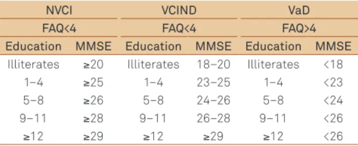
A total of 50 patients with vascular subcortical lesions (in-cluding white periventricular matter, thalamus and basal gan-glia lesions) were selected from medical iles of an outpatient memory clinic of the Santa Marcelina Hospital and matched (according to age, gender and education) with 50 healthy elder-ly controls. he patients who had subcortical lesions accord-ing to medical records were invited to participate in the study and classiied by an experienced neurologist. All patients were at a chronic phase more than three months after conirmed stroke. Medical history, neurological examination and MRI were performed in all subjects. Patients were classiied into three groups according to scores obtained on the Mini-Mental State Examination (MMSE), based on the education level for the adapted Brazilian scale16, and on the Functional Activity
Questionnaire (FAQ)17 (Table 1). his classiication was later
conirmed by the results of neuropsychological tests: no vascu-lar cognitive impairment (NVCI): FAQ<4 and MMSE scores ≥
median value for education; VCIND: FAQ<4 and MMSE score between – 1.5 SDs and mean value for educational level; and VaD: patients with probable VaD according to NINDS-AIREN criteria16, FAQ>4 and MMSE<1.5SDs from the mean.
he exclusion criteria for the patients were: aphasia; se-vere paresis in dominant hand; uncorrected visual or hear-ing deicit; use of medications that directly afect the central nervous system, such as benzodiazepines, anticholinergics drugs and neuroleptics; orthopedic or rheumatologic distur-bances that could afect the exam; presence of areas of corti-cal ischemia on magnetic resonance imaging.
Control group
Healthy control subjects were matched by age and educa-tional level to each group of vascular patients. Controls were selected based on FAQ scores of less than 4 points and MMSE scores above the mean for each educational level. he control group included individuals drawn from the community, spouses and patients of the other specialties who fulilled the inclusion criteria. Controls had no psychiatric or neurological diseases; no use of drugs with central nervous system action; no risk factors for cerebrovascular disease; no uncorrected sensory impairment; and no motor compromise due to orthopedic or rheumatologic conditions. Controls were matched with each patient group.
After assessment of the functional activities using the FAQ and brief cognitive evaluation through the MMSE, pa-tients and controls were submitted to a neuropsychological examination with a psychologist blinded to their condition.
he demographic data (gender, age and education) of the study are shown in Table 2, with baseline MMSE and FAQ scores. We found no signiicant diferences among the vari-ables age, education and gender.
he study was approved by the Ethics Committees of the Federal University of São Paulo and of Santa Marcelina Hospital. All subjects gave prior written consent for partici-pation in the study.
Neuropsychological assessment
Neuropsychological assessment included digit span18:
attention span, mental control and capacity to retain and manipulate verbal information; logical memory18: episodic
memory in immediate and delayed recall; Wisconsin Card Sorting Test (WCST)18: the capacity to form concepts and
mental lexibility; Trail Making Test (TMT) A and B18: speed
of visuospatial search, attention and mental lexibility; Rey Complex Figure – copy and delayed recall18: planning,
non-verbal memory, visual and space perception and praxis. It was applied the Clock Test – CLOX19, in which subjects are
asked to “draw a clock showing 1.45” on the back of a score sheet, through which a black circle is displayed in the bottom right-hand corner (CLOX 1). Once completed, the examiner draws the clock as required in front of the subject, placing 12, 6, 3 and 9 irst, then setting the hands to ‘1.45’ and making them into arrows. he subject then has to copy the examiner’s clock (CLOX 2).
Table 1. Classification into three groups according to scores obtained on the Mini-Mental State Examination (MMSE), based on the education level for the adapted Brazilian scale.
NVCI VCIND VaD
FAQ<4 FAQ<4 FAQ>4 Education MMSE Education MMSE Education MMSE
Illiterates ≥20 Illiterates 18–20 Illiterates <18
1–4 ≥25 1–4 23–25 1–4 <23
5–8 ≥26 5–8 24–26 5–8 <24
9–11 ≥28 9–11 26–28 9–11 <26
≥12 ≥29 ≥12 ≥29 ≥12 <26
Moreover, in the assessment were include Stroop Test18:
inhibition of a habitual answer rather than an unusual an-swer, and to mental lexibility, impulsiveness and inhibitory control; verbal luency: tests were accomplished by phonemic category – FAS18 is requested to utter as many words as
pos-sible starting with the letter ‘F’, ‘A’ and ‘S’; semantic category – animals18, it is expected that the patient state, within 60
sec-onds, as many names of animals as possible; test of excluding letters20: assesses executive function while requiring control of
impulsive answers. It consists of the generation of words for one minute, excluding a letter (in this case, letter E).
Functional evaluation of the activities of daily living
Participants were evaluated according to the FAQ scores.
STATISTICAL ANALYSIS
he Statistical Package for the Social Sciences (SPSS) – version 13.0 was used for data analysis. Non-parametric tests were used to evaluate non-categorical variables among groups. he Mann-Whitney test veriied possible diferences among VaD, VCIND and NVCI groups and their respective controls. he Kruskal-Wallis Test was applied to compare VaD, VCIND and NVCI groups for performance on neuropsychological tests. Diferences
among the groups were analyzed using the Mann-Whitney Test, and Spearman’s Correlation was performed to verify the relation-ship between ADLs and the neuropsychological tests. he level of signiicance was set at 0.05. A forward stepwise logistic regression analysis (diagnosis of yes/no cognitive impairment as dependent variable) was performed to establish the independent predic-tive ability of tests that presented p values less than 0.05 after the Mann-Whitney test. In order to perform the logistic regression analysis, we used the Mann-Whitney test to compare groups (yes or no cognitive impairment), to determine which tests could be elected for the regression model. To this, it was considered that a variable could be eligible if p<0.200. We applied the logistic regres-sion analysis in function on the dependent variable ‘group’. Once we found the variables (neuropsychological tests) eligible to the modeling process through the application of logistic regression analysis, we constructed a regression model with four variables, which achieved the balance of the regression model.
RESULTS
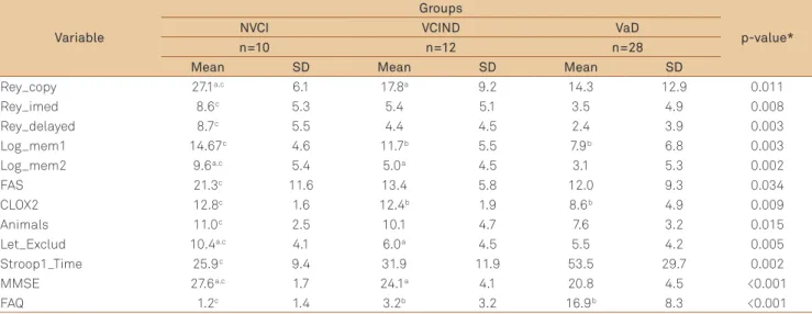
Table 3 shows the mean and standard deviations on those tests revealing a signiicant diference among patient groups, with diferences between each group. We found poorer performance associated with severity of clinical
Table 2. Demographic data of study and control groups.
Variable
Groups
p-value
Controls NVCI VCIND VaD
n=50 n=12 n=10 n=28
Sex F=25 M=25 F=9 M=3 F=4 M =6 F=12 M=16 0.2651
Age Mean (SD) 66.1 (9.1) 65.3 (10.3) 62.3 (9.2) 67.8 (8.1) 0.3782
Education Mean (SD) 5.9 (4.5) 7.8 (4.1) 4.9 (3.9) 5.3 (5.0) 0.1932
MMSE Mean (SD) 28.5 (2.1) 27.6 (1.7) 24.1 (4.1) 20.8 (4.5) <0.0012
1Chi-Square; 2Kruskal-Wallis Test; MMSE: Mini-Mental State Examination; NVCI: no vascular cognitive impairment; VCIND: vascular cognitive impairment with
dementia; VaD: vascular dementia.
Table 3. Scores on neuropsychological tests and statistical differences among patient groups.
Variable
Groups
p-value*
NVCI VCIND VaD
n=10 n=12 n=28
Mean SD Mean SD Mean SD
Rey_copy 27.1a.c 6.1 17.8a 9.2 14.3 12.9 0.011
Rey_imed 8.6c 5.3 5.4 5.1 3.5 4.9 0.008
Rey_delayed 8.7c 5.5 4.4 4.5 2.4 3.9 0.003
Log_mem1 14.67c 4.6 11.7b 5.5 7.9b 6.8 0.003
Log_mem2 9.6a.c 5.4 5.0a 4.5 3.1 5.3 0.002
FAS 21.3c 11.6 13.4 5.8 12.0 9.3 0.034
CLOX2 12.8c 1.6 12.4b 1.9 8.6b 4.9 0.009
Animals 11.0c 2.5 10.1 4.7 7.6 3.2 0.015
Let_Exclud 10.4a.c 4.1 6.0a 4.5 5.5 4.2 0.005
Stroop1_Time 25.9c 9.4 31.9 11.9 53.5 29.7 0.002
MMSE 27.6a.c 1.7 24.1a 4.1 20.8 4.5 <0.001
FAQ 1.2c 1.4 3.2b 3.2 16.9b 8.3 <0.001
*Kruskal-Wallis Test; NVCI: no vascular cognitive impairment; VCIND: vascular cognitive impairment with no dementia; VaD: vascular dementia; MMSE: mini-mental state examination; FAQ: functional activity questionnaire. aStatistically significant differences between NVCI and VCIND groups (Mann-Whitney test). bSignificant difference
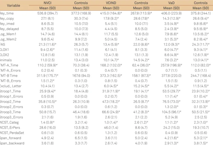
Table 4. Comparison among VaD, VCIND and NVCI groups with their respective controls.
Variable NVCI Controls VCIND Controls VaD Controls Mean (SD) Mean (SD) Mean (SD) Mean (SD) Mean (SD) Mean (SD)
Rey_time 536.8 (394.7) 377.3 (168.9) 415.0 (164.8)# 267.8 (113.9)# 406.0 (223.9) 375.8 (244.1)
Rey_copy 27.1 (6.1) 30.3 (7.4) 17.8 (9.2)# 28.6 (7.9)# 14.3 (12.9)# 26.6 (9.4)#
Rey_imed 8.6 (5.3) 10.5 (7.0) 5.4 (5.1) 10.0 (7.1) 3.5 (4.9)# 9.8 (6.8)#
Rey_delayed 8.7 (5.5) 10.0 (7.3) 4.4 (4.5) 8.5 (5.0) 2.4 (3.9)# 9.5 (6.9)#
Log_Mem1 14.7 (4.6) 14.4 (6.1) 11.7 (5.5) 12.6 (5.0) 7.9 (6.8)# 13.5 (6.2)#
Log_Mem2 9.6 (5.4) 9.9 (7.2) 5.0 (4.5) 7.4 (2.4) 3.1 (5.3)# 8.2 (6.4)#
FAS 21.3 (11.6)# 26.3 (5.7) 13.4 (5.9)# 22.0 (8.8)# 12.0 (9.3)# 24.3 (11.7)#
CLOX1 9.4 (2.6)# 11.4 (1.6) 6.1 (4.1) 8.1 (3.3) 6.0 (4.7)# 9.3 (4.1)#
CLOX2 12.8 (1.6) 13.1 (1.9) 12.4 (1.9) 13.1 (1.3) 8.6 (4.9)# 12.6 (1.9)#
Animals 11.0 (2.5) 13.4 (3.0) 10.1 (4.7)# 14.5 (4.2)# 7.6 (3.2)# 13.0 (4.1)#
TMT-A_Time 110.2 (59.9)# 70.3 (38.4) 168.2 (132.0)# 62.4 (36.0)# 257.9 (196.9)# 112.0 (82.0)#
TMT-A_Errors 0.2 (0.4) 0.1 (0.3) 0.4 (0.7) 0.0 (0.0) 0.7 (1.1) 0.5 (1.1)
TMT-B Time 311.9 (175.7)# 167.6 (94.0) 373.3 (162.5)# 158.1 (87.3)# 377.8 (220.0) 244.7 (168.4)
TMT-B_Errors 1.5 (1.2)# 0.3 (1.0) 0.8 (1.5) 0.4 (0.7) 1.5 (1.5) 0.9 (1.2)
Exclud_ Letter 10.4 (4.1) 13.4 (2.7) 6.0 (4.5)# 15.2 (4.5)# 5.5 (4.2)# 11.5 (4.5)#
Stroop1_Time 25.9 (9.4)# 18.4 (4.8) 31.9 (11.9)# 19.1 (4.1)# 53.5 (29.7)# 23.9 (10.2)#
Stroop1_Errors 0.5 (0.9) 0.0 (0.0) 0.2 (0.7) 0.0 (0.0) 1.1 (1.4)# 0.1 (0.4)#
Stroop2_Time 35.8 (10.5)# 26.3 (10.9) 47.3 (18.2)# 26.5 (8.7)# 76.5 (73.0)# 32.3 (13.9)#
Stroop2_Errors 0.3 (0.7) 0.0 (0.0) 0.6 (1.2) 0.0 (0.0) 1.3 (2.0)# 0.1 (0.3)#
Stroop3_Time 50.8 (15.7) 46.4 (18.6) 66.6 (24.9)# 41.2 (8.3)# 104.5 (96.1)# 50.5 (21.5)#
Stroop3_Errors 2.1 (1.6) 1.9 (1.6) 2.6 (2.1) 2.1 (2.2) 5.2 (4.9) 2.5 (2.5)
WCST_Categ 1.4 (0.9)# 3.2 (1.4) 1.0 (1.4)# 2.6 (1.2)# 1.1 (1.2)# 2.3 (1.0)#
WCST_ErPers 28.6 (16.0) 13.5 (8.2) 46.0 (1.4) 8.6 (4.7) 24.2 (15.0) 19.3 (10.7)
WCST_PerdaSet 0.6 (1.0) 0.6 (0.5) 1.3 (1.2) 0.6 (0.5) 0.4 (0.9) 0.5 (0.6)
Span_forward 4.9 (2.4) 6.5 (1.5) 3.6 (1.1) 5.0 (2.4) 4.2 (1.6)# 5.3 (2.1)#
Span_backward 3.6 (1.8) 3.3 (1.7) 2.6 (1.4) 4.0 (1.9) 2.9 (1.3)# 3.8 (1.5)#
*Mann-Whitney test; (p<0.05); VaD: vascular dementia; VCIND: vascular cognitive impairment with no dementia; NVCI: no vascular cognitive impairment; TMT: trail making test; WCST: wisconsin card sorting test. #significant difference between control and patient group.
condition. here was no diference among groups on the following tests: trail making test, WCST, Stroop Test, for-ward and backfor-ward digit span.
Table 4 depicts results of each group and their con-trols, matched by age and educational level, on neu-ropsychological tests. We observed significant statisti-cal differences among VaD patients and their respective controls on verbal and nonverbal memory tests (imme-diate and delayed recall), on visuospatial perception and construction tasks, on verbal fluency tests, speed pro-cessing, categorization, abstract thinking, attention and working memory.
Comparison among VCIND patients and their controls revealed statistical diferences on tests measuring visuoper-ception and construction, verbal luency, sustained atten-tion, processing speed, categorization and abstraction.
Comparisons between NVCI and the control group showed diferences on tests of phonemic verbal luency, vi-sual construction, sustained attention, speed of processing, categorization and abstract thinking.
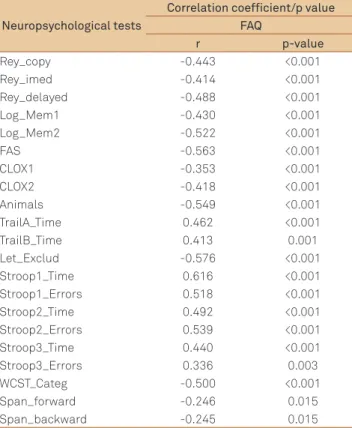
As expected, a negative correlation between FAQ scores (r=-0.387, p<0.001) was found. Correlations were performed to determine whether these measures (FAQ) were associated with neuropsychological functions (Table 5).
Based on results of the Mann-Whitney test, Animals, WCST_categ and Span_OI were selected for input to the lo-gistic regression model. In general and concomitantly, the lower the value of Animals, of WSCT_Categ and of Span_OI and the larger the value on the FAQ, then the higher the prob-ability of having the cognitive impairment when FAQ yields medium to high values, independently of the other variables. (Table 6).
hese results showed that neuropsychological proiles difer among vascular severity groups.
DISCUSSION
Vascular cognitive impairment can be deined as cog-nitive alterations due to cerebrovascular lesions, in which cognition is impaired at various degrees of severity21. We
have classiied patients with subcortical ischemic lesions by cognitive level based on a screening test (MMSE) and a functional activities questionnaire, with vascular dementia ranging from no evidence of cognitive impairment to vas-cular dementia.
impairment or owing to the inability to distinguish slight dif-ferences among VCI proiles, but largely due to an expected overlap of cutof scores. Notwithstanding, the groups pre-sented clear diferences on many cognitive measures, show-ing a clear-cut division.
Our results showed progressive impairment in many ex-ecutive functions from the NVCI to the probable VaD group, in phonemic luency, visuospatial skills and sustained at-tention, strategy and constructional praxis, divided atten-tion, categorization and abstract thinking tests, as well as in verbal and nonverbal memory tests. It is important to emphasize that TMT-B was greatly compromised in the Probable VaD group, with many patients unable to com-plete the task, proving to be a diicult type of test for low educated subjects.
Signiicant diferences were found between the studied groups and their controls, with the probable VaD group be-ing most impaired in relation to their controls. As observed, the NVCI group had poorer performance than their controls on many cognitive measures, particularly in relation to verbal
Table 6. Logistic regression.
Variable Coefficient S.E. Z Odds Ratio 95%CI p-value Lower Upper
Animais -0.421 0.274 2.361 0.656 0.383 1.123 0.124
WCST_categ -2.071 1.087 3.634 0.126 0.015 1.060 0.057
Span_OI 1.556 0.820 3.601 4.740 0950 23.650 0.058
WCST: wisconsin card sorting test; SE: standard error.
luency, visuospatial skills and sustained attention, and on strategy and constructional praxis, divided attention, catego-rization and abstract thinking. his indicates that early eval-uation of these functions could prevent more serious condi-tions, as patients were functionally adequate. According to Laukka22, cognitive deicits could be presented in a
preclini-cal phase of VaD, resulting from circulatory disturbances that could afect cerebral function before dementia is diagnosed. In our study, we found poorer results in patients with no cog-nitive impairment on the screening test and with preserved ADLs (NVCI group), perhaps suggesting that these tests are efective at screening for very mild cognitive impairments in patients with ischemic subcortical lesions.
All patient groups performed worse than their controls on tests of verbal luency, processing speed and abstract thinking akin to other reports19 which compared healthy
con-trols to VCI and VaD patients.
he VCIND group had impaired performance in rela-tion to their controls on tests related to cortico-subcortical pathways, although demonstrated intact functional ADL. hese patients presented depressive symptoms that did not correlate with neuropsychological performance. his fact allows us to make some inferences in VCIND proile subjects, for example, that there is early compromise of ex-ecutive functions which cannot be explained by depressive symptoms alone. Pantoni and colleagues12 showed that the
VCIND group demonstrated signiicant diiculties on mea-sures of cognitive lexibility, verbal retrieval and verbal rec-ognition memory, but not on measures of confrontational naming or verbal luency. he probable VaD group was im-paired on all cognitive measures assessed, suggesting that poor cognitive lexibility and verbal retrieval in the context of preserved function in other domains may characterize the prodromal stage of VaD.
Our indings comparing VCIND and probable VaD groups were similar to those of the study by Stephens et al.23 who
described compromise of executive functions in both these patient groups ascribing this to early damage in VCI. Other studies have reported similar indings, in addition to decline in ADLs and frequent depressive symptoms24,25.
However, we noted that in mild stages of impairment, such as in VCIND, there is signiicant compromise of visual construction, verbal luency, visuospatial skills, attention, ab-straction thinking, inhibitory control and processing speed.
he VCIND group presented impairment in mental lex-ibility whereby this group committed the largest number of
Neuropsychological tests
Correlation coefficient/p value FAQ
r p-value
Rey_copy -0.443 <0.001
Rey_imed -0.414 <0.001
Rey_delayed -0.488 <0.001
Log_Mem1 -0.430 <0.001
Log_Mem2 -0.522 <0.001
FAS -0.563 <0.001
CLOX1 -0.353 <0.001
CLOX2 -0.418 <0.001
Animals -0.549 <0.001
TrailA_Time 0.462 <0.001
TrailB_Time 0.413 0.001
Let_Exclud -0.576 <0.001
Stroop1_Time 0.616 <0.001
Stroop1_Errors 0.518 <0.001
Stroop2_Time 0.492 <0.001
Stroop2_Errors 0.539 <0.001
Stroop3_Time 0.440 <0.001
Stroop3_Errors 0.336 0.003
WCST_Categ -0.500 <0.001
Span_forward -0.246 0.015
Span_backward -0.245 0.015
*Spearman’s correlation; FAQ: functional activity questionnaire; WCST: wisconsin card sorting test.
perseverative errors on the WCST. his inding is in agree-ment with the study of Garret et al.9 who observed
sig-niicant diiculties in mental lexibility in their sample of VCIND patients.
In the study by Stephens et al.23, deficits in attention
and executive function were observed in patients with stroke, but without cognitive impairment (NVCI), com-pared to controls. Comparing these patients with a group of patients with vascular cognitive impairment without dementia, they observed that the latter had greater im-pairment in tests of executive functions, memory and lan-guage. Our comparison between NVCI and VCIND groups yielded the same results as the study by Stephens, since our VCIND group was significantly more impaired on tests of memory and language. In our study, we also detected significant differences between the NVCI group and con-trols on the tests evaluating attention and executive func-tions (TMT-B, Stroop and WCST).
Comparison of our VCIND and probable VaD groups yielded results similar to those found by Stephens. Our pa-tients difered in episodic memory, visuoconstructive ability and ADLs.
We observed a pattern of decreased cognitive functions according to the degree of early impairment in executive functions and verbal luency, and deicits in visual-spatial skills, divided attention and abstract thinking in individuals with vascular lesions. hese deicits were greater in subjects with VCIND than in ones with NVCI, and we also observed compromise in planning and mental control, even in indi-viduals with VaD in that all the tests of executive functions were impaired, indicating early impairment of dopaminer-gic and cholinerdopaminer-gic pathways that traverse the subcortical white matter26.
Depressive symptoms are common in patients with cere-brovascular disease,and cognitive impairment is common in geriatric depression. Also, depressed individuals with co-morbid cognitive impairment are at increased risk for a num-ber of adverse medical, psychiatric and cognitive outcomes27.
herefore, it is always important to diferentiate executive dysfunction and depressive symptoms for adequate thera-peutic conduct.
We also noted signiicant correlation between FAQ scores and neuropsychological measures. he results of our study re-veal that patients with subcortical vascular lesions have cog-nitive impairment, mainly in executive functions, diiculties in ADLs and presence of depressive symptoms, indicating
that searching for these types of signs and symptoms proves to be efective for early diagnosis of VCI.
Some patients presented very mild cognitive impair-ment, that did not result in signiicant ADL diiculties, and few depressive symptoms. Such cases in our survey were de-noted as NVCI. In these subjects, only a neuropsychological evaluation veriied decreased performance. hese indings could point to a need for a more detailed evaluation in pa-tients with subcortical lesions for early diagnosis. In these cases, it is possible that neuropsychological evaluation could represent a gold standard method for diagnosis at basal measurement in order to follow cognitive outcome.
Some limitations should be observed in our study. hese concern the severity of subcortical ischemic lesions, since no ranking scale was used to evaluate their presence at the time of evaluation, precluding correlation of lesion severity with cognitive impairments. Unfortunately, we did not use quan-titative scales of white matter hyperintensities or quantii-cation of brain volume loss. Nevertheless, our patients were carefully selected with respect to the presence of subcorti-cal lesions, seen on magnetic resonance imaging. All patients had been at a chronic phase for more than three months af-ter conirmed stroke. he sample size was relatively small in the VCI and NVCI groups, but patients were highly selected. We believe that limitations regarding the number of patients were minimized by matching each type of patient group with its own control group, providing a neuropsychological pro-ile with considerable consistency. Possible education inlu-ence on majority of the tests was minimized homogenizing the samples by matching for schooling.
Our results allow us to conclude that there is a continu-um in cognitive proiles, from mild signs of cognitive impair-ment to incontestable deimpair-mentia, despite the fact this was a transversal study. he pattern of cognitive deicit of VCIND and VaD are similar, except that in the probable VaD group the deicits tend to be greater than in the VCIND group. All patient groups presented diiculties on tests evaluating ex-ecutive functions (planning, mental control, abstraction, per-severation, inhibitory control and processing speed), albeit to diferent degrees.
he results showed that executive impairments mani-fest early in mild impairment among subcortical ischemic lesion patients, who also present an increased frequency of depressive symptoms, as well as some diiculties in ADLs. Evaluation of functional activity seems to be a valuable ap-proach for early detection of vascular cognitive impairment.
References
1. Bowler JV. Modern concept of vascular cognitive dementia impairment. Br Med Bull 2007;83:291-305.
2. Vas CJ, Pinto C, Panikker D, et al. Prevalence of dementia in an urban Indian population. Int Psychogeriatr 2001;13:439-450.
3. Rocca WA, Hofman A, Brayne C, et al. The prevalence of vascular
dementia in Europe: facts and fragments from 1980-1990 studies. EURODEM-Prevalence Research Group. Ann Neurol 1991;30:817-824. 4. Herrera Jr. E, Caramelli P, Nitrini R. Population epidemiologic study of
5. Silva DW, Damasceno BP. Demência na população de pacientes do Hospital das Clínicas da UNICAMP. Arq Neuropsiquiatr 2002; 60:996-999.
6. Vale FA, Miranda SJ. Clinical and demographic features of patients with dementia attended in a tertiary outpatient clinic. Arq Neuropsiquiatr 2002;60:548-552.
7. Fujihara S. Brucki SMD, Rocha MSG, Carvalho AA, Piccolo AC. Prevalence of presenile dementia in a tertiary outpatient clinic. Arq Neuropsiquiatr 2004;62:592-595.
8. Wentzel C, Rockwood K, MacKnight C, et al. Progression of impairment in patients with vascular cognitive impairment without dementia. Neurology 2001;57:714-716.
9. Garret KD, Browndyke JN, Whelihan W, et al. The neuropsychological profile of vascular cognitive impairment—no dementia: comparisons to patients at risk for cerebrovascular disease and vascular dementia. Arch Clin Neuropsychol 2004;19:745-757.
10. Jager CA, Hogervorst E, Combrinck M, Budge MM. Sensitivity and specificity of neuropsychological tests for mild cognitive impairment, vascular cognitive impairment and Alzheimer’s disease. Psychol Med 2003;33:1039-1050.
11. Chui H. Dementia due to subcortical ischemic vascular disease. Clin Cornerstone 2001;3:40-51.
12. Pantoni L, Leys D, Fazekas F, et al. Role of the white matter lesions in cognitive impairment of vascular origin. Alzheimer Dis Assoc Disord 1999;13(Suppl 3):S49-S54.
13. Rockwood K, Bowler J, Erkinjuntti T, Hachinski V, Wallin A. Subtypes of vascular dementia. Alzheimer Dis Assoc Disord 1999;13(Suppl 3): S59-S65.
14. Pohjasvaara T, Mäntylä R, Ylikoski R, Kaste M, Erkinjuntti T. Comparison of different clinical criteria (DSM-III, ADDTC, ICD-10, NINDS-AIREN, DSM-IV) for the diagnosis of vascular dementia. National Institute of Neurological Disorders and Stroke-Association Internationale pour la Recherche et l’Enseignement en Neurosciences. Stroke 2000;31:2952-2957.
15. Román GC. Defining dementia: clinical criteria for the diagnosis of vascular dementia. Acta Neurol Scand Suppl 2002;17806(Suppl 178): S6-S9.
16. Brucki SMD, Nitrini R, Caramelli P, Bertolucci PHF, Okamoto IH. Sugestões para o uso do Mini-Exame do Estado Mental no Brasil. Arq Neuropsiquiatr 2003;61:771-781.
17. Pfeffer RI, Kurosaki TT, Harrah CH, Chance JM, Filos S. Measurement of functional activities in older adults in the community. J Gerontol 1982;37:323-329.
18. Spreen O, Strauss E. A Compendium of Neuropsychological Tests. New York. Oxford University Press; 1991.
19. Royall DR, Cordes J, Polk M. CLOX: an executive clock drawing task. J Neurol Neurosurg Psychiatry 1998;64:588-594.
20. Bryan J, Luszcz MA, Crawford JR. Verbal knowledge and speed of information processing as mediators of age differences in verbal fluency performance among older adults. Psychol Aging1997;12:473-478. 21. Yesavage JA, Brink TL, Rose TL, et al. Development and validation of a
geriatric depression screening scale: a preliminary report. J Psychiatr Res 1982-1983;17:37-49.
22. Laukka EJ, Jones S, Fratiglioni L, Bäckman L. Cognitive functioning in preclinical vascular dementia a 6-year follow-up. Stroke 2004;35:1805-1809.
23. Stephens S, Kenny RA, Rowan E, et al. Neuropsychological characteristics of mild vascular cognitive impairment and dementia after stroke. Int J Geriatr Psychiatry 2004;19:1053-1057.
24. Bartrés-Faz D, Clemente IC, Junqué C. White matter changes and cognitive performance in aging. Rev Neurol 2001;33:347-353. 25. Steffens DC, Potter GG. Geriatric depression and cognitive impairment.
Psychol Med 2008;38:163-175.
26. Engelhardt E, Moreira DM, Laks J. Vascular dementia and the cholinergic pathways. Dement Neuropsychol 2007;1:2-9.