Cigarette Smoke Disturbs the Survival of
CD8
+
Tc/Tregs Partially through Muscarinic
Receptors-Dependent Mechanisms in Chronic
Obstructive Pulmonary Disease
Gang Chen☯, Mei Zhou☯, Long Chen, Zhao-Ji Meng, Xian-Zhi Xiong*, Hong-Ju Liu, Jian-Bao Xin, Jian-Chu Zhang
Department of Respiratory and Critical Care Medicine, Key Laboratory of Pulmonary Diseases of Health Ministry, Union Hospital, Tongji Medical College, Huazhong University of Science and Technology, Wuhan, Hubei, China
☯These authors contributed equally to this work. *xxz0508@hust.edu.cn
Abstract
Background
CD8+T cells (Cytotoxic T cells, Tc) are known to play a critical role in the pathogenesis of smoking related airway inflammation including chronic obstructive pulmonary disease (COPD). However, how cigarette smoke directly impacts systematic CD8+T cell and regu-latory T cell (Treg) subsets, especially by modulating muscarinic acetylcholine receptors (MRs), has yet to be well elucidated.
Methods
Circulating CD8+Tc/Tregs in healthy nonsmokers (n = 15), healthy smokers (n = 15) and COPD patients (n = 18) were evaluated by flow cytometry after incubating with anti-CD3, anti-CD8, anti-CD25, anti-Foxp3 antibodies. Peripheral blood T cells (PBT cells) from healthy nonsmokers were cultured in the presence of cigarette smoke extract (CSE) alone or combined with MRs agonist/antagonist for 5 days. Proliferation and apoptosis were eval-uated by flow cytometry using Ki-67/Annexin-V antibodies to measure the effects of CSE on the survival of CD8+Tc/Tregs.
Results
While COPD patients have elevated circulating percentage of CD8+T cells, healthy
smok-ers have higher frequency of CD8+Tregs. Elevated percentages of CD8+T cells correlated
inversely with declined FEV1 in COPD. CSE promoted the proliferation and inhibited the apoptosis of CD8+T cells, while facilitated both the proliferation and apoptosis of CD8+
Tregs. Notably, the effects of CSE on CD8+Tc/Tregs can be mostly simulated or attenuated
by muscarine and atropine, the MR agonist and antagonist, respectively. However, neither muscarine nor atropine influenced the apoptosis of CD8+Tregs.
OPEN ACCESS
Citation:Chen G, Zhou M, Chen L, Meng Z-J, Xiong X-Z, Liu H-J, et al. (2016) Cigarette Smoke Disturbs the Survival of CD8+Tc/Tregs Partially through Muscarinic Receptors-Dependent Mechanisms in Chronic Obstructive Pulmonary Disease. PLoS ONE 11(1): e0147232. doi:10.1371/journal.pone.0147232
Editor:Yunchao Su, Georgia Regents University, UNITED STATES
Received:October 16, 2015
Accepted:December 30, 2015
Published:January 25, 2016
Copyright:© 2016 Chen et al. This is an open access article distributed under the terms of the Creative Commons Attribution License, which permits unrestricted use, distribution, and reproduction in any medium, provided the original author and source are credited.
Data Availability Statement:All relevant data are within the paper and its Supporting Information files.
Conclusion
The results imply that cigarette smoking likely facilitates a proinflammatory state in smokers, which is partially mediated by MR dysfunction. The MR antagonist may be a beneficial drug candidate for cigarette smoke-induced chronic airway inflammation.
Introduction
Cigarette smoke-induced pulmonary inflammation, such as chronic obstructive pulmonary
disease (COPD), is the fourth leading cause of death worldwide [1–3]. Many lines of evidence
indicate that adaptive immunity plays important roles in the development of COPD, and the systemic inflammation may be a result of overspill of inflammatory mediators from the lungs [4]. However, the immune regulation in smoking related airway inflammation has not yet been fully illuminated.
Previous studies have shown that CD8+T cells in both lung parenchyma and pulmonary
arteries are inversely correlated with pulmonary function [5], which suggest that CD8+T cells
(Cytotoxic T cells, Tc) may play a critical role in the pathogenesis of smoking related
pulmo-nary inflammation. While much attention has been given to the subsets of CD4+regulatory T
cells (Tregs) for their role in the maintenance of immune homeostasis, recent findings have
demonstrated that CD8+Tregs display immune regulation functions as well [6,7]. Meanwhile,
our previous study indicated that there was an inverse correlation between the circulating
CD8+Tregs and smoking index [8]. However, the precise role of CD8+ Tc/Treg cells in the
pathogenesis of chronic airway inflammation remains unclear. Although some in vitro research has indicated that soluble components extracted from cigarette smoke can signifi-cantly reduce the proliferation and activation of T cell [9,10], the influence of cigarette smoke
on CD8+T cells remains unclear.
T cells express cholinergic receptors, including muscarinic acetylcholine receptors (mAChRs or MRs) and nicotinic acetylcholine receptors (nAChRs), and represent a cellular source of Ach [11]. In particular, our published data [12] and that of others [13] have reported that MR expression on human peripheral blood T cells (PBT cells) was mostly restricted to MR3, MR4 and MR5. COPD is a type of chronic inflammatory disease characterized by the hyperfunctioning of the cholinergic system [14] coupled with increased MR3 expression and
CD8+-ACh binding on PBT cells [15], indicating that MRs play important regulatory roles in
CD8+T cell survival. In addition, our previous study indicated that the MR antagonist
tiotro-pium bromide increased the number of CD8+Tregs in cigarette smoke-induced COPD
patients [16,17]. We therefore hypothesized that the cholinergic system may be involved in the pathogenesis of cigarette smoke-induced chronic inflammation by regulating T cells,
particu-larly the CD8+Tc/Tregs subsets.
In the present study, we attempted to investigate the effects of cigarette smoke extract (CSE)
and MR signaling on the proliferation and apoptosis of CD8+T cells and CD8+Tregs. Our
cur-rent data showed that cigarette smoking facilitates the formation of pro-inflammatory milieu
and the development of COPD by causing an imbalance between CD8+T and Treg cells, which
is skewed toward CD8+T cell-dominant phenotype. To the best of our knowledge, this is the
first report showing that cigarette smoke may disturb the survival of CD8+Tc/Tregs partially
through MRs. Competing Interests:The authors have declared
Materials and Methods
Subjects and sample collection
This study was approved by the Ethics Committee of Union Hospital, Tongji Medical School, Huazhong University of Science and Technology, and informed written consent was obtained from each donor. Based on the criteria supplied by the Global Initiative for Chronic Obstruc-tive Lung Disease (GOLD) guidelines [18], we recruited patients with stable COPD (n = 18) who were free of exacerbation for at least 4 weeks at the time of blood draw. Meanwhile, healthy smokers (HS, n = 15) and healthy nonsmoker controls (HC, n = 15) with normal lung functions were also collected and were free of any lung or systemic disease. COPD patients and healthy smokers had a smoking history of 10 pack years or more. Exclusion criteria for the study included the following characteristics: other respiratory diseases apart from COPD, sys-temic autoimmune diseases, and treatment with anticholinergics, glucocorticoids or immuno-modulators within 4 weeks prior the research. Peripheral blood samples were collected in heparin-treated tubes. As mentioned before [12], the peripheral blood T cells (PBT cells) were harvested and resuspended in complete RPMI 1640 medium plus 10% fetal bovine serum
(FBS) and placed in an incubator at 37°C in 5% CO2for subsequent experiments, depending
on the availability of cells.
Flow cytometric analysis
The expression of surface marker and transcription factors by T cells was assessed by flow cytometry as previously described [12] after surface or intracellular staining with peridinin
chlorophyll protein (PerCP)-Cy5.5–conjugated anti-CD3 (eBioscience, San Diego, CA, USA),
fluorescein isothiocyanate (FITC)-conjugated anti-CD8 (BD Biosciences, San Jose, CA, USA), phycoerythrin (PE)-Cy7-conjugated CD25 (BD Biosciences), and PE-conjugated anti-Foxp3 (eBioscience) antibodies. Isotype controls were generated to perform compensation and to confirm antibody specificity. Flow cytometry was performed on a FACS Canto II (BD Bio-sciences) and analyzed using BD FCSDiva Software and FCS Express 4 software (De Novo
Soft-ware, Los Angeles, CA, USA).Fig 1showed the gating strategy.
CSE preparation
CSE was prepared using the method of Blue and Janoff [19]. The CSE concentrations used in the experiments were chosen in consistent with our previous report [12]. Briefly, cigarette smoke was drawn into a 50 ml plastic syringe, and smoke was then slowly bubbled into a tube containing 5 ml of sterile RPMI 1640 medium. A total of 5 ml of CSE was produced from two cigarettes (Huang Helou, Wuhan, Hubei, China). Next, the CSE solution was filtered through 0.22-μm filters. To ensure the vitality of the substances in the CSE, all of the CSE was prepared within a half hour before each experiment.
Proliferation and apoptosis of T cells
Proliferation and apoptosis assays in vitro were performed as described in detail previously [12]. In brief, PBT cells were maintained in the presence of CSE, MR agonist muscarine (Mus, 50μM, Sigma-Aldrich, St. Louis, MO, USA), and MR antagonist atropine (Atro, 100μM,
Sigma-Aldrich), either alone or in various combinations. In the proliferation assays, 1μg/ml
PHA and 50 ng/ml PMA (both from Sigma-Aldrich) were added to the medium to stimulate T cell proliferation. After 5 days, the cells were harvested and the proportions of proliferating
anti-human Ki-67 (eBioscience), allophycocyanin (APC)-conjugated Annexin V and propi-dium iodide (PI) (Annexin V Apoptosis Detection Kit APC; eBioscience).
Statistical analysis
Data are expressed as the mean ± SEM (unless otherwise indicated). Multiple comparisons between different groups were performed using the nonparametric Kruskal-Wallis test
fol-lowed by Dunn’s post hoc test. Correlations between variables were determined using the
Spearman rank test. Data analysis was performed using GraphPad Prism v.5.01 software (GraphPad Software, La Jolla, CA, USA), and two-tailed P values of less than 0.05 were consid-ered statistically significant.
Results
Subject Demographics
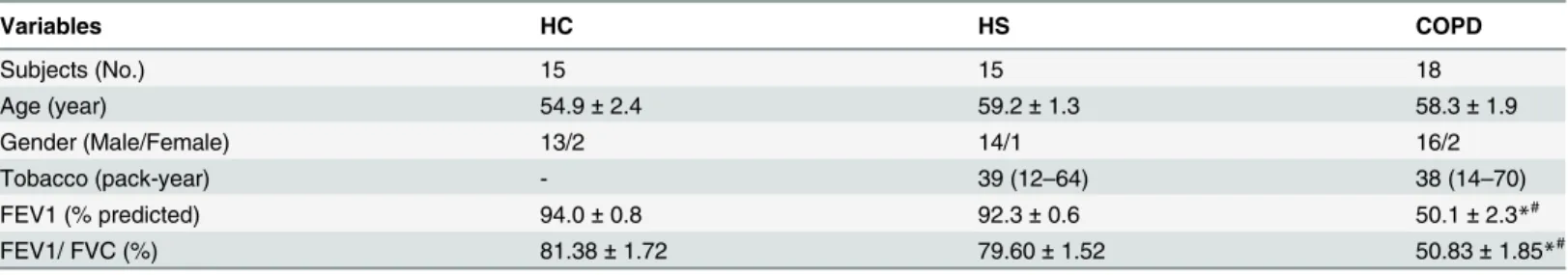
The main clinical characteristics of the three groups studied are presented inTable 1. The
unequal sex ratio was consistent with the much higher prevalence of COPD in males than in females. The smoking history of smokers with normal lung function and patients with COPD was similar. Patients with COPD showed moderate airflow obstruction, whereas the other two groups have normal spirometry.
Fig 1. Representative dot plots show the gating strategy of the assays.In the proliferation assays, CD8+
Tregs were gated from CD8+T cells (CD3+CD8+) based on their positive expression of Foxp3, and the
expression of Ki-67 by CD8+T cells and CD8+Tregs was then further analyzed. However, in the apoptosis assays, CD8+Tregs were gated from CD8+T cells (CD3+CD8+) based on their high expression of CD25, and
the apoptosis of CD8+T cells and CD8+Tregs gated on propidium iodide-negative and Annexin V-positive
cells was then further analyzed.
doi:10.1371/journal.pone.0147232.g001
Table 1. Demographics and clinical characteristics of all participants.
Variables HC HS COPD
Subjects (No.) 15 15 18
Age (year) 54.9±2.4 59.2±1.3 58.3±1.9
Gender (Male/Female) 13/2 14/1 16/2
Tobacco (pack-year) - 39 (12–64) 38 (14–70)
FEV1 (% predicted) 94.0±0.8 92.3±0.6 50.1±2.3*#
FEV1/ FVC (%) 81.38±1.72 79.60±1.52 50.83±1.85*#
The data are represented as the mean±SEM or median (range). FEV1: forced expiratory volume in one second; FVC: forced vital capacity.
*P<0.05 vs. the HC group; #
P<0.05 vs. the HS group.
Elevated percentages of CD8
+T cells correlated inversely with declined
FEV1 in COPD
We first investigated the frequencies of CD8+Tc/Treg cells in peripheral blood of different
groups. The percentage of CD8+T cells was significantly higher in patients with COPD
(mean = 26.54%) than in HS (mean = 20.53%, p<0.05) or HC (mean = 19.11%, p<0.05)
sub-jects. The levels of CD8+T cells in HS subjects were slightly higher than those in the HC
sub-jects, although the difference did not reach statistical significance (Fig 2A). Furthermore, the
frequency of CD8+Tregs was markedly higher in HS donors (mean 1.46%) than in COPD
patients (mean = 0.98%, p<0.05) and HC groups (mean = 0.96%, p<0.05) (Fig 2B).
Single regression analysis between lymphocytes and airflow obstruction in COPD patients
was performed. We observed that the levels of CD8+T cells displayed a negative correlation
with FEV1 (% predicted) (r = - 0.54, p<0.05) (Fig 2C), supporting a possible role for CD8+T
cells in the airway inflammation. However, frequency of CD8+Tregs did not correlate with
pulmonary function tests (r = 0.09, p = 0.72) (Fig 2D).
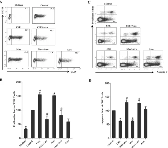
Impacts of CSE and muscarinic receptors on CD8
+T cells survival
To investigate the effects of cigarette smoke on the proliferation and apoptosis of CD8+T cells
(excluding CD8+Tregs), we performed proliferation and apoptosis assays using PBT cells
obtained from healthy nonsmokers. As shown inFig 3A and 3B, Ki-67+CD8+T cells were
sig-nificantly more abundant in CSE and Mus treated group, which can be abolished by Atro, a MR antagonist.
Fig 2. Imbalance of circulating CD8+Tc/Tregs in patients with chronic obstructive pulmonary disease
(COPD).Comparisons of percentages of CD8+T cells (A) and CD8+Tregs (B) in peripheral blood from
healthy controls (HC, n = 15), healthy smokers (HS, n = 15) and COPD patients (n = 18). Correlation of CD8+
T cells (C) and CD8+Tregs (D) with FEV1% predicted value in COPD patients (n = 18).
*P<0.05.
Additionally, CSE reduced the apoptosis of CD8+T cells (excluding CD8+Tregs), whereas Atro treatment completely abrogated this anti-apoptotic effect. Indeed, both CSE and Mus
reduced the apoptosis of CD8+T cells, which can be fully aboished by Atro (Fig 3C and 3D).
Impacts of CSE and muscarinic receptors on CD8
+Tregs survival
We further analyzed the effects of cigarette smoke on CD8+Treg. The proliferation of CD8+
Tregs was analyzed using CD3+CD8+Foxp3+as the Treg phenotypic marker set (Fig 1).
Con-sidering that the cell membrane must be kept intact for the detection of apoptotic cells by PI
and Annexin V (thus, Foxp3 staining cannot be achieved), CD3+CD8+CD25hiwas used as the
CD8+Treg phenotypic marker profile (Fig 1). As expected, the percentages of CD8+CD25hiT
cell subset overlapped with CD8+FoxP3+cells (data not shown).
Fig 3. Effects of CSE and muscarinic receptors on proliferation and apoptosis of CD8+T cells.PBT cells from healthy nonsmokers were cultured for 5 days in the presence of CSE and MRs agonist/antagonist, either alone or in various combinations. (A) In the proliferation assays, medium combined with PHA and PMA was considered as control treated wells. Ki-67+CD8+T cells (excluding CD8+Tregs) were examined by flow cytometry, and the representative flow cytometric dot plots are shown. (B) The proliferation index of control wells was considered to be 100, and data are expressed as fold increase relative that in the control wells (n = 5). (C) In the apoptosis assays, medium alone was considered as control treated wells. Apoptotic CD8+T
cells (excluding CD8+Tregs) were examined by flow cytometry, and the representative flow cytometric dot plots are shown. (D) The apoptosis index of control wells was considered to be 100, and data are expressed as fold increase relative that in the control wells (n = 6). The results are reported as the mean±SEM.*P<0.05 compared with the control wells; #P<0.05 indicates CSE plus Atro compared with CSE or Mus plus Atro compared with Mus.
As indicated inFig 4A and 4B, for CD8+Tregs from healthy nonsmokers, both CSE and Mus significantly facilitated proliferation, which can be blocked by Atro. Unexpectedly, CSE
robustly promoted CD8+Treg apoptosis in healthy nonsmokers (Fig 4C and 4D). However,
neither MR agonist nor antagonist influenced apoptosis, indicating that CD8+Treg apoptosis
was less sensitive to MR modulation.
Discussion
Previous studies have mainly focused on bronchodilation of MR-based treatments for chronic airway inflammation. However, the potential for anti-inflammatory benefits in addition to bronchodilation is not well understood. To the best of our knowledge, the present study shows
for the first time that cigarette smoke disturb the survival of CD8+T/CD8+Tregs partially
through MRs-dependent mechanisms.
Fig 4. Effects of CSE and muscarinic receptors on proliferation and apoptosis of CD8+Treg cells.PBT cells from healthy nonsmokers were cultured for 5 days in the presence of CSE and MRs agonist/antagonist, either alone or in various combinations. (A) In the proliferation assays, medium combined with PHA and PMA was considered as control treated wells. Ki-67+CD8+Treg cells were examined by flow cytometry, and the
representative flow cytometric dot plots are shown. (B) The proliferation index of control wells was considered to be 100, and data are expressed as fold increase relative that in the control wells (n = 5). (C) In the
apoptosis assays, medium alone was considered as control treated wells. Apoptotic CD8+Treg cells were
examined by flow cytometry, and the representative flow cytometric dot plots are shown. (D) The apoptosis index of control wells was considered to be 100, and data are expressed as fold increase relative that in the control wells (n = 6). The results are reported as the mean±SEM.*P<0.05 compared with the control wells;
#P<0.05 indicates CSE plus Atro compared with CSE or Mus plus Atro compared with Mus.
Many studies have reported increased percentages of CD8+T cells confined to the lung of COPD patients, such as pulmonary parenchyma and lung arteries [5], central and peripheral airways [20,21], and bronchoalveolar lavage (BAL) [22]. Remarkably, our findings extend these
observations by showing that the number of CD8+T cells is also increased in the peripheral
blood of COPD patients. The presented results agree with a previous study in blood [15],
sug-gesting that the origin of the abnormal CD8+T cell infiltration seen in the lungs of COPD
patients is a systemic rather than a local event. In support of these findings [5,20], we have
dem-onstrated that the increased frequency of circulating CD8+T cells in patients with COPD shows
an inverse correlation with the declined lung function. There is growing interest in the potential
mechanisms by which CD8+T cells worsen lung function in cigarette smoke-induced COPD.
Hamid and his coworkers demonstrated that exposure of CSE lead to the activation of CD8+T
cells, resulting in the production of IL-1β, IL-6, IL-10, IL-12p70, TNF-αand IFN-γ, which was
associated Toll-like receptor (TLR)-4 and TLR9 [23]. And, in keeping with these findings, the lung function of COPD patients was inversely associated with the percentage of BAL and
intrae-pithelial CD8+T cells producing TNF-α[24], as well as the frequency of CD8+T cells activation
and CD8 IFN-γproduction [25]. We could speculate this increased cytokines can cause the
aggregation of other inflammatory cells to the lung, ultimately perpetuating the inflammation and damage observed in COPD patients, even in individuals who have quitted smoking. Obvi-ously, much work remains to be done before our understanding of emphysema pathogenesis.
Interestingly, we showed that cigarette smoking increased the number of circulating CD8+
Tregs in healthy smokers, but not in somkers with COPD. The increased CD8+Foxp3+Tregs
observed in healthy smokers is in agreement with the results of previous report [26], which
revealed a prominent upregulation of CD8+γδ+Tregs in bronchoalveolar lavage from smokers
with normal lung function. Given the relevance of Foxp3+andγδ+T-lymphocytes in tissue
homeostasis, these observations are compatible with a physiological response aimed to protect the lungs from the injury caused by cigarette smoking.
According to genetic knockout experiments, the cholinergic pathway is involved in the
regula-tion of CD8+T cells [27], and increased ACh-binding to CD3+CD8+T cells in the peripheral
blood occurs in COPD patients [15]. However, the exact effects of MRs on CD8+T cells during
the development of COPD remain unknown. In this present study, cells were incubated with CSE or Mus (with or without Atro) to discern whether smoking and MRs affected the survival of CD8 + T cells by using the proliferation assay. Our present data indicated that both CSE and Mus
sig-nificantly promoted the proliferation of CD8+T cells from healthy nonsmokers. Our data are
con-sistent with previous research showing that MR activation could promote CD8+cytolytic T cell
generation [27]. Considering that CSE could promote the proliferation of human fetal lung
fibro-blasts by increasing MR1 and MR3 expression and ERK1/2 and NF-κB activation [2], combing
with our finding that MR antagonist Atro could abrogate the pro-proliferative effect of CSE, we
presumed that the effect of CSE on CD8+T cells may function through MRs. Our study is the first
to show that CSE could promote CD8+T cell proliferation by activating the muscarine system,
thereby contributing to the increased numbers of CD8+T cells in the airways of COPD patients.
CSE may exert effects on apoptosis of CD8+T cells through a variety of substances, as
ciga-rette smoke comprises more than 4,000 components, including endotoxin, reactive oxygen
spe-cies and free radicals [3]. We showed that CSE and Mus can promote apoptosis of CD8+T
cells, which can be completely abrogated by Atro, indicating that the anti-apoptotic role of CSE is MR dependent. Our results were consistent with the previous study showing that the
apopto-sis of CD8+T cells in the peripheral blood of COPD patients can be induced by anticholinergic
drugs in a caspases3/8 and IκB-dependent manner [15].
Notably, we also have shown for the first time that CSE paradoxically played both
smoking on CD8+Tc/Tregs is linked to population susceptibility: in smokers with normal lung
function, pro-proliferative role on CD8+Tregs is in a dominant position, which successfully
delays the inflammatory cells (including CD8+and CD4+lymphocytes) amplification and
pro-tects the lungs from the injury caused by current tobacco smoking; In smokers with COPD,
however, pro-apoptotic role on CD8+Tregs takes precedence, and chronic cigarette smoke
exposure in COPD patients results in sustained cholinergic signaling activation and CD8+T
cell infiltration, eventually impairing the immunity of COPD patients.
We speculate that certain master switch controls the different effects of CSE on proliferative
and apoptotic level of CD8+Treg cells. On the one hand, it is reported that the effects of CSE
on T cells development depend on inflammatory milieu and differentiation condition [28]. It is also known that the cytokine milieu of COPD differs from that of healthy controls, with IL-12 being the predominant cytokine and IL-4 being the deficient cytokine [29,30]. Therefore, the difference in the inflammatory milieu could trigger the dichotomous effects of CSE treatment
on healthy- vs COPD- smokers. On the other hand, a deficiency ofα-1-antitrypsin (α-1AT) is
the only known gene linked to the early onset of emphysema, and there is a large phenotypic
variability even among carriers of different forms ofα-1AT proteins [31,32]. In particular,
scanning the entire genome for SNPs has been used to identify several new risk genes involved
in human COPD, such as theα- nAChR (CHRNA 3/5) [33]. These findings further indicate
that genetic susceptibility may be the master switch that controls different proliferative and apoptotic level. Predictably, clarifying distinct clinical phenotypes and identifying the suscepti-ble genotypes will not only provide personalized treatment to smoke-induced emphysema, but also will provide methods to predict smokers who are at a higher risk of developing COPD.
Additionally, although MR agonist had pro-proliferative effect on CD8+Tregs resembling
that of CSE, MRs did not seem to affect CD8+Treg apoptosis, indicating that CSE promotes
the apoptosis of CD8+Treg cells in a MR-independent signaling pathway: Firstly, since we
have previously reported that the expression of anti-inflammatoryα7 nAChR by circulating
CD4+T cells was reduced in smokers with COPD [12], CSE may disrupt a signaling pathway
of nicotinic but not muscarinic acetylcholine receptor, contributing to the apoptosis of CD8+
Tregs in susceptible populations. Secondly, as cigarette smoke is a potent source of oxidative stress and apoptosis for human lung fibroblasts (HFL-1) [34], CSE may similarly contribute to
the apoptosis of CD8+Treg cells by the pathway of oxidative stress. Lastly, numerous
sub-stances besides nicotine were contained in CSE, so further works will be necessary to clarify the mechanisms of action of CSE in vivo.
Conclusions
Our experiments demonstrated that cigarette smoke may directly cause an imbalance of
pro-and anti-inflammatory response via its effects on CD8+T cell/CD8+Treg survival, which may
explain the sustained impairment of immune defenses in COPD patients who continue to
smoke. Furthermore, MR activation is involved in the effect of CSE on CD8+ T cells’
prolifera-tion/apoptosis and CD8+ Tregs’proliferation. Thus, targets on MRs-dependent mechanisms
may abrogate the effects of cigarette smoke to some extent and thereby regulate abnormal acti-vation of the inflammatory response. These findings raise the possibility that MR antagonists may restore a balance of pro-/anti-inflammation, which are therapeutically beneficial in addi-tion to bronchodilaaddi-tion for chronic airway inflammaaddi-tion.
Supporting Information
S1 File. The underlying data points for the graphs reported in the figures.
Acknowledgments
The authors wish to thanks Dr. Ming-Qiang Zhang for his comments on the manuscript and AJE (www.aje.com) for its linguistic assistance during the preparation of this manuscript.
Author Contributions
Conceived and designed the experiments: XZX. Performed the experiments: GC MZ. Analyzed the data: LC ZJM. Contributed reagents/materials/analysis tools: HJL JBX JCZ. Wrote the paper: GC MZ XZX.
References
1. Zhou H, Hua W, Jin Y, Zhang C, Che L, Xia L, et al. (2015) Tc17 cells are associated with cigarette smoke-induced lung inflammation and emphysema. Respirology 20: 426–433. doi:10.1111/resp. 12486PMID:25677967
2. Profita M, Bonanno A, Siena L, Bruno A, Ferraro M, Montalbano AM, et al. (2009) Smoke, choline acet-yltransferase, muscarinic receptors, and fibroblast proliferation in chronic obstructive pulmonary dis-ease. J Pharmacol Exp Ther 329: 753–763. doi:10.1124/jpet.108.145888PMID:19190237
3. Lee J, Taneja V, Vassallo R (2012) Cigarette smoking and inflammation: cellular and molecular mecha-nisms. J Dent Res 91: 142–149. doi:10.1177/0022034511421200PMID:21876032
4. Sinden NJ, Stockley RA (2010) Systemic inflammation and comorbidity in COPD: a result of 'overspill' of inflammatory mediators from the lungs? Review of the evidence. Thorax 65: 930–936. doi:10.1136/ thx.2009.130260PMID:20627907
5. Saetta M, Baraldo S, Corbino L, Turato G, Braccioni F, Rea F, et al. (1999) CD8+ve cells in the lungs of smokers with chronic obstructive pulmonary disease. Am J Respir Crit Care Med 160: 711–717. PMID: 10430750
6. Dinesh RK, Skaggs BJ, La Cava A, Hahn BH, Singh RP (2010) CD8+ Tregs in lupus, autoimmunity, and beyond. Autoimmun Rev 9: 560–568. doi:10.1016/j.autrev.2010.03.006PMID:20385256
7. Suzuki M, Konya C, Goronzy JJ, Weyand CM (2008) Inhibitory CD8+ T cells in autoimmune disease. Hum Immunol 69: 781–789. doi:10.1016/j.humimm.2008.08.283PMID:18812196
8. Xiong XZ, Jin Y, Zhou Q, Zhang XJ, Du W, Liu W, et al. (2008) [Correlation between FoxP3(+) regula-tory T cells and chronic obstructive pulmonary disease]. Zhonghua Yi Xue Za Zhi 88: 471–474. PMID: 18642789
9. Glader P, Moller S, Lilja J, Wieslander E, Lofdahl CG, von Wachenfeldt K (2006) Cigarette smoke extract modulates respiratory defence mechanisms through effects on T-cells and airway epithelial cells. Respir Med 100: 818–827. PMID:16242311
10. Lambert C, McCue J, Portas M, Ouyang Y, Li J, Rosano TG, et al. (2005) Acrolein in cigarette smoke inhibits T-cell responses. J Allergy Clin Immunol 116: 916–922. PMID:16210070
11. Fujii T, Takada-Takatori Y, Kawashima K (2008) Basic and clinical aspects of non-neuronal acetylcho-line: expression of an independent, non-neuronal cholinergic system in lymphocytes and its clinical sig-nificance in immunotherapy. J Pharmacol Sci 106: 186–192. PMID:18285654
12. Zhang MQ, Wan Y, Jin Y, Xin JB, Zhang JC, Xiong XZ, et al. (2014) Cigarette smoking promotes inflam-mation in patients with COPD by affecting the polarization and survival of Th/Tregs through up-regula-tion of muscarinic receptor 3 and 5 expression. PLoS One 9: e112350. doi:10.1371/journal.pone. 0112350PMID:25375131
13. Tayebati SK, El-Assouad D, Ricci A, Amenta F (2002) Immunochemical and immunocytochemical characterization of cholinergic markers in human peripheral blood lymphocytes. J Neuroimmunol 132: 147–155. PMID:12417445
14. Gross NJ, Skorodin MS (1984) Role of the parasympathetic system in airway obstruction due to emphy-sema. N Engl J Med 311: 421–425. PMID:6749189
15. Profita M, Riccobono L, Montalbano AM, Bonanno A, Ferraro M, Albano GD, et al. (2012) In vitro anti-cholinergic drugs affect CD8+ peripheral blood T-cells apoptosis in COPD. Immunobiology 217: 345–
353. doi:10.1016/j.imbio.2011.07.013PMID:21855166
16. Zhang J, Deng L, Xiong X, Wang P, Xin J, Ma W (2011) Effect of tiotropium bromide on expression of CD(8) (+)CD (25) (+)FoxP (3) (+) regulatory T cells in patients with stable chronic obstructive pulmonary disease. J Huazhong Univ Sci Technolog Med Sci 31: 463–468. doi:10.1007/s11596-011-0474-4
17. Zhang JC, Wang P, Xiong XZ, Deng L, Xin JB, Ma WL, et al. (2010) [Muscarinic cholinergic receptor antagonists enhanced the expression of CD(8)(+)CD(25)(+)Foxp(3)(+) regulatory T cells in stable COPD patients]. Zhonghua Jie He He Hu Xi Za Zhi 33: 33–36. PMID:20368022
18. Vestbo J, Hurd SS, Agusti AG, Jones PW, Vogelmeier C, Anzueto A, et al. (2013) Global strategy for the diagnosis, management, and prevention of chronic obstructive pulmonary disease: GOLD execu-tive summary. Am J Respir Crit Care Med 187: 347–365. doi:10.1164/rccm.201204-0596PPPMID: 22878278
19. Blue ML, Janoff A (1978) Possible mechanisms of emphysema in cigarette smokers. Release of elas-tase from human polymorphonuclear leukocytes by cigarette smoke condensate in vitro. Am Rev Respir Dis 117: 317–325. PMID:637413
20. O'Shaughnessy TC, Ansari TW, Barnes NC, Jeffery PK (1997) Inflammation in bronchial biopsies of subjects with chronic bronchitis: inverse relationship of CD8+ T lymphocytes with FEV1. Am J Respir Crit Care Med 155: 852–857. PMID:9117016
21. Saetta M, Di Stefano A, Turato G, Facchini FM, Corbino L, Mapp CE, et al. (1998) CD8+ T-lymphocytes in peripheral airways of smokers with chronic obstructive pulmonary disease. Am J Respir Crit Care Med 157: 822–826. PMID:9517597
22. Mikko M, Forsslund H, Cui L, Grunewald J, Wheelock AM, Wahlstrom J, et al. (2013) Increased intrae-pithelial (CD103+) CD8+ T cells in the airways of smokers with and without chronic obstructive pulmo-nary disease. Immunobiology 218: 225–231. doi:10.1016/j.imbio.2012.04.012PMID:22652413
23. Nadigel J, Prefontaine D, Baglole CJ, Maltais F, Bourbeau J, Eidelman DH, et al. (2011) Cigarette smoke increases TLR4 and TLR9 expression and induces cytokine production from CD8(+) T cells in chronic obstructive pulmonary disease. Respir Res 12: 149. doi:10.1186/1465-9921-12-149PMID: 22070100
24. Hodge G, Nairn J, Holmes M, Reynolds PN, Hodge S (2007) Increased intracellular T helper 1 proin-flammatory cytokine production in peripheral blood, bronchoalveolar lavage and intraepithelial T cells of COPD subjects. Clin Exp Immunol 150: 22–29. PMID:17614970
25. Zhu X, Gadgil AS, Givelber R, George MP, Stoner MW, Sciurba FC, et al. (2009) Peripheral T cell func-tions correlate with the severity of chronic obstructive pulmonary disease. J Immunol 182: 3270–3277.
doi:10.4049/jimmunol.0802622PMID:19234225
26. Pons J, Sauleda J, Ferrer JM, Barcelo B, Fuster A, Regueiro V, et al. (2005) Blunted gamma delta T-lymphocyte response in chronic obstructive pulmonary disease. Eur Respir J 25: 441–446. PMID: 15738286
27. Zimring JC, Kapp LM, Yamada M, Wess J, Kapp JA (2005) Regulation of CD8+ cytolytic T lymphocyte differentiation by a cholinergic pathway. J Neuroimmunol 164: 66–75. PMID:15913791
28. Turowska A, Weissmann N, Baumgartl N, Garn H (2015) Effects Of Cigarette Smoke Extract-Condi-tioned Medium On T Helper Cell Development And Differentiation In Vitro. Am J Respir Crit Care Med 191: A2733.
29. Kalathil SG, Lugade AA, Pradhan V, Miller A, Parameswaran GI, Sethi S, et al. (2014) T-regulatory cells and programmed death 1+ T cells contribute to effector T-cell dysfunction in patients with chronic obstructive pulmonary disease. Am J Respir Crit Care Med 190: 40–50. doi: 10.1164/rccm.201312-2293OCPMID:24825462
30. Majori M, Corradi M, Caminati A, Cacciani G, Bertacco S, Pesci A (1999) Predominant TH1 cytokine pattern in peripheral blood from subjects with chronic obstructive pulmonary disease. J Allergy Clin Immunol 103: 458–462. PMID:10069880
31. Laurell CB, Eriksson S (1965) The Serum Alpha-L-Antitrypsin In Families with Hypo-Alpha-L-Antitrypsi-nemia. Clin Chim Acta 11: 395–398. PMID:14347984
32. Kheradmand F, Shan M, Xu C, Corry DB (2012) Autoimmunity in chronic obstructive pulmonary dis-ease: clinical and experimental evidence. Expert Rev Clin Immunol 8: 285–292. doi:10.1586/eci.12.7
PMID:22390492
33. Pillai SG, Ge D, Zhu G, Kong X, Shianna KV, Need AC, et al. (2009) A genome-wide association study in chronic obstructive pulmonary disease (COPD): identification of two major susceptibility loci. PLoS Genet 5: e1000421. doi:10.1371/journal.pgen.1000421PMID:19300482