Tribology in Industry
www.tribology.fink.rs
AFM Surface Roughness and Topography Analysis of
Lithium Disilicate Glass Ceramic
M.
Pantić
a, S.
Mitrović
a, M.
Babić
a, D.
Jevremović
b, T. Kanjevac
c, D.
Džunić
a, D.
Adamović
aaFaculty of Engineering, University of Kragujevac, Kragujevac, Serbia, bUniversity Business Academy, School of Dentistry, Pančevo, Serbia,
cDepartment for Preventive and Pediatric Dentistry, Faculty of Medical Sciences, University of Kragujevac, Kragujevac, Serbia.
Keywords:
Lithium-disilicate glass ceramics AFM analysis
3D topography Surfaces roughness Roughness parameters Polishing
Glazing Grinding
A B S T R A C T
The aim of this study is presenting AFM analysis of surface roughness of Lithium disilicate glass ceramic (IPS e.max CAD) under different finishing procedure (techniques): polishing, glazing and grinding. Lithium disilicate glass ceramics is all-ceramic dental system which is characterized by high aesthetic quality and it can be freely said that properties of material provide all prosthetic requirements: function, biocompatibility and aesthetic. Experimental tests of surface roughness were investigated on 4 samples with dimensions: 18 mm length, 14 mm width and 12 mm height. Contact surfaces of three samples were treated with different finishing procedure (polishing, glazing and grinding), and the contact surface of the raw material is investigated as a fourth sample. Experimental measurements were done using the Atomic Force Microscopy (AFM) of NT-MDT manufacturers, in the contact mode. All obtained results of different prepared samples are presented in the form of specific roughness parameters (Rа, Rz, Rmax, Rq) and
3D surface topography.
© 5 Published by Faculty of Engineering Corresponding author:
Marko Pantić
Faculty of Engineering, University of Kragujevac, Kragujevac, Serbia. E-mail: [email protected]
1. INTRODUCTION
In dentistry, it has always strived to finding a new restorative material, whose properties would precisely change natural structure of teeth. With the development of science in general and the development of dentistry through various scientific and technical researches, restorative dental materials have been developed to a high level of aesthetic quality. One of these advanced materials in aesthetic dentistry is all-ceramic systems. Their
properties of material provide all prosthetic requirements: function, biocompatibility and aesthetic. Today the big and enduring problem of every man is tooth loss and excessive wear of contact surface (tooth or ceramic restoration). Studying the surface roughness of material is of great importance in dentistry, so that today the world's attention is focused on tribological investigations in that direction.
One of the essential problems of all-ceramics in aesthetic dentistry is the surface roughness under
R
E
S
E
A
R
C
different finishing procedure of materials [1]. Surface roughness has a large impact on aesthetic appearance of the surface of material and discoloration of restorations, secondary caries and gingival irritation and wear of opposing and adjacent teeth or ceramic restauration. It is known that if the dental restoration has a surface roughness of 0.2 mm or more increasing the risk for accumulation of dental plaque and caries [2-3]. On the other side, the best oral comfort and hygiene patient has with smoother surface of intraoral structures. [4-6]. Moreover, irregular polished surface can lead to residual surface roughness which impairs the mechanical and aesthetic properties of the surface material [7-9].
In recent years, as the most reliable technology for experimental determination of the surface roughness of material has been used Atomic Force Microscopy (AFM). AFM was developed by Binning and Quate in 1986 [10], as the first technology for high-precision measurement and displaying images in real-time. Its application until today is largely present in all scientific fields, including and aesthetic dentistry. AFM enables 3D scanning topography of the contact surfaces of different materials at micro and nano level, in the form of 3D images with high-resolutions [11-15]. Due to the sharper tip and small loading force, the lateral resolution in AFM is extremely improved in comparison with the conventional profilometer. However, the maximum measuring range of AFM is limited to the surface of x μm.
Based on above mentioned, aim of this study is presenting AFM analysis of surface roughness under different finishing procedure (polishing, glazing and grinding) of Lithium disilicate glass ceramic (IPS e.max CAD, Ivoclar Vivadent, Schaan, Liechtenstein). All obtained results of different prepared samples are presented in the form of specific roughness parameters (Rа, Rz, Rmax, Rq) and
3D surface topography.
2. MATERIALS AND METHODS
First breakthroughs in development of all-ceramic systems have done the company Ivoclar Vivadent AG from Liechtenstein, with IPS Empress system (1991 year). After that, the company Ivoclar Vivadent was developed a new all-ceramic system IPS e.max that is used in
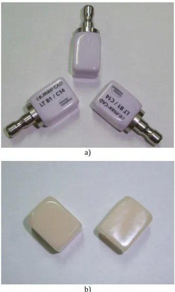
PRESS and CAD/CAM technology. The blocks are cast in one piece and they are partially crystallized. As a raw material, the value of their strength is small. Partial crystallization ensures that the blocks can be easily processed in an intermediate crystalline phase, enabling rapid machining with CAD/CAM systems, Fig. 1a. In crystallization process, blocks getting the final state of lithium disilicate crystals (Li2Si2O5) and
high strength of 400 MPa, Fig. 1b [16-18].
a)
b)
Fig. 1. Lithium disilicate blocks (IPS e.max CAD), a) partially crystallized blocks, b) final state of crystallized blocks.
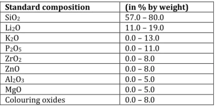
the restoration of high load areas. Table 1 shows the chemical composition of lithium disilicate glass ceramic (IPS e.max CAD).
Table 1. Chemical composition of IPS e.max CAD [15]. Standard composition (in % by weight)
SiO2 57.0 – 80.0
Li2O 11.0 – 19.0
K2O 0.0 – 13.0
P2O5 0.0 – 11.0
ZrO2 0.0 – 8.0
ZnO 0.0 – 8.0
Al2O3 0.0 – 5.0
MgO 0.0 – 5.0
Colouring oxides 0.0 – 8.0
2.1Samples preparation
Experimental tests of surface roughness were investigated on 4 samples of commercial all-ceramic lithium disilicate glass all-ceramic (IPS e.max CAD, Ivoclar Vivadent, Schaan, Liechtenstein). All samples are in the shape of a block, with dimensions: 18 mm length, 14 mm width and 12 mm height (Figs. 1a and 1b). Contact surfaces of three samples were treated with different finishing procedure (techniques): polishing (Fig. 2a), glazing (Fig. 2b) and grinding (Fig. 2c), while the contact surface of the raw material is investigated as a fourth sample (Fig. 2d). All contact surfaces of the 4 samples (Fig. 2) were obtained by using the optical microscope CSM with 5x magnification, at the Tribology Center on the Faculty of Engineering in Kragujevac.
All samples, except raw material, are firstly crystallized in a furnace at a prescribed temperature according to manufacturer's instructions (Ivoclar Vivadent).
а)
b)
c)
d)
Fig. 2. Contact surfaces of lithium disilicate glass ceramic (IPS e.max CAD) treated with different finishing procedure: a) polished surface, b) glazed surface, c) ground surface, and d) contact surface of raw materials.
Polishing procedure was done by using diamond sandpaper with different grits (280, 400, 600, 800, 1200, 2000) under the water. Fine polishing was done on the end of procedure, by using the liquid emulsion with grain size of 6 and . µm (DP-suspension, and the O-M In
Suspension). The contact surface of the second
sample was grinded by using diamond borer (Medin, ISO: 8 , μm – max). On the end, contact surface of the third samples was glazed according to the manufacturer’s recommendations (Ivoclar Vivadent).
2.2 Atomic Force Microscopy (AFM)
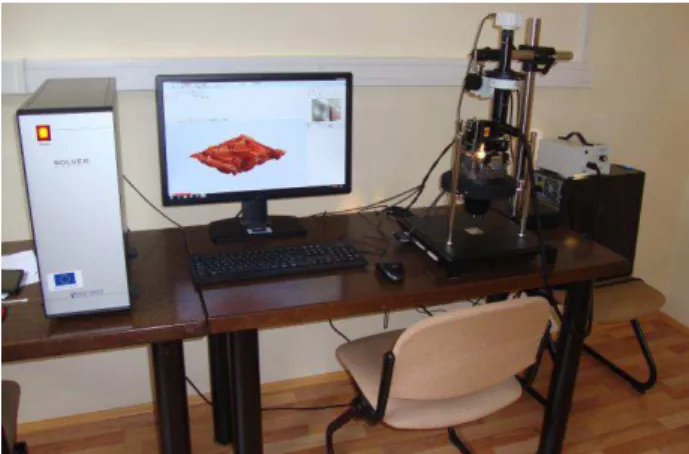
All experimental measurements of 3D surface topography and roughness parameters were obtained using the Atomic Force Microscopy (AFM) of NT-MDT manufacturers, which is located at the Tribology Center on the Faculty of Engineering in Kragujevac (Fig. 3).
Fig. 3. NT-MDT Atomic Force Microscopy (AFM), at the Tribology Center on the Faculty of Engineering in Kragujevac.
Prior to the AFM analysis the surfaces of the samples were cleaned with 70 % alcohol, and allowed to dry at room temperature.
AFM can operate in several different modes, depending on the measurement. The experiment was performed in the contact mode at room temperature, where the top of the AFM cantilever (radius of 10 nm) is in constant contact with the surface of the lithium disilicate sample. At the end of the cantilever is placed a sensor. Sensor detects movement of the cantilever when changing forces acting between the cantilever and the contact surface of the sample. Software all time of the process measured and monitored bending and/or twisting the cantilever over the tops unevenness of surface roughness. On that way, the 3D surfaces topographies of the different prepared samples are reconstructed on a very precision method.
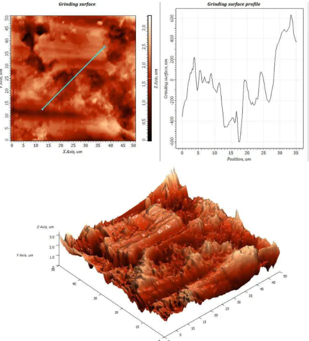
3. RESULTS AND DISCUSSION
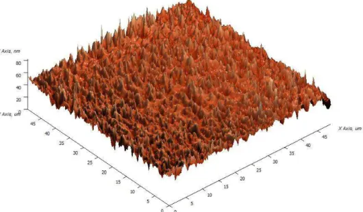
Fig. 4. 3D topography and roughness profile of polished surface of the lithium disilicate glass ceramic.
Fig. 6. 3D topography and roughness profile of raw surface of the lithium disilicate glass ceramic.
Table 2. Comparative display ofroughness parameters (Rа, Rz, Rmax, Rq) under different finishing procedure of lithium disilicate glass ceramic (IPS e.max CAD).
Measuring range, 50x μm Roughness parameters
Rа Rz Rmax Rq
The polished surface 2.049 nm 8.382 nm 11.201 nm 2.532 nm
The glazed surface 17.195 nm 76.242 nm 113.565 nm 21.788 nm
The grinding surface . µm . µm . µm . µm
The surface of raw material 2.365 nm 11.592 nm 21.119 nm 3.086 nm
Also, following roughness parameters are being presented as obtained results of AFM analysis (Table 2):
Arithmetic average roughness (Rа),
Mean height of roughness in ten points (Rz), Maximum height of the profile (Rmax), Root mean square roughness (Rq).
From presented results in the Table 2, it can be said that lowest values of roughness parameters have polished finishing procedure, as expected. Up to now, a few studies have done comparison of different finishing procedure (polishing and glazing) [19-22]. It should be noted that in aesthetic dentistry is always strived to a finer finishing surface procedures of ceramic restorations. Moreover, a well-polished surface can improve the overall strength of ceramic restorations [23].
In addition to polishing process, as a form of coatings glazing procedure is used on the surface of all-ceramic restorations like finishing procedure. Primarily, glazing is procedures that reduce porosity, reduce the surface roughness of
the material and improves the aesthetic appearance of all-ceramic restauration in the form of aesthetic shine [1,24-26]. It should emphasize that the glazing process on surface of material does not improve the strength of the all-ceramic restauration [27]. Obtained results of roughness parameters for the glazed surface (presented in Table 2), can be considered as good because the roughness values are slightly higher in comparison with the polished surface.
surface: accumulation of dental plaque, retention of bacteria, discoloration of restorations, secondary caries and gingival irritation and wear of opposing and adjacent teeth or ceramic restauration [3-5,13]. Therefore, there is general agreement among dentists that roughened ceramic surfaces must be polished to prevent or at least minimize numerous negative effects, as well as to enhance aesthetics and restoration longevity, by removing the defects produced after surface grinding [27].
4. CONCLUSIONS
AFM analysis provides very detailed information about the structural details of the materials surface. Based on this, the comparative display of surface roughness of lithium disilicate (IPS e.max CAD) using AFM was shown in this experiment under different finishing procedure.
Based on the obtained results, it can be concluded that surface roughness mostly depends on the finishing procedure (techniques). In order to avoid numerous negative effects, that are consequence of the high surface roughness, it should be strived to a finer finishing surface technique of ceramic restorations.
Acknowledgement
Research presented in this paper was supported by Ministry of Education, Science and Technology Development of Republic of Serbia, Grant TR-35021.
REFERENCES
[1] G. Aksoy, H. Polat, M. Polat and G. Coskun, 'Effect of various treatment and glazing (coating) techniques on the roughness and wettability of ceramic dental restorative surfaces', Colloids Surf B Biointerfaces, vol. 53, no. 2, pp. 254-259, 2006.
[2] S.C. Hanganu, A.O. Armencia, A.M. Murariu and L.C. Hanganu, 'Surface and Depth Modification Assessment sor Biomaterials used in Restorative Dentistry', Digest Journal of
Nanomaterials and Biostructures, vol. 8, no. 2, pp. 885 - 898, 2013.
[3] C.M. Bollen, P. Lambrechts and M. Quirynen, 'Comparison of surface roughness of oral hard materials to the threshold surface roughness for bacterial plaque retention: a review of the literature', Dent Mater, vol. 13, pp. 258-269, 1997. [4] L.M. Cavalcante, K. Masouras, D.C. Watts, L.A. Pimenta and N. Silikas, 'Effect of nanofillers’ size on surface properties after toothbrush abrasion', Am J Dent, vol. 22, pp. 60-64, 2009. [5] M. Ono, T. Nikaido and M. Ikeda, 'Surface
properties of resin composite materials relative to biofilm formation', Dent Mater J, vol. 26, pp. 613-622, 2007.
[6] S.R. Jefferies, 'Abrasive finishing and polishing in restorative dentistry: a state-of-the-art review',
Dent Clin North Am, vol. 51, pp. 379-397, 2007. [7] K.Z. Kantorski, R. Scotti and L.F. Valandro,
'Surface roughness and bacterial adherence to resin composites and ceramics', Oral Health Prev Dent, vol. 7, pp. 29-32, 2009.
[8] H. Lu, L.B. Roeder and L. Lei,'Effect of surface roughness on stain resistance of dental resin composites', J Esthet Restor Dent, vol. 17, pp. 102-108, 2005.
[9] M. Morgan, 'Finishing and polishing of direct posterior resin restorations', Pract Proced Aesthet Dent, vol. 16, pp. 211-217, 2004.
[10] G. Binnig, C.F. Quate and C. Gerber,'Atomic force microscope', Phys Rev Lett, vol. 56, no. 9, pp. 930-933, 1986.
[11] A. Kakaboura, M. Fragouli and C. Rahiotis, 'Evaluation of surface characteristics of dental composites using profilometry, scanning electron, atomic force microscopy and gloss-meter', J Mater Sci Mater Med, vol. 18, pp. 155-163, 2007.
[12] H.C. Ko, J.S. Han and M. Bächle, 'Initial osteoblast-like cell response to pure titanium and zirconia/alumina ceramics', Dent Mater, vol. 23, pp. 1349-1355, 2007.
[13] U. Covani, L. Giacomelli and A. Krajewski, 'Biomaterials for orthopedics:a roughness analysis by atomic force microscopy', J Biomed Mater Res A, vol. 82, pp. 723-730, 2007.
[15] M. Pustan and O. Belcin, 'Aplication of Atomic Force Microscope for Mechanical and Tribological Characterization of Teeth and Biomaterials', Tribology in Industry, vol. 31, no. 1-2, pp. 43-46, 2009.
[16] Ivoclar – Vivadent. Manual instruction for IPS e.max system. Ivoclar-Vivadent AG, Schaan, 2005.
[17] M. Bošković, S. Stanković, Z. Ajduković and N. Krunić,'SHORT REVIEW OF Non - metal ceramic systems', Acta Stomatologica Naissi, vol. 24, no. 57, pp. 767-774, 2008.
[18] Ivoclar – Vivadent. Scientific Documentation IPS e.max. Ivoclar-Vivadent AG, Schaan, 2011. [19] M.C. Bottino, L.F. Valandro, K.Z. Kantorski, J.C.
Bressiani and M.A. Bottin, 'Polishing methods of an alumina-reinforced feldspar ceramic', Braz Dent J, vol. 17, no. 4, pp. 285-289, 2006.
[20] D. Sarac, Y.S. Sarac, E. Yuzbasioglu and S. Bal, 'The effects of porcelain polishing systems on the color and surface texture of feldspathic porcelain', J Prosthet Dent, vol. 96, no. 2, pp. 122-128, 2006.
[21] Tholt de Vasconcellos B, Miranda-Junior WG, Prioli R, Thompson J and Oda M, 'Surface roughness in ceramics with different finishing techniques using atomic force microscope and profilometer', Oper Dent, vol. 31, no. 4, pp. 442-449, 2006.
[22] Brentel AS, Kantorski KZ, Valandro LF, Fucio SB, Puppin- Rontani RM and Bottino MA, 'Confocal laser microscopic analysis of biofilm on newer
feldspar ceramic', Oper Dent, vol. 36, no. 1, pp. 43-51, 2001.
[23] F.C. Chu, N. Frankel and R.J. Smales, 'Surface roughness and flexural strength of self-glazed, polished, and reglazed In-Ceram/Vitadur Alpha porcelain laminates', Int J Prosthodont, vol. 13, no. 1, pp. 66-71, 2000.
[24] K.C. Cheung and B.W. Darvell, 'Sintering of dental porcelain: effect of time and temperature on appearance and porosity', Dent Mater, vol. 18, no. 2, pp. 163-173, 2002.
[25] K. Mehulic, V. Svetlicic, S Segota, D. Vojvodic, I. Kovacic, D. Katanec, N. Petricevic, D. Glavina and A. Celebic, 'A Study of the Surface Topography and Roughness of Glazed and Unglazed Feldspathic Ceramics', Coll. Antropol, vol. 34, pp. 235–238, 2010.
[26] S. Štefančić, L. Ćurković, G. Baršić, M. Majić -Renjo and K. Mehulić, 'Investigation of Glazed Y-TZP Dental Ceramics Corrosion by Surface Roughness Measurement', Acta stomatol Croat, vol. 47, no. 2, pp. 163-168, 2013.
[27] T. Asai, R. Kazama, M. Fukushima and T. Okiji, 'Effect of overglazed and polished surface finishes on the compressive fracture strength of machinable ceramic materials', Dent Mater J, vol. 29, no. 6, pp. 661- 7, 2010.