Two new species of Myrmedonota Cameron
(Staphylinidae, Aleocharinae) from Mexico
Quiyari J. Santiago-Jiménez1
1 Museo de Zoología, Facultad de Biología-Xalapa, Universidad Veracruzana, Zona Universitaria, Circuito Gonzalo Aguirre Beltrán s/n, Xalapa, Veracruz, C.P. 91090, MÉXICO
Corresponding author:Quiyari J. Santiago-Jiménez (qsantiago@uv.mx)
Academic editor: J. Klimaszewski | Received 3 September 2014 | Accepted 12 November 2014 | Published 16 December 2014
http://zoobank.org/B809DD17-B7FA-4836-BDE2-A8E4CA3CDDC0
Citation: Santiago-Jiménez QJ (2014) Two new species of Myrmedonota Cameron (Staphylinidae, Aleocharinae) from Mexico. ZooKeys 464: 49–62. doi: 10.3897/zookeys.464.8549
Abstract
Two new species of Myrmedonota are described from Mexico. Illustrations and a distribution map are provided, as are keys to identify Myrmedonota known from the Nearctic and Neotropics. Specimens were collected by means of mercury vapor light traps or light interception traps.
Keywords
Lomechusini, false Lomechusini, Nearctic, Neotropical
Introduction
Recently, Hlaváč et al. (2011) cataloged 207 genera and 2,205 species belonging to the Lomechusini tribe. Although the Lomechusini tribe is a polyphyletic group distributed around the world, a clade of false Lomechusini, distributed exclusively in the Neotrop-ics, has been identiied using molecular markers (Elven et al. 2010, 2012, pers. obs.).
he genus Myrmedonota was described originally by Cameron in 1920. It only included species distributed in Asia and was recently expanded, when Maruyama et al. (2008) redescribed the genus, to include two new species from North America. Later, Eldredge (2010) described another species from Kansas, U.S.A. he latter author in-cluded a key to the species distributed in the America, north of Mexico. Later, Mathis
http://zookeys.pensoft.net
Copyright Quiyari J. Santiago-Jiménez. This is an open access article distributed under the terms of the Creative Commons Attribution License (CC BY 4.0), which permits unrestricted use, distribution, and reproduction in any medium, provided the original author and source are credited.
and Eldredge (2014) described two other species of Myrmedonota from Mexico and included commentary on the taxonomy and behavior of this genus. Now, I am add-ing another two species to this genus, one each from the states of Veracruz and Jalisco in Mexico. he specimens match the generic characters outlined by Maruyama et al. (2008) in their redescription.
Materials and methods
Between 2004 and 2006, on two ield trips to Jalisco and Veracruz in Mexico speci-mens were collected using mercury vapor light traps or light interception traps. he samples were preserved in 96% ethanol, and some of the specimens were identiied as belonging to Myrmedonota.
he specimens were observed using a Stemi DV4 stereomicroscope. Photographs from slides were taken using an image processing system (VELAB microscope model VE–633, with Digital LCD model DMS-153). Whereas, habitus photographs were taken using an Stemi 2000-C, with digital camera Canon PowerShot G10. Images were merged using the image stacking software Combine ZP. Illustrations were made based on those photographs of the structures. Permanent microscope slides were pre-pared using the techniques described by Santiago-Jiménez (2010). he terminology used here follows Santiago-Jiménez (2010), and in some cases Ashe (1984). Holotypes and paratypes were deposited at the Museo de Zoología, Universidad Veracruzana, Xalapa, Veracruz, Mexico. Some paratypes will be deposited in IEXA.
Taxonomy
Myrmedonota Cameron, 1920
he genus was redescribed by Maruyama et al. (2008). More recently, Eldredge (2010) and Mathis and Eldredge (2014) proposed a diagnosis for the genus, based only par-tially on the characters used by Maruyama et al. (2008). For example, they didn’t mention nothing about pronotum transverse or medial projection on apodeme that used Maruyama et al. (2008) on their diagnosis. Eldredge (2010) made a key to the species of North America, but it has some problems (e.g. body length in the key does not coincide with body length in the descriptions). To date, the species from Mexico have not been included in any key.
Taxonomic comments
However, there is a misunderstanding about the phylogenetic relationships, because the tree was not completely resolved to support the conclusion that Myrmedonota
belongs to Athetini. However, there is evidence that false (i.e., misidentiied) Lome-chusini from the Neotropics are a completely diferent clade from those included in the true Lomechusini and Athetini based on molecular analysis with more taxa and more characters (Santiago-Jiménez and Gusarov, in prep.).
Diagnosis. Maruyama et al. (2008) characterized the genus as follows: 1) body sur-face inely punctate; 2) head with occipital suture; 3) pronotum transverse, >1.5 wider than long; 4) setation on abdomen sparse to moderate; 5) cardo of maxilla covers bases of stipes and lacinia; 6) lacinia extremely narrowed and parallel-sided; 7) mentum al-most as long as wide; 8) apodeme of labium with medial projection; 9) 1st segment of
labial palpus longer than 2nd segment; 10) each lobe of ligula with 2 setulae.
Key to Myrmedonota species from the Nearctic and the Neotropics
1 Length of body 3.0 mm or less ...2
– Length of body more than 3.0 mm (maximum 4.2 mm) ...6
2 Pronotum yellowish; spermatheca with proximal end curved over itself (Fig. 20 in Eldredge 2010) ...M. heliantha Eldredge
– Pronotum reddish brown, dark brown or black; spermatheca with proximal end not curved over itself ...3
3 Abdominal segments unicolored, black; spermatheca V–shaped (Fig. 21 in Maruyama et al. 2008)...M. lewisi Maruyama & Klimaszewski
– Abdominal segments bicolored, usually II–IV or only anterior half of IV paler than V–VIII; spermatheca S–shaped, or if V–shaped, then abdominal tergites bicolored, with II–III and base of IV dark brown, and posterior half of IV to VIII black ...4
4 Abdominal tergites II–IV dark brown, and V–VIII black; spermatheca V–shaped (Fig. 4 in Mathis and Eldredge 2014) ...M. shimmerale Mathis & Eldredge
– Abdominal tergites II–IV yellowish to reddish brown, with at most a dark brown spot on each one, and tergites V–VIII darker; spermatheca S–shaped ...5
5 Abdominal tergites II–IV yellowish with a dark spot on medial area of tergites III–IV (Fig. 2), tergites V–VIII black; apex of median lobe, short, slightly curved ventrally (Fig.15); spermatheca with apex of the neck plain, as in Fig. 18 ...M. jaliscensissp. n.
– Abdominal tergites II–IV reddish brown and V–VIII blackish brown (some-times medial areas of tergite IV and V blackish brown); apex of median lobe, long, looking more sharply curved ventrally (Fig. 8 in Maruyama et al. 2008); spermatheca with apex of the neck concave (Fig. 12 in Maruyama et al. 2008) ...M. aidani Maruyama & Klimaszewski
V–VIII dark brown to black (except basal region of tergite V is yellowish); apex of median lobe, slightly curved ventrally (Fig. 6 in Mathis and Eldredge 2014); spermatheca without accessory gland (Fig. 8 in Mathis and Eldredge 2014) ...M. xipe Mathis & Eldredge
– Pronotum dark brown to black; elytra not bicolored, humeral region not yellow, elytra entirely brown; abdominal tergites III–V with apical region yellowish brown, appearing paler than the rest (Fig. 1); apex of median lobe, more sharply curved ventrally (Fig. 7); spermatheca with accessory gland close to the neck as in Fig. 10 ...M. cordobensis sp. n.
Myrmedonota cordobensis sp. n.
http://zoobank.org/0DC18D13-34C6-45A4-9978-7BE0C0095556
Figures 1, 3–10, 19
Type locality. Mexico, Veracruz: Córdoba, Matlaquiahuitl, 1570m, 18°59'41"N, 96°53'35.1"W, cloud forest, light trap, 6.VII.2006, J. Asiain, J. Márquez, L. Delgado and Q. Santiago leg.
Type material. Holotype male, pinned. Original label: “MÉXICO: Veracruz, Córdoba, Matlaquiahuitl. 6.VII.2006. Bosque Mesóilo de Montaña perturbado, 1,570m, 18°59'41"N, 96°53'35.1"W, ex. trampa de luz. J. Asiain, J. Márquez, L. Del-gado y Q. Santiago”/“MUZ-UV-COL-00000065”/”HOLOTYPE Myrmedonota cor-dobensis Santiago-Jiménez, 2014” [red label].
Other material. Paratypes, same data as holotype (42 males, 14 females MUZ-UV, IEXA).
Description. Body length: 3.5–4.1 mm. Most of body black to dark brown; elytra and legs brown; apical region of abdominal segments III–V, usually brown. Pubes-cence dense to sparse on head, pronotum and elytra, denser on elytra; dorsal surface of abdomen almost glabrous, dense pubescence on ventral surface of abdomen.
Head: Transverse, with or without impression on disc; without protuberance or carinae. Antennal articles 1–3 brown, 4–11 black, tip of 11 brown. Antennal articles 1–2 very elongate, 3–9 elongate, 10 slightly elongate, and 11 very elongate.
Figure 1. Habitus of Myrmedonota cordobensis Santiago-Jiménez, sp. n., male.
short); without medial spines. Prementum with two medial setae, insertions widely separated; medial pseudopore ield present; lateral pseudopore ield composed of one setose pore, and two asetose pores, with setae on aboral margin of hypoglossa, adoral margin also with setae. Mentum without microsculpture on surface; with scarcely distributed pores on mentum (around 30 pores on each side of the midline), more densely distributed toward the apex.
horax: Pronotum transverse, wider on anterior third; surface inely punctured, moderately dense; without reticulate microsculpture; setae moderately dense on sur-face; with 4 macrosetae along lateral margins, 3 macrosetae on each side of the mid-line, 2 macrosetae between lateral and medial macrosetae, distributed on anterior half. Scutellum with surface smooth, moderately covered with short setae. Elytra slightly wider on apical area; surface inely punctured, moderately dense; without reticulate microsculpture; setae moderately dense, covering the surface; with 6 macrosetae: 3 on lateral margin, and 3 diagonally placed starting from the base of midline outward. Hind wings well developed, labellum with 16–17 spines. Mesocoxal acetabula com-pletely margined posteriorly. Mesocoxal cavities moderately separated (approx. 0.20 mm) by meso- and metaventral processes; mesoventral process short (approx. 0.18 mm) with apex truncated; metaventral process medium-sized (approx. 0.56 mm), marginate and with apex acuminate; isthmus distinctly present (approx. 0.09 mm). Legs short, tarsal formula 4–5–5, every leg with an empodium, one seta on empodium and a pair of tarsal claws, each claw with a subbasal tooth.
Abdomen: Subparallel-sided, narrower than elytra, wider around segments IV–V; surface smooth, tergites III–VII almost glabrous, but with a row of 3 macrosetae along posterior margins on each side of midline of every segment and one macroseta closer to the meso-lateral region; tergite VIII (Figs 3–4) with 5 macrosetae on each side of the midline; tergite IX with 4 macrosetae on each side of the midline; tergite X with 4 macrosetae on each side of the midline. Other conspicuous characters are: tergites III–VI with basal impression; sternite IV with a central and transverse reservoir, with-out glands on basal region, withwith-out striae or cuticle vesicles on anterocentral region, without spiracles on basal region, without transversal cuticular impressions on basal region, without pseudopores on basal region.
Secondary sexual structures: Sternite VII of male with external gland on basal region and pseudopores on posterior margin of gland. Tergite VIII of male (Fig. 3) with posterior margin truncate and crenate (around 6–7 denticles), and one lateral protrusion on each external margin. Tergite VII of female without external gland or pseudopores. Tergite VIII of female (Fig. 4) not crenate and without lateral protru-sion. Sternite VIII of male and female as illustrated in Figures 5 and 6, respectively.
para-merite clearly articulated anterior to edge of velum; condylite with a line of sensory pores; velum short (less than one half of the length of the paramere). Apical lobe with 4 macrosetae visible (see Eldredge 2010).
Spermatheca: Basal bulb simple, rounded at base; tube S–shaped; internal tube of neck with denticles; with accessory gland (Fig. 10).
Remarks. It is very similar in size to M. xipe, but M. cordobensis sp. n. is easy to distinguish because it is darker, the elytra are not bicolored, the apical region of tergites III–V is brown–yellowish, and the spermatheca is diferent in shape.
Etymology. he name makes reference to the municipality where the specimens were collected, Córdoba in the state of Veracruz.
Habitat. Unknown. he adult specimens were collected with mercury vapor light traps. he larval habitat is not known.
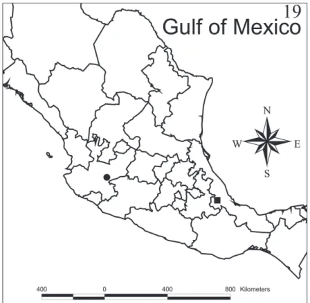
Distribution.Myrmedonota cordobensis sp. n. is only known from the type locality in the central region of the state of Veracruz, Mexico. his locality is 1,570 m above sea level, in a disturbed cloud forest. Matlaquiahuitl is the highest mountain in the municipality of Córdoba, Veracruz (Fig. 19).
Myrmedonota jaliscensis sp. n.
http://zoobank.org/BA2F7DF8-089F-4ED8-8CE6-DF555594BBA2
Figures 2, 11–18, 19
Type locality. Mexico, Jalisco: Chapala, 4 Km. Ajijic–Chapala, 20°17'48.8"N,
103°12'55.5"W, dry deciduous forest (Acacia sp.), light interception trap, 17.IX.2004, S. Gámez, A. López and Q. Santiago leg.
Type material. Holotype male, pinned. Original label: “MÉXICO: Jalisco, Chapala, 4 Km. Ajijic–Chapala. 15–17.IX.2004. Huizache, 1,620 m, 20°17'48.8"N, 103°12'55.5"W, ex. trampa de intercepción de vuelo. S. Gámez, A. López y Q. Santi-ago”/“ MUZ-UV-COL-00000603”/”HOLOTYPE Myrmedonota jaliscensis Santiago-Jiménez, 2014” [red label].
Other material. Paratypes, same data as holotype (15 males, 5 females MUZ-UV, IEXA).
Description. Body length: 2.6–3.0 mm. Most of body black to dark brown; an-terior edge of elytra, abdominal segments III–IV, and legs (except apical half of meso- and metafemur darker) yellowish brown. Densely pubescent on head, pronotum and elytra; dorsal surface of abdomen almost glabrous, densely pubescent on ventral surface of abdomen.
Head: Transverse, with or without impression on disc; without protuberance or carinae. Antennal articles 1–3 brown, 4–11 black, but tip of 11 is brown. Antenno-meres 1–3 very elongate, 4–10 elongate, and 11 very elongate.
on mesolateral region. Mandibles: asymmetrical; right mandible with medial tooth on dorsal position; left mandible without tooth; without incisor tooth; with serration between apex and medial area of mandibles; with large velvety patch, wider than half of mandible base, composed of small denticles; prostheca with short hairs along entire length, except base, which has a ctenidium; prosthecal hairs not bifurcated on medial area. Maxilla: with a row of seven spines and two rows of large setae contiguous with the apical spines on apical third of the lacinia, between two rows of setae there is a gla-brous area; the two rows of setae continue with numerous setae on middle third of the lacinia; scarcely distributed setae present on basal third of the lacinia; with pseudopores on cardo. Labium: with a short ligula and divided to near the base; with a small pair of setulae on each lobe of the ligula (one very short on the apex); without medial spines. Prementum with two medial setae, insertions widely separated; medial pseudopore ield present; lateral pseudopore ield composed of one setose pore, and two asetose pores; with setae on aboral margin of hypoglossa, adoral margin with setae too. Men-tum without microsculpture on surface; with scarce pores on menMen-tum (around 20–22 pores on each side of midline), more densely toward the apex.
horax: Pronotum transverse, wider on anterior third; surface inely punctured, moderately dense; without reticulate microsculpture; setae moderately dense on sur-face; with 4 macrosetae along lateral margins, 3 macrosetae on each side of the mid-line, 2 macrosetae between lateral and medial macrosetae distributed on anterior half. Scutellum with reticulate microsculpture, moderately covered with short setae. Elytra slightly wider on apical area; surface inely punctured, moderately dense; without re-ticulate microsculpture; covered moderately with setae; with 8 macrosetae: 3 on lat-eral margin, 3 on mesal area, and 2 in diagonal closer to inner border. Hind wings well developed, labellum with 15 spines (one female had only 10 spines). Mesocoxal acetabula completely margined posteriorly. Mesocoxal cavities moderately separated (approx. 0.16 mm) by meso- and metaventral processes; mesoventral process short (approx. 0.17 mm) with apex truncated; metaventral process medium-sized (approx. 0.56 mm), marginate and with apex acuminate; isthmus distinctly present (approx. 0.07 mm). Legs short, tarsal formula 4–5–5, every leg with an empodium, one seta on empodium and a pair of tarsal claws, each claw with a subbasal tooth.
Abdomen: Subparallel-sided, narrower than elytra, wider around segments IV–V; surface smooth, tergites III–VII almost glabrous, but with a row of 3 macrosetae along posterior margins on each side of the midline of every segment and one macroseta closer to the meso-lateral region; tergite VIII (Figs 11–12) with 5 macrosetae on each side of midline; tergite IX with 4 macrosetae on each side of midline; tergite X with 4 macrosetae on each side of midline. Other conspicuous characters are: tergites III–VI with basal impression; sternite IV with a central and transverse reservoir sac; without glands in basal region; without striae or cuticle vesicles on anterocentral region; with-out spiracles in basal region; withwith-out transversal cuticular impressions in basal region; without pseudopores in basal region.
Figures 3–10.Myrmedonota cordobensis Santiago-Jiménez, sp. n. male (3, 5, 7–9) and female (4, 6, 10).
3 tergite VIII 4 tergite VIII 5 sternite VIII (note that macrosetae were lost, only pores were illustrated)
6 sternite VIII (note that macrosetae were lost, only pores were illustrated) 7 median lobe, lateral view
Figure 19. Collection sites of Myrmedonota cordobensis Santiago-Jiménez, sp. n. (black square) and M. jaliscensis Santiago-Jiménez, sp. n. (black circle).
(around 6 denticles), and one lateral protrusion on each side of the midline. Tergite VIII of female (Fig. 12) is not crenate and it has a lateral protrusion. Sternite VIII of male and female as illustrated in Figures 13 and 14, respectively.
Aedeagus: Median lobe pear-shaped (Figs 15–16); with internal sac of median lobe with many spinules; medial lobe with short, well deined compressor plate; apical lobe curved to the ventral side (visible in lateral view), and pointed; basal ridge convex. Paramere as in Fig. 17; anterodorsal margin of paramerite with prominent sensory pores present beneath the velar sac; hinge zone of paramerite faint, extended from dorsal surface to near articulation between condylite and paramerite; apical process of paramerite clearly articulated anterior to edge of velum; condylite with a line of sensory pores; velum short (less than one half of the length of the paramere). Apical lobe with 3 macrosetae visible.
Spermatheca: Basal bulb simple, rounded at base; tube S–shaped; internal tube of neck with denticles; without accessory gland (Fig. 18).
is not curved over itself; from M. lewisi because the abdomen is bicolored; from M. shimmerale because the spermatheca is S–shaped; and inally, from M. aidani because tergites II–IV are yellowish with a dark spot on medial area of tergites III–IV, and the diferently shaped spermatheca.
Etymology. he name makes reference to the state of Jalisco, Mexico, where the specimens were collected.
Habitat. Unknown. he adult specimens were collected with interception light
traps. he larval habitat is not known.
Distribution.Myrmedonota jaliscensis sp. n. is only known from the type locality around Lake Chapala in Jalisco state, Mexico (Fig. 19). his locality is 1,620 m above sea level, where it is common to ind Acacia sp. trees, the common name of which is Huizache.
Discussion
More species of Myrmedonota are being described from the Nearctic and Neotropical regions, and here I have described two new species, and it is possible that more species will be discovered in the future. Although Eldredge (2010) and Mathis and Eldredge (2014) presented a new diagnosis of Myrmedonota, it is not clear what specimens they used to select their diagnostic characters. Specimens reviewed here matched with di-agnostic characters proposed by Eldredge (2010) and Mathis and Eldredge (2014); however, as mentioned above, they didn’t mention nothing about pronotum transverse or medial projection on apodeme that used Maruyama et al. (2008) on their diagnosis. Moreover, there is an inconsistency about labial palpomeres from diagnosis by Mathis and Eldredge (2014) compared to previous diagnosis by Eldredge (2010). I think it should be labial palpomeres I and III subequal in length, not II and III as mentioned by Mathis and Eldredge (2014). herefore, I suggest we follow the redescription pro-posed by Maruyama et al. (2008) because they reviewed the type species of Myrme-donota and it has been useful to diagnose Nearctic and Neotropical species. Diagnostic characters should be proposed in a future analysis by mean of synapomorphies on a phylogenetic context.
Misunderstandings in Elven et al. (2010) about the limits of the Lomechusini-Athetini complex are causing confusion for people working with both tribes. hat phylogeny was not completely resolved, and the main conclusion is that the species of false Lomechusini from the Neotropics belong to a diferent clade, but it was not pos-sible to conclude whether they should be part of Athetini.
Acknowledgments
he author is very grateful to colleagues for their support at the Facultad de Biología, Universidad Veracruzana and Instituto de Ecología, A.C., particularly M. Morales, R. Ortega and R. Novelo for the use of the microscopes and to C. Álvarez-Castillo for preparing the illustrations. J. Asiain, J. Márquez, L. Delgado, S. Gámez, and A. López assisted on collection trips. I thank B. Delfosse and two anonymous reviewers for their comments, which helped improve the manuscript.
References
Ashe JS (1984) Generic revision of the subtribe Gyrophaenina (Coleoptera: Staphylinidae: Aleo-charinae) with a review of described subgenera and major features of evolution. Quaestiones Entomologicae 20(3): 129–349.
Cameron M (1920) New species of Staphylinidae from Singapore. Part III. Transactions of the Entomological Society of London 1920(1–2): 212–284.
Eldredge KT (2010) A new species of Myrmedonota Cameron from eastern Kansas (Coleoptera, Staphylinidae, Aleocharinae). ZooKeys 53: 17–24. doi: 10.3897/zookeys.53.493
Elven H, Bachmann L, Gusarov VI (2010) Phylogeny of the tribe Athetini (Coleoptera: Staphylinidae) inferred from mitochondrial and nuclear sequence data. Molecular Phylo-genetics and Evolution 57: 84–100. doi: 10.1016/j.ympev.2010.05.023
Elven H, Bachmann L, Gusarov VI (2012) Molecular phylogeny of the Athetini-Lomechusini-Ecitocharini clade of aleocharinae rove beetles (Insecta). Zoologica Scripta 41(6): 617–636. doi: 10.1111/j.1463-6409.2012.00553.x
Hlaváč P, Newton AF, Maruyama M (2011) World catalogue of the species of the tribe Lome-chusini (Staphylinidae: Aleocharinae). Zootaxa 3075: 1–151.
Maruyama M, Patrick LB, Klimaszewski J (2008) First record of the genus Myrmedonota Cameron (Coleoptera, Staphylinidae) from North America, with descriptions of two new species. Zootaxa 1716: 35–43.
Mathis KA, Eldredge T (2014) Descriptions of two new species of Myrmedonota Cameron (Staphylinidae: Aleocharinae) from Mexico with comments on the genus taxonomy and behavior. Zootaxa 3768(1): 95–100. doi: 10.11646/zootaxa.3768.1.7
Motschulsky MV (1858) Énumération des nouvelles espèces de Coléoptères rapportés de ses voyages. 2-d. Article (continuation). Bulletin de la Société Impériale des Naturalistes de Moscou 31(3): 204–264.