___________________________
Corresponding author: Seiied Mojtaba Mohaddes Ardebili, Head department of medical genetics,Faculty of medicine,Tabriz university of medical sciences, Tabriz-Iran Tel/Fax: ++98-411-3371587,e-mail: mohaddesmo@yahoo.com
UDC 575 DOI: 10.2298/GENSR1302503G
Original scientific paper
INVESTIGATION OF FIVE POLYMORPHIC DNA MARKERS ASSOCIATED WITH LATE ONSET ALZHEIMER DISEASE
Jalal GHARESOURAN1, Maryam REZAZADEH1,2, Seiied MOJTABA MOHADDES
ARDEBILI1
1Department of Medical Genetics, Faculty of Medicine, Tabriz University of Medical Sciences, Tabriz, Iran.
2Genetics Research Center, University of Social Welfare and Rehabilitation Sciences, Tehran, Iran.
Gharesouran J, M. Rezazadeh, S. Mojtaba Mohaddes Ardebili (2013):
Investigation of five polymorphic DNA markers associated with late onset Alzheimer disease. Genetika, Vol 45, No. 2, 503-514.
Alzheimer's disease is a complex neurodegenerative disorder characterized by memory and cognitive impairment and is the leading cause of dementia in the elderly. The aim of our study was to examine the polymorphic DNA markers CCR2 (+190 G/A), CCR5 32, TNF- (-308 G/A), TNF- (-863 C/A) and CALHM1 (+394 C/T) to determine the relationship between these polymorphisms and the risk of late onset Alzheimer's disease in the population of Eastern Azerbaijan of Iran. A total of 160 patient samples and 163 healthy controls were genotyped by PCR-RFLP and the results confirmed using bidirectional sequencing.
Statistical analysis of obtained data revealed non-significant difference between frequency of CCR5 32 in case and control groups. The same result was observed for TNF- (-863 C/A) genotype and allele frequencies. Contrary to above results, significant differences were detected in frequency of TNF- (-308 G/A) and CCR2-64I genotypes between the cases and healthy controls. A weak significant difference observed between allele and genotype frequencies of rs2986017 in CALHM1 (+394 C/T; P86L) in patient and control samples. It can be concluded that the T allele of P86L variant in CALHM1 & +190 G/A allele of CCR2 have a protective role against abnormal clinical features of Alzheimer's disease.
Key words: Alzheimer disease; CCR2; CCR5; CALHM1; TNF- ; Eastern
Azerbaijan.
INTRODUCTION
generally differentiated: Early-onset (<60 years) familial AD (EOFAD) following Mendelian inheritance and late-onset AD (LOAD) or so-called ‘‘sporadic’’ cases with less apparent or no familial aggregation usually occurring later in life ( 60 years). However this traditional dichotomization is overly simplistic as there are cases of early-onset AD without evidence for Mendelian transmission while, conversely, LOAD is frequently observed with a strong familial clustering, sometimes resembling a Mendelian pattern (BERTRAM et al. 2010; RUBIO-PEREZ and MORILLAS-RUIZ 2012).
Neurodegenerative disorders such as AD and also for many of the other neurodegenerative diseases, familial aggregation was already recognized as a salient feature decades before any of the underlying molecular genetics and biochemical properties were known. The cause of AD is principally genetic but also environmental, all mostly unknown. As a matter of fact, it was often only the identification of specific, disease-segregating mutations in previously unknown genes that directed the attention of molecular biologists to certain proteins and pathways that are now considered crucial in the development of the various neurodegenerative diseases (BERTRAM et al. 2012; ZLOKOVIC 2011).
The etiology of LOAD is complex and it has strong genetic heterogeneity (PASTOR and GOATE 2004; TUPPO and ARIAS 2005).Furthermore, the genetic component of this form is itself complex and heterogeneous: complex because there is no single or simple model explaining the mode of disease transmission and heterogeneous because the gene mutations or polymorphisms may interact with one another and with environmental factors (LAMBERT and AMOUYEL 2011). Therefore studying of any candidate gene can help to identify the heterogeneous nature of LOAD in different populations.
The genetics of LOAD has taken impressive steps forwards in the last few years. To date, more than six-hundred genes have been linked to the disorder. However, only a minority of them are supported by a sufficient level of evidence (OLGIATI et al. 2011). These studies
identified several new susceptibility genes includingTumor necrosis factor-alpha (TNF- ), C-C chemokine receptor type 2(CCR2), C-C chemokine receptor type 5 (CCR5) and Calcium homeostasis modulator 1 (CALHM1).
TNF plays a critical role in brain development, brain physiology, synaptic plasticity, sleep, circadian rhythm and normal behavior. Serum concentrations of TNF- were found to be elevated in Alzheimer patients (BONOTIS et al. 2008). The gene product is an important pro inflammatory cytokine and activates the nuclear factor kappa B (NF-KB), a protein which activates the secretion of Apo lipoprotein E (APOE). APOE gene located on chromosome 19 is the only recognized susceptibility gene for this form of Alzheimer (BALES et al. 2000; SESHADRI et al. 2010). Polymorphisms in the promoter region of TNF- gene have been reported to
increase the transcription rate of the gene and thus might influence the risk of AD(FELDMANN and MAINI 2003; TEDDE et al 2008).Amongst them the polymorphisms located on 308bp and
-836bp have been shown to be associated with an increased and decreased transcriptional activity of the gene respectively(FARGION et al. 2004).
CCR2 signaling can mediate accumulation of microglia at sites affected by neuroinflammation. CCR2 and its main ligand CCL2 (MCP-1) might also be involved in the altered metabolism of A (HARRIES et al. 2012; NAERT and RIVEST 2012; WESTIN et al. 2012).
There is growing evidence that chemokines and their receptors are upregulated in reactive microglial cells in the brain tissue of affected individuals, and they may play a role in the recruitment and accumulation of microglial cells at A sites in senile plaques. Indeed, some immunohistochemical studies have shown that the activated microglial cells in both control and patients present an increased expression of chemokine receptor CCR5, indicating to important role of CCR5 receptor in regulation of brain immune response in AD. Recently it has been suggested that CCR5 32 polymorphism has a protective effect towards some inflammatory diseases such as AD.
Tissue Info studies (SKRABANEK and CAMPAGNE 2001) and the Alzgene database (BERTRAM et al. 2007) have introduced the gene Calcium homeostasis modulator 1 (CALHM1;
MIM# 612234), located about 1.6 Mb apart from LOAD marker D10S1671 on chromosome 10, as one of the candidate genes for LOAD (Bertram et al. 2000). CALHM1 is preferentially expressed in the hippocampus, a brain region which is affected early in the course of AD. The multipass transmembrane glycoprotein encoded by CALHM1, controls cytosolic Ca2+
concentrations in brain cells. In in vitro experiments, CALHM1 expression promotes calcium
influx, and triggers a decrease in amyloid levels, along with an elevation of soluble APP levels that appears to be regulated by calcium influx (DRESES-WERRINGLOER et al. 2008).
Substitution of proline with leucine at codon 86 (P86L) results in the SNP rs2986017 in
CALHM1. A recent study has reported that this variant of CALHM1 increases the risk of AD by 40% (DRESES-WERRINGLOER et al. 2008). Functional studies have shown that the rs2986017 SNP
results in decreased permeability to calcium ions and lowered calcium ion levels, ultimately leading to an increase in Aß peptide (AQDAM et al. 2010).
Although many LOAD associated genes have been identified, the replication studies play a critical role in confirming the reported data, especially within different ethnic populations. The present study was conducted to reveal the possible association of polymorphisms of above mentioned genes in the population of eastern Azerbaijan of Iran. Similar results had been reported for a number of ethnicities when the present study was started; however no published data was present about the population investigated in our study.
MATERIALS AND METHODS
Design, Setting, and Participant
Reported information from investigations of genetic association of LOAD with SNPs located in the candidate gene regions was collected from a recent large genome-wide association study (GWAS) conducted by the Alzheimer Disease Genetics Consortium (ADGC). Our study included 160 AD patients (94 women & 66 men) and 163 healthy controls (95 women & 68 men). The age of patient group at onset was ranged within 65-99 years (mean age 76.06 ±7.75 years).
Gene selection
We tested the genetic association of LOAD with three genes encoding the inflammatory pathway factors and with one other Calcium homeostasis modulator gene. The Tumor necrosis factor alpha, CCR2, CCR5 and CALHM1 genes have included in this study.
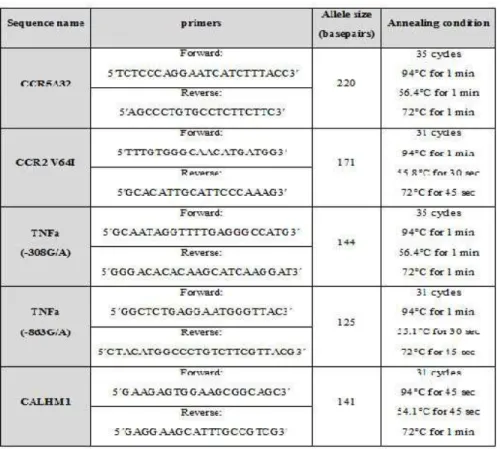
Table 1. PCR primers complementary to the studied variation
Sample Preparation, Genotyping and quality control of genotype data
PCR conditions for each of the polymorphic DNA fragments have been summarized in table 1. The PCR or RFLP products were fractionated on an 8% acrylamide gel and visualized flowing to staining by AgNO3.
Association analysis
The CCR5 32 (32 bp deletion, nt794; chromosome 3p21) genotype was determined by PCR without RFLP. The PCR produced a 220bp product from the wild type allele and an 188bp product from the deleted allele. The conditions of RFLP reactions for each of the remaining polymorphisms have been shown in table 2.
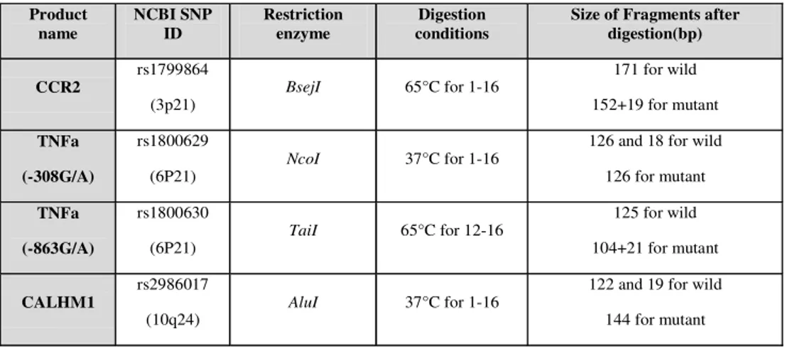
Table 2: Digestion conditions of amplified DNA fragments
Product name
NCBI SNP ID
Restriction enzyme
Digestion conditions
Size of Fragments after digestion(bp)
CCR2
rs1799864
(3p21) BsejI 65°C for 1-16
171 for wild 152+19 for mutant
TNFa
(-308G/A)
rs1800629
(6P21) NcoI 37°C for 1-16
126 and 18 for wild 126 for mutant
TNFa
(-863G/A)
rs1800630
(6P21) TaiI 65°C for 12-16
125 for wild 104+21 for mutant
CALHM1
rs2986017
(10q24) AluI 37°C for 1-16
122 and 19 for wild 144 for mutant
Heterogeneity study and gene-based multiple testing correction
The allelic and genotypic frequencies were obtained by direct counting. Data analysis was performed using SPSS16. Chi square and Fisher’s exact test were used to compare the genotypes. Statistical significance was set at P< 0.05. The odds ratio (OR) was calculated at 95% CI.
RESULTS
Case-control analysis, full sample
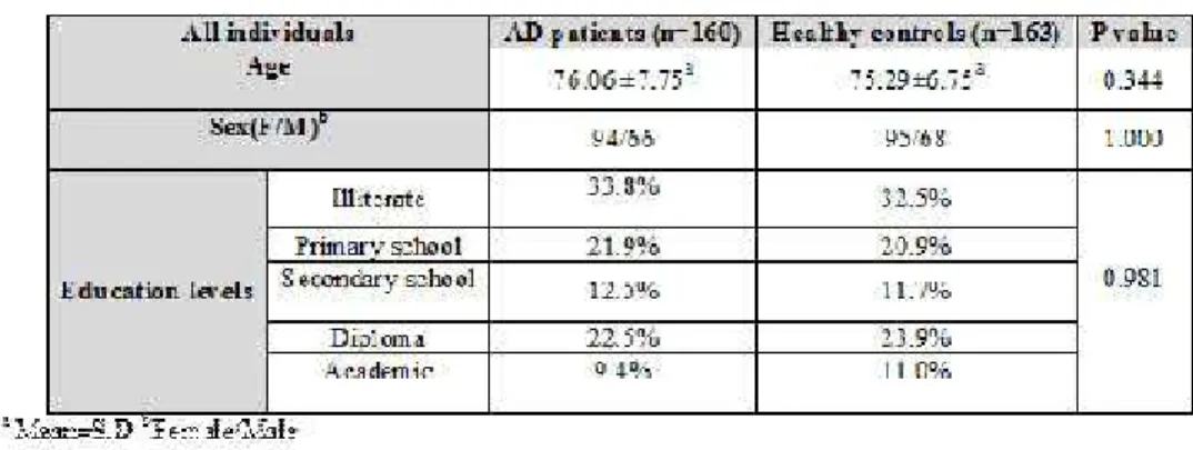
In this study the control group was matched by age, gender, race and education to the subjects. There was no major departure of the genotypes from the Hardy–Weinberg equilibrium. Table 3 shows the results obtained from statistical analysis of mentioned variables, indicating to non-significant differences between the two groups.
Table 3. Comparasion of mean age, sex and education levels between AD cases and control subjects using t-test and 2 test anylysis
Table4. Statically analysis results for association of AD with SNPs in CCR2, CCR5, TNFa and CALHM1 in Individuals
Gene/SNP MA MAF MA-OR(95% CI)
w/m + m/m frequencies
OR (95% CI) P Value a CCR2
rs1799864 A P < 0.001
4.5
(2.78-7.29)
4.78
(2.83-7.94) P < 0.001
CCR5
rs333 - 0.26
0.67
(0.33-1.35)
0.65
(0.32-1.35) 0.25
TNFa
rs1800629 A P < 0.001
0.08
(0.04-0.13)
0.067
(0.03-0.11) P < 0.001
TNFa
rs1800630 A 0.27
0.68
(0.34-1.35)
0.65
(0.32-1.35) 0.25
CALHM1
rs2986017 T 0.003
0.4
(0.22-0.74)
0.41
(0.22-0.79) 0.008
Abbreviations:AD, Alzheimer disease;MA, minor allele; MAF, w, wild type; m, mutant; weighted-average minor allele frequency;OR, odds ratio; SNP, single-nucleotide polymorphism.
a: P values and ORs estimated under an additive model using logistic regression with covariates (adjusted for age, and
Amplification of CCR5 ( 32) by PCR demonstrated a single band of 220bp in individuals homozygous for the wild-type allele and two bands of 220 and 188bp in heterozygotes. No homozygous mutant subject was detected in this study. Table 4 shows non-significant difference between the case and control groups for the polymorphic region of the CCR5 gene.
The statistical analysis of patients, genotypes and allele frequencies (Table 4) revealed a significant difference in TNF- , -308 G/A genotype and allele frequencies between the AD patients and healthy subjects. Non-significant difference was detected between TNF- -863 C/A genotype and allele frequencies between AD patients and healthy controls (Table 4).
The allele and genotype frequency distribution of P86L genotype were weakly significant between the two study groups (Table 4). As the difference between distribution of T allele (mutant) in case and control groups is significant, it indicates to linkage of this allele with AD.
DISCUSSION
On the basis of the data obtained in previous studies, the recruitment of microglial cells in senile plaques is induced by chemokines and their up regulated receptors in AD brain (AKIYAMA
et al. 2000; BAJETTO et al. 2002; FELDMANN and MAINI 2003). A deposition is associated with a
local inflammatory response, which is initiated by the activation and migration of microglia in inflammatory sites (DEAN et al. 2000, SHARMA et al. 2012).
As CCR2 is located about 19Kb apart from CCR5 gene on chromosome 3, the co-segregation or independent assortment of the two markers was studied in case and control groups. CCR2-64I carriers were never found to be homozygous for the CCR5 32, confirming almost complete linkage disequilibrium of the two markers.
In our study population, occurrence of CCR2-64I polymorphism is decreased in AD patients indicating to association with low risk of developing the disease (P <0.001; OR=4.5, 95% CI:
2.78-7.29). The low frequency of the genotype 64I/64I in AD patients proves real protective effect of this polymorphism on AD (P<0.001; OR=4.78, 95% CI: 2.83-7.94). Our finding about
this polymorphism covered the results of Galimberti et al carried out on Italian population (P=0.037; OR=0.65, 95% CI: 0.41–1.03). The conclusion drawn from this study is also
comparable to those obtained by Huerta et al. performed on Spanish population (P>0.05; OR=1.06, 95% CI: 0.57–1.96) (HUERTA et al. 2004).
The statistical analysis of the data obtained in our study revealed non-significant difference in genetic distribution of CCR5 polymorphism between the case and control groups (P=0.25;
OR=0.67, 95% CI: 0.33-1.35). Also, the haplotypes of CCR5 32/ 32 were never observed either in AD or in control group. These observations are in agreement with the results reported for Italic ethnicity (P>0.05; OR=1.54, 95% CI: 0.97–2.45) (GALIMBERTI et al. 2004).
The–308 polymorphism has been associated with an increased transcriptional activity and TNF- release, whereas the –863 polymorphism have been associated with a reduced transcriptional activity. In the present study the -308G/A polymorphism was shown to be a genetic marker for susceptibility to AD in Azeri Turk population (P<0.001; OR=0.067, 95% CI:
0.03-0.11). Our results are similar to the results came out for the other ethnic groups (MOHADDES
et al. 2011). A report about the population of Southern China suggests that the -308G/A polymorphism of TNF- gene might be a risk factor for AD in this ethnicity. Similar results have been reported for the Chilean population (DI et al. 2004). The association of -863 polymorphism
with AD was not approved in the present study. This finding had been previously reported by others (P=0.24; OR=1.24, 95% CI: 0.86–1.85) (DI et al. 2004). Studies in Spain and Italy have
also reported that there is no association between TNF- -863C/A and AD.
Regarding to the different roles of -308 G/A and -863 C/A polymorphisms on the levels of TNF- transcription, a negative association is expected between Alzheimer’s disease and -863 C/A polymorphism. However our results did not reveal such an association.
CALHM1 gene encodes a multipass transmembrane glycoprotein that controls cytosolic Ca2+ concentrations. CALHM1 expression controls APP processing by interfering with extracellular A accumulation. Also CALHM1 controls APP proteolysis in a Ca2+ dependent manner. According to Calcium hypothesis of brain aging and Alzheimer’s disease neuropathological changes associated with AD is caused by changes in Ca2+ homeostasis (KHACHATURIAN et al. 1989).
It had been shown that rs2986017 SNP in CALHM1 gene (P86L) is associated with
increased risk of LOAD, significant dysregulation of Ca2+ homeostasis and APP metabolism (DRESES-WERRINGLOER et al. 2008; SHOJI et al. 2005). In a US study, the impact of rs2986017 on
the risk of developing AD in 2043 AD cases and 1361 controls Distribution of the T allele was increased in AD cases as compared to controls in all studies, with odds ratios (ORs) ranging from 1.29 to 1.99 (DRESES-WERRINGLOER et al. 2008). In a similar study a significant association
was detected between CALHM1-P86L polymorphism and AD in the ethnic Chinese (CUI et al.
2010). In a study carried out in Spain a weak evidence of association between P86L mutation and LOAD susceptibility was observed when a recessive model was applied (P=0.24; OR=1.38,
CI: 1.01-1.89) (BOADA et al. 2010). However some other investigations performed on
populations from other ethnic origins have reported that the difference between the above mentioned polymorphism and LOAD is non-significant (BERTRAM et al. 2008; INOUE et al.
2010; NACMIAS et al. 2010).
Our study corroborates the findings by different groups (P=0.008; OR=0.41, CI: 0.22-0.79)
(BOADA et al. 2010; CUI et al. 2010; DRESES-WERRINGLOER et al. 2008). Even though our sample
was much smaller than the original study, it showed an association with LOAD cases.
ACKNOWLEDGEMENT
We are grateful for valuable cooperation of Danesh laboratory in providing the control samples. We thank Dr. Parvin Javadi (Khuban old people’s home, Tabriz) and Dr. Shoukoh Mosavipoor (Mehr old people’s home, Tabriz) for their kind collaboration. The study was supported by a grant from Deputy for Research, Tabriz University of Medical Sciences.
Received February 18th, 2013
REFERENCES
AQDAM, M.J, K KAMALI, M RAHGOZAR, M OHADI, M MANOOCHEHRI, A TAHAMI, L BOSTANSHIRIN, HR KHORRAMKHORSHID (2010): Association of CALHM1 Gene Polymorphism with Late Onset Alzheimer’s Disease in Iranian Population. AJMB. 2: 153-157.
AKIYAMA, H, S BARGER, S BARNUM, B BRADT, J BAUER, GM COLE, NR COOPER, P EIKELENBOOM, M EMMERLING, B.L FIEBICH, CE FINCH, S FRAUTSCHY, WS GRIFFIN, H HAMPEL M, HULL, G LANDRETH, , L LUE, R MRAK, IR MACKENZIE, PL MCGEER, MK O'BANION, J PACHTER, G PASINETTI, C PLATA-SALAMAN, J ROGERS, R RYDEL, Y SHEN, W STREIT, R STROHMEYER, I TOOYOMA, FL VAN MUISWINKEL, R VEERHUIS, D WALKER, S WEBSTER, B WEGRZYNIAK, G WENK, T WYSS-CORAY (2000): Inflammation and Alzheimer's disease. Neurobiol Aging. 21(3): 383-421.
BAJETTO, A, R BONAVIA, S BARBERO, G SCHETTINI (2002: Characterization of chemokines and their receptors in the central nervous system: physiopathological implications. J Neurochem. 82(6): 1311-1329.
BALES, KR, Y DU, D HOLTZMAN, B CORDELL, SM PAUL (2000): Neuroinflammation and Alzheimer's disease: critical roles for cytokine/Abeta-induced glial activation, NF-kappaB, and apolipoprotein E. Neurobiol Aging. 21(3): 427-32.
BERTRAM, L, BM SCHJEIDE, B HOOLI, K MULLIN, M HILTUNEN, H SOININEN, M INGELSSON, L LANNFELT, D BLACKER, RE TANZI (2008: No Association between CALHM1 and Alzheimer’s disease risk. Cell. 135: 993-996. BOADA, M, C ANTÚNEZ, J LÓPEZ -ARRIETA, JJ GALÁN, FJ MORÓN, I HERNÁNDEZ, J MARÍN, MARTÍNEZ- P LAGE, M ALEGRET,
JM CARRASCO, C MORENO, LM REAL, A GONZÁLEZ-PÉREZ, L TÁRRAGA, A RUIZ (2010): CALHM1 P86L polymorphism is associated with late-onset Alzheimer's disease in a recessive model. J Alzheimer’s Dis., 20: 247-251.
BONOTIS, K, E KRIKKI, V HOLEVA, C AGGOURIDAKI, V COSTA, S BALOYANNIS (2008): Systemic immune aberrations in Alzheimer's disease patients. J. Neuroimmunol. 193, 183–187.
CUI, PJ, L ZHENG, L CAO, Y WANG, YL DENG, G WANG, W XU, HD TANG, JF MA, T ZHANG, JQ DING, Q CHENG, SD CHEN (2010): CALHM1 P86L polymorphism is a risk factor for Alzheimer's disease in the Chinese population. J Alzheimer’s Dis. 19: 31-35.
DEAN, M, M CARRINGTON, SJ O’BRIEN (2002): Balanced polymorphism selected by genetic versus infectious human disease. Annu. Rev. Genomics Hum. Genet. 3: 263–292.
DI BD, , G CANDORE, C FRANCESCHI, F LICASTRO, G COLONNA-ROMANO, C CAMMA, et al. (2009) Systematic review by metaanalyses on the possible role of TNF- lpha polymorphisms in association with Alzheimer's disease. Brain Res Rev. 61(2): 60-8.
DRESES-WERRINGLOER, U, JC LAMBERT, V VINGTDEUX, H ZHAO, H VAIS, A SIEBERT, A JAIN, J KOPPEL, , A ROVELET-LECRUX, D HANNEQUIN, F PASQUIER, D GALIMBERTI, E SCARPINI, D MANN, C LENDON, D CAMPION, P AMOUYEL, P DAVIES, JK FOSKETT, F CAMPAGNE, P MARAMBAUD (2008): A polymorphism in CALHM1 influences Ca2+ homeostasis, Abeta levels, and Alzheimer's disease risk. Cell. 133(7): 1149-61.
FARGION, S, L VALENTI, P DONGIOVANNI, AL FRACANZANI (2004): TNFalpha promoter polymorphisms. Methods Mol Med. 98: 47-58.
FAVOROVA, OO, TV ANDREEWSKI, AN BOIKO, MA SUDOMOINA, AD ALEKSEENKOV, OG KULAKOVA, et al. (2002): The chemokine receptor CCR5 deletion mutation is associated with MS in HLA-DR4-positive Russians. Neurology. 59:1652–5.
FELDMANN, M, RN MAINI (2003) :Lasker Clinical Medical Research Award. TNF defined as a therapeutic target for rheumatoid arthritis and other autoimmune diseases. Nat Med. 9(10): 1245-50.
HARRIES, LW, RM BRADLEY-SMITH, DJ LLEWELLYN, LC PILLING, A FELLOWS, W HENLEY, D HERNANDEZ, JM GURALNIK, S BANDINELLI, A SINGLETON, L FERRUCCI, D MELZER (2012): Leukocyte CCR2 Expression Is Associated with Mini-Mental State Examination Score in Older Adults. Rejuvenation Res. 4:395-404. Epub.
HUERTA, C, V ALVAREZ, IF MATA, E COTO, R RIBACOBA, C MARTÍNEZ, M BLÁZQUEZ, LM GUISASOLA, C SALVADOR, CH LAHOZ, J PEÑA (2004): Chemokines (RANTES and MCP-1) and chemokine-receptors (CCR2 and CCR5) gene polymorphisms in Alzheimer's and Parkinson's disease. Neurosci Lett. 370(2-3):151-4.
INOUE, K, N TANAKA, F YAMASHITA, Y SAWANO, T ASADA, Y GOTO (2010): The P86L common allele of CALHM1 does not influence risk for Alzheimer disease in Japanese cohorts. Am J Med Genet B Neuropsychiatr Genet., 1532: 532-535.
JILLIAN, J (2009): Alzheimer disease: Alzheimer’s disease neuropathology in the oldest old. Nat RevNeurol., 5: 411-412. KHACHATURIAN, ZS (1989): Calcium, membranes, aging, and Alzheimer’s disease.Introduction and overview.Ann. N.Y.
Acad. Sci., 568: 1–4.
KHORRAM KHORSHID, HR, M MANOOCHEHRI, L NASEHI, M OHADI, M RAHGOZAR, K KAMALI (2011): Ccr2-64i and Ccr5 32 Polymorphisms in Patients with Late-Onset Alzheimer’s disease; A Study from Iran (Ccr2-64i And Ccr5 32 Polymorphisms in Alzheimer’s disease). J Basic Med Sci., 15(4): 937–944.
MOHADDES, SM, M REZAZADEH, J GHARESOURAN, T YEGHANEH, M, ARHOUDI H AYROMLOU, M TALEBI, M GHOJAZADEH (2011) Association of CCR2 and CCR5 gene polymorphisms with Alzheimer’s disease. JSCi. 22: 11-116. ARDEBILI, SM, T YEGHANEH, J GHARESOURAN, M REZAZADEH, M FARHOUDI, H AYROMLOU, M TALEBI, M GHOJAZADEH
(2011). Genetic association of TNF- -308 G/A and -863 C/A polymorphisms with late onset Alzheimer's disease in Azeri Turk population of Iran. J Res Med Sci., 16(8):1006-13.
NACMIAS, B, A TEDDE, S BAGNOLI, E LUCENTEFORTE, E CELLINI, I PIACERI, BM GUARNIERI, V BESSI, L BRACCO, S SORBI (2010): Lack of implication for CALHM1 P86L common variation in Italian patients with early and late onset Alzheimer's disease. J Alzheimer's Dis., 20: 37-41.
NAERT, G, S RIVEST (2012): Hematopoietic CC-chemokine receptor 2 (CCR2) competent cells are protective for the cognitive impairments and amyloid pathology in a transgenic mouse model of Alzheimer's disease. Mol Med. 18(1):297-313. doi: 10.2119/molmed.2011.00306.
OLGIATI, P, AM POLITIS, GN PAPADIMITRIOU, DD RONCHI, A SERRETTI (2011): Genetics of Late-Onset Alzheimer’s Disease: Update from the Alzgene Database and Analysis of Shared Pathways.
PASTOR, P, AM GOATE (2004): Molecular genetics of Alzheimer's disease. Curr Psychiatry Rep., 6: 125-133. PERRY, VH, JA NICOLL, C HOLMES (2010): Microglia in neurodegenerative disease. Nat Rev Neurol 6: 193–201. RUBIO-PEREZ, JM, JM MORILLAS-RUIZ (2012): A review: inflammatory process in Alzheimer's disease, role of cytokines.
Scientific World Journal 75,6357.
SESHADRI, S, AL FITZPATRICK, MA IKRAM, AL DESTEFANO, V GUDNASON, M BOADA, JC BIS, AV SMITH, MM CARASSQUILLO, JC LAMBERT, D HAROLD, EM SCHRIJVERS, R RAMIREZ-LORCA, S DEBETTE, WT LONGSTRETH, AC JANSSENS, VS PANKRATZ, JF DARTIGUES, P HOLLINGWORTH, T ASPELUND, I HERNANDEZ, A BEISER, LH KULLER, PJ KOUDSTAAL, DW DICKSON, C TZOURIO, R ABRAHAM, C ANTUNEZ, Y DU, JI ROTTER, YS AULCHENKO, TB HARRIS, RC PETERSEN, C BERR, MJ OWEN, J LOPEZ-ARRIETA, BN VARADARAJAN, JT BECKER, F RIVADENEIRA, MA NALLS, NR GRAFF-RADFORD, D CAMPION, S AUERBACH, K RICE, A HOFMAN, PV JONSSON, H SCHMIDT, M LATHROP, TH MOSLEY, R AU, BM PSATY, AG UITTERLINDEN, LA FARRER, T LUMLEY, A RUIZ, J WILLIAMS, P AMOUYEL, SG YOUNKIN, PA WOLF, LJ LAUNER, OL LOPEZ, CM VAN DUIJN, MM BRETELER (2010): Genome-wide analysis of genetic loci associated with Alzheimer disease. JAMA.,303: 1832-1840.
SHARMA, V, V. THAKUR, SN SINGH, R. GULERIA (2012): Tumor Necrosis Factor and Alzheimer’s Disease: A Cause and Consequence Relationship. Bulletin of Clinical Psychopharmacology, 22(1):86-97.
proposal for diagnostic and clinical assessment criteria for Alzheimer's disease.RinshoShinkeigaku., 45: 128-137.
TEDDE, A, AL PUTIGNANO, .NACMIAS, .BAGNOLI, .CELLINI, S. SORBI (2008): Lack of association between TNF-alpha polymorphisms and Alzheimer's disease in an Italian cohort. Neurosci Lett. Dec 3;446(2-3):139-42. TUPPO, EE, HR ARIAS (2005): The role of inflammation in Alzheimer’s disease.Int J Biochem Cell Biol.,37: 289-305. WANG, FS, WG HONG, Y CAO, MX LIU, L,JIN LP HU, et al. (2003: Population survey of CCR5 D32, CCR5 m303, CCR2b
64I, and SDF1 3VA allele frequencies in indigenous Chinese Healthy individuals, and in HIV-1 infected and HIV-1 uninfected individuals in HIV-1 risk groups. J Acquir Immune Defic Syndr 32:124– 30.
ISPITIVANJA PET POLIMORFNIH DNK MARKERA VEZANIH SA KASNIM PO ETKOM ALCHAJMEROVE BOLESTI
Jalal GHARESOURAN1, Maryam REZAZADEH1,2, Seiied MOJTABA MOHADDES
ARDEBILI1
1 Odelenje za medicinsku genetika, medicinski fakultet, Tabriz Universitet medicinskih nauka Tabriz, Iran.
2 Istraživa ki centar za geneti ka istraživanja, Univerzitet za socijalnu pomo i nauke za rehabitilaciju, Tehran, Iran.
Izvod
Cilj istraživanja je bio ispitivanje pet polimorfnih DNK markera: CCR2 (+190 G/A), CCR5 32, TNF- (-308 G/A), TNF- (-863 C/A) i CALHM1 (+394 C/T I utvr ivanje odnosa polimorfnosti markera i rizika kasne pojave Alchajmerove bolesti u populaciji Irana – isto nog dela Azerbejdžana. U ispitivanja je uklju eno ukupno 160 uzoraka obolelih pacijenata i 163 uzorka zdravih osoba. Genotipiziranje je vršeno ispitivanjem polimorfizma (PCR – RFLP) a rezultati su potvr eni dvosmernim sekvenciranjem. Statisti kom obradom rezultata nisu utvr ene zna ajne razlike u estalosti CCR5 32 unutar controlne grupe. Isti rezultat je dobijen za TNF- (-863 C/A) genotip i frekvece alela. Za razliku od navedenog utvr ene su statisti ki zna ajne razlike u frekvenci TNF- (-308 G/A) i CCR2-64I genotipova bolesnika i kontrolnih uzoraka. Utvr ena je niska statisti ki zna ajna razlika izme u alela i frekvence genotipa rs2986017 u CALHM1 (+394 C/T; P86L) izme u pacijenata i kontrolnih uzoraka. Zaklju eno je da T alel P86L varijante u CALHM1 & +190 G/A alela CCR2 ima zaštitnu ulogu za pojavu abnormalnih osobina Alzhajmerove bolesti
Primljeno 18. II 2013.